


Web Stories


Latest Blogs

X-Ray: Uses, Types, Procedure, Side Effects, And Safety Information
If your doctor has advised an x ray, you may have questions about what it shows, how it works, and whether it is safe. That is completely understandable. An x ray is one of the quickest and most common imaging tests used in healthcare. It is painless, usually takes only a short time, and can help doctors look at bones, joints, the chest, and other parts of the body. In many cases, it gives fast answers and helps guide the next steps in your care. What Is An X-Ray? An x ray is a medical imaging test that uses a small amount of radiation to create pictures of the inside of your body. If you have ever wondered what is x ray, the simple answer is that it helps doctors see structures that cannot be checked from the outside. The x ray full form is X-radiation. In healthcare, the term usually refers to both the radiation itself and the image created during the test. An x ray creates black, white, and grey images. Dense structures such as bones absorb more radiation, so they appear white. Soft tissues absorb less and appear grey. Air absorbs the least, so areas such as the lungs appear black. This is why an x ray is often the first test used when your doctor wants to check for Bone Fractures, joint changes, or certain chest problems. How Does An X-Ray Work? To understand how an x ray works, think of it as a quick beam passing through your body. During the test, an x ray machine sends a small, focused amount of radiation through the body part being examined. A detector placed behind or near that part captures the image. Different tissues absorb the x ray beam in different ways. Bone absorbs more of it. Soft tissues absorb less. This difference creates the final image that your doctor and radiologist can review. The process is fast, and you do not feel the radiation passing through your body. What Are X Rays Used For? There are many x rays uses in everyday medical care. Doctors may recommend an x ray to look for injury, infection, changes in bone structure, or problems in the chest. Common x ray uses include: Checking for broken bones after a fall, accident, or sports injury Looking for joint changes linked to arthritis Examining the chest for signs of infection or other concerns Assessing a lung x ray when you have cough, chest pain, or breathing symptoms Reviewing a leg x ray after pain, swelling, or suspected fracture Performing a head x ray in selected injury or bone-related situations Looking for dental problems such as cavities or jaw issues Finding swallowed objects in children Using contrast studies to look at the digestive tract or blood vessels In some situations, a body x ray may be used to assess a larger area, depending on your symptoms and the doctor’s concern. When Might You Need An X-Ray? Your doctor may advise an x ray when your symptoms suggest that imaging could help make the diagnosis clearer. You may need an x ray if you: Have pain after an injury Cannot move a joint normally Have swelling or tenderness in a bone or limb Have a cough that is not improving Have chest pain or breathlessness Need a follow-up image after treatment Need a head x ray or body x ray for a specific medical concern An x ray does not replace a full medical evaluation, but it often gives useful early information. Types Of X-Rays There are different types of x ray tests, and each one serves a specific purpose. Bone X-Ray: Used to check for fractures, infections, arthritis, or bone changes. Chest X-Ray: Helps assess the lungs and chest. A lung x ray is often done when you have fever, cough, breathlessness, or chest discomfort. Head X-Ray: May be used in selected cases to assess the skull or certain bone-related conditions. Dental X-Ray: Helps detect cavities, gum problems, and jaw concerns. Abdominal X-Ray: Used to look for issues such as some types of stones, bowel gas patterns, or swallowed objects. Mammogram: A specialised x ray used to screen breast tissue. Bone Density Scan: A special type of x ray that helps measure bone strength. Contrast X-Ray Studies: These use a contrast material such as barium or iodine to make certain organs or blood vessels easier to see. A leg x ray, lung x ray, or head x ray is named after the part of the body being examined. Preparing For An X-Ray In most cases, very little preparation is needed. Before your x ray, you may be asked to: Remove jewellery, glasses, hearing aids, or metal objects Wear comfortable clothing or change into a gown Tell the technician if you are pregnant or think you may be pregnant Share details about your medicines, allergies, or recent procedures if relevant If your test involves contrast material, you may be given extra instructions. These may include temporary fasting or drinking the contrast before the scan. X-Ray Procedure: What To Expect If you feel anxious about the procedure, it may help to know that an x ray is usually simple and quick. You Will Be Positioned Correctly You may be asked to stand, sit, or lie down, depending on the body part being examined. The Detector Will Be Placed Near The Area A plate or digital detector will be positioned behind or under the body part. You May Need To Move Slightly The technician may adjust your arm, leg, chest, or head to get the right view. If you are having a leg x ray or head x ray, the angle matters for a clear image. You Will Need To Stay Still Staying still helps prevent a blurred image. In some cases, you may be asked to hold your breath for a few seconds. The Image Will Be Taken The actual image capture usually takes only a few seconds. Additional Views May Be Needed Sometimes more than one image is taken to assess the area properly. A simple x ray is often completed within minutes. More detailed studies may take longer. What Happens After An X-Ray? After an x ray, you can usually return to your normal routine straight away. The images are reviewed by a radiologist, who prepares a report. Your doctor then explains what the x ray shows and what it means for your care. In urgent situations, the results may be reviewed quickly. In routine cases, the timing may vary depending on the centre and the type of x ray done. If contrast was used, you may be advised to drink fluids afterwards, depending on the type of study. X-Ray Side Effects And Risks Many people worry about x ray side effects. This is a common concern, but for most people, an x ray is considered safe when used appropriately. A standard x ray uses a small amount of radiation. The risk from a single x ray is usually very low. However, doctors still avoid unnecessary exposure, especially if repeat imaging is not needed. If contrast material is used, some people may notice mild side effects such as: Warmth or flushing A metallic taste in the mouth Nausea Mild itching Rarely, a stronger allergic reaction can happen with contrast material. This is why it is important to tell your healthcare team about any previous contrast reactions or allergies. X-Ray Safety Information X ray safety is based on using the lowest amount of radiation needed to get a clear image. Modern x ray machines are designed to limit exposure while still producing accurate images. Doctors do not advise x rays casually. They recommend them when the expected benefit is greater than the very small risk. You can improve x ray safety by: Sharing your medical history clearly Informing the team if you are pregnant Bringing previous imaging records if asked Following instructions carefully to avoid repeat images A body x ray, lung x ray, or leg x ray is done with the same overall focus on safety, though the radiation dose may vary depending on the body part and the reason for the test. Are X-Rays Safe For Children? Yes, x rays are generally safe for children when they are medically needed. Children are more sensitive to radiation than adults, so extra care is taken to keep exposure as low as possible. The technician may use careful positioning and appropriate settings based on your child’s age and size. If a child finds it hard to stay still, gentle support may be used to reduce the chance of repeat images. Are X-Rays Safe During Pregnancy? If you are pregnant or think you may be pregnant, tell your doctor and the technician before the x ray. In many cases, your healthcare team may postpone the test, protect the area carefully, or choose another imaging method such as ultrasound if it is suitable. The decision depends on the body part being examined and how urgent the test is. The key point is not to panic. Simply inform the team so they can make the safest choice for you and your baby. How Often Can You Have An X-Ray? There is no single rule that applies to everyone. How often you can have an x ray depends on your symptoms, your medical condition, the body part being examined, and your previous imaging history. Your doctor will consider the value of the test before advising repeat imaging. If you need follow-up care for a fracture, chest condition, or chronic joint problem, more than one x ray may sometimes be necessary. The aim is always to get useful information while avoiding unnecessary exposure. What Are The Limitations Of An X-Ray? An x ray is very useful, but it does have limitations. It is best for showing bones and some chest findings. It is less effective for many soft tissue problems. For example, some ligament injuries, early organ changes, or small abnormalities may not show clearly on a plain x ray. An x ray can sometimes suggest a problem, but it may not confirm the exact cause. In such cases, your doctor may advise another test such as ultrasound, CT, or MRI. This is also why a body x ray is not a complete test for every health concern. Frequently Asked Questions Is An X-Ray Harmful? A single x ray usually involves very low radiation exposure and is generally considered safe. The larger concern is repeated unnecessary exposure over time, which is why doctors only advise x rays when needed. Can X-Rays Detect Cancer? An x ray can sometimes show a tumour or an abnormal area, but it is not the main test used to diagnose most cancers. In many cases, further imaging or other tests are needed for confirmation. How Long Does An X-Ray Take? A simple x ray often takes only a few minutes. The actual image capture usually lasts only a few seconds. More complex tests, especially those using contrast, may take longer. Do You Need To Fast Before An X-Ray? Most routine x ray tests do not require fasting. If your test includes contrast material or focuses on the digestive tract, you may be given special instructions beforehand. Can You Eat And Drink After An X-Ray? Yes, in most cases you can eat and drink normally after an x ray. If you had a contrast study, your doctor may advise you to drink extra fluids. What Is The Difference Between A Chest X-Ray And A Body X-Ray? A chest x ray focuses on the lungs, heart area, and surrounding chest structures. The term body x ray is broader and may refer to imaging of another region or a larger part of the body, depending on the medical need. The Bottom Line An x ray is a quick, painless, and widely used imaging test that helps doctors assess bones, joints, the chest, and other parts of the body. It can play an important role in diagnosing injuries, checking symptoms, and guiding treatment. If you have been advised to get an x ray, it is natural to have questions. The good news is that x rays are generally safe, fast, and carefully used when they are truly needed. Looking after your health also means staying proactive, not just seeking care when symptoms appear. Metropolis Healthcare supports you in that journey with 4,000+ tests, full body checkups, speciality testing, accurate reports, and easy booking through the website, app, call, and WhatsApp. For many lab tests, you can also choose home sample collection through its wide network of touchpoints, making preventive healthcare and regular wellness monitoring simpler and more convenient. References MedlinePlus. X-Rays. U.S. National Library of Medicine. U.S. Food and Drug Administration. Medical X-Ray Imaging. RadiologyInfo.org. X-ray (Radiography). American College of Radiology and Radiological Society of North America. National Institute of Biomedical Imaging and Bioengineering. X-rays. National Institutes of Health. U.S. Environmental Protection Agency. Radiation And Medical X-Rays.

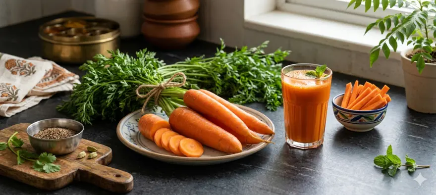
9 Black Carrot Benefits And Simple Recipes
Seeing black carrots in the market can make you curious. Their deep purple-black colour stands out, but their appeal is not just visual. Black carrot is a nutrient-rich root vegetable valued for its anthocyanins, fibre, and beta-carotene. These compounds are linked with antioxidant activity and may support heart health, digestion, blood sugar balance, and overall wellness when included as part of a balanced diet. What Is Black Carrot? Black carrot is a coloured variety of carrot with a dark purple to almost black outer appearance. Its colour comes largely from anthocyanins, which are plant pigments with antioxidant properties. Black carrots have been grown and used in regions such as India, the Middle East, and parts of Asia, and they are especially popular in seasonal winter foods and drinks. Compared with regular orange carrot, black carrot is known more for its anthocyanin content, while orange carrot is usually richer in carotenoids. That is why black carrot is often discussed for its antioxidant value, while still offering fibre and useful micronutrients. You can eat it raw, juice it, cook it, or use it in traditional preparations such as kanji. Nutritional Value Of Black Carrot Nutritional Value of Black Carrots Black carrots are low in calories but rich in essential nutrients. Here is what a 100g serving typically provides: Nutrient Amount Calories 35 kcal Carbohydrates 8.2g Dietary Fibre 2.9g Protein 1g Vitamin A 334% of RDI Vitamin C 6% of RDI Potassium 7% of RDI Source: USDA Beyond the numbers, black carrots are particularly rich in anthocyanins, beta-carotene, lutein, Vitamin C, and Vitamin K. This combination makes them a genuinely powerful addition to a balanced diet. 9 Health Benefits Of Black Carrot 1. Rich In Antioxidants One of the most talked-about black carrot benefits is its antioxidant content. The anthocyanins in black carrot help protect cells from oxidative stress, which is one reason deeply coloured vegetables are often included in a healthy diet. 2. Supports Heart Health Anthocyanin-rich foods have been studied for their possible role in supporting cardiovascular health. Reviews suggest anthocyanins may help improve lipid markers and lower inflammation-related markers, which makes black carrot a smart addition to a heart-aware eating pattern. It should support, not replace, your overall healthy lifestyle. 3. Helps Support Digestion Black carrot contains fibre, and fibre is important for bowel regularity and gut health. A fibre-rich diet supports stool formation, gut transit, and the gut microbiome. This makes black carrot a simple way to add more plant food to your meals. 4. May Help With Blood Sugar Control Black carrot is often discussed in the context of metabolic health because anthocyanins have been studied for their role in glucose regulation and insulin sensitivity. This does not mean black carrot is a treatment for diabetes, but it can be a useful part of a balanced, fibre-rich diet. 5. Promotes Eye Health Like other carrots, black carrot contains beta-carotene, which your body can convert into vitamin A. Vitamin A is important for normal vision, especially in dim light. That makes black carrot a useful food to include in a diet that supports eye health. 6. Supports Skin Health The mix of antioxidants and provitamin A carotenoids in black carrot may help support healthy skin as part of an overall nutritious diet. Foods rich in vitamin A precursors are important for skin and epithelial tissue health, so black carrot can be a helpful seasonal vegetable to include regularly. 7. May Help Strengthen Immunity Vitamin A and antioxidant-rich foods play a role in normal immune function. Since black carrot provides carotenoid compounds and other plant nutrients, it can contribute to a diet that supports your body’s natural defences. 8. May Support Healthy Weight Management Black carrot is naturally low in calories and gives you fibre, which can help you feel fuller and make meals more satisfying. It is not a magic food for weight loss, but it fits well into a balanced diet focused on whole vegetables, fruit, legumes, and grains. 9. May Help Reduce Inflammation Anthocyanins are widely studied for their anti-inflammatory potential. Since black carrot is one of the notable vegetable sources of anthocyanins, it may help you increase your intake of these protective plant compounds through everyday meals. Health Benefits Of Black Carrot For Different Age Groups For Children And Teenagers Children and teenagers need a varied diet that supports growth, immunity, and eye health. Black carrot can add colour, fibre, and carotenoid-rich nutrition to salads, soups, or fresh juice in age-appropriate portions. For Adults As an adult, you may look for foods that are simple, filling, and nutrient-dense. Black carrot fits well into that routine. Its fibre supports digestion, and its anthocyanins make it a useful addition to a diet aimed at long-term heart and metabolic health. For Older Adults For older adults, foods that support eye health, digestion, and overall antioxidant intake can be especially useful. Soft cooked black carrot, soup, or lightly fermented preparations can be practical ways to include it in meals. Simple Black Carrot Recipes To Try 1. Black Carrot Juice This is one of the easiest ways to enjoy black carrot. Ingredients 3 to 4 black carrots 1 small piece of ginger 1 teaspoon lemon juice Water as needed Method Wash and peel the black carrots. Chop them into small pieces. Blend or juice with ginger and a little water. Strain if needed. Add lemon juice and serve fresh. If you already read about carrot juice benefits, black carrot juice can feel like a colourful seasonal variation. It also works well for readers interested in beetroot and carrot juice benefits, especially when you want a more earthy and antioxidant-rich option. 2. Black Carrot Kanji Kanji is a traditional fermented drink often made with black carrots, water, and mustard. Research on black carrot kanji has shown probiotic potential and high antioxidant activity, which explains its continued popularity in seasonal home kitchens. Ingredients 4 black carrots, cut into sticks 1 litre water 2 teaspoons crushed mustard seeds Salt to taste A pinch of red chilli powder, optional Method Add all ingredients to a clean glass jar. Cover loosely and keep in sunlight for 3 to 4 days. Stir once daily with a clean spoon. Once it turns tangy, refrigerate and serve chilled. 3. Black Carrot Salad Ingredients 2 black carrots, grated or julienned 1 cucumber, sliced 1 tablespoon roasted seeds Lemon juice Salt and pepper Method Combine the vegetables in a bowl. Add seeds, lemon juice, salt, and pepper. Toss well and serve immediately. This recipe keeps the texture crisp and fresh, and it is a simple way to enjoy black carrot without much preparation. 4. Black Carrot Soup Ingredients 3 black carrots, chopped 1 small onion, chopped 2 garlic cloves 2 cups vegetable stock Salt and pepper 1 teaspoon olive oil Method Heat oil and sauté onion and garlic. Add chopped black carrots and stock. Simmer until soft. Blend until smooth. Season and serve warm. 5. Black Carrot Halwa Ingredients 2 cups grated black carrot 1 cup milk 1 to 2 teaspoons ghee 1 to 2 teaspoons jaggery or a small amount of sugar Chopped nuts Method Heat ghee in a pan and sauté the grated black carrot. Add milk and cook on low heat. Stir until the mixture thickens. Add jaggery or sugar and mix well. Garnish with nuts and serve warm. Black carrot halwa is best enjoyed as an occasional dessert. It lets you use this seasonal vegetable in a familiar and comforting way. Are There Any Side Effects Or Precautions? Black carrot is generally safe when eaten as a regular food. Still, moderation matters. If you have diabetes, kidney stones, food allergies, or a medically restricted diet, it is sensible to check with your doctor or dietitian before making major changes to your intake. With fermented drinks like kanji, clean preparation and proper storage are important. How To Add Black Carrot To Your Daily Diet You do not need a complex plan to include black carrot in your meals. You can: Add it to salads Blend it into juice or smoothies Make kanji during the season Add it to soup Roast it with other vegetables Use it in halwa once in a while If you enjoy reading about beetroot and carrot juice benefits, you can also combine black carrot with beetroot, ginger, or amla for a seasonal drink. The key is to keep the recipe simple and balanced. FAQs About Black Carrot What Are The Nutritional Benefits Of Black Carrots? Black carrots provide anthocyanins, fibre, beta-carotene, and useful vitamins and minerals. Their dark colour is a sign of their anthocyanin content, while their carrot base still gives you familiar carotenoid-related benefits. Can Black Carrots Help With Weight Loss? They can support a weight-conscious diet because they are low in calories and contain fibre. They are not a quick fix, but they can help make meals more filling. Are There Any Side Effects Of Consuming Black Carrots? Most people can eat black carrots safely in normal food amounts. You should be a little more careful if you have a history of kidney stones, digestive sensitivity, or a condition that needs a special diet. How Do Black Carrots Compare To Regular Carrots? Regular orange carrots are best known for beta-carotene, while black carrots stand out for anthocyanins and antioxidant activity. Both can be part of a healthy diet, but black carrots bring a different phytochemical profile. What Is The Best Way To Incorporate Black Carrots Into Your Diet? You can start with juice, salad, soup, or kanji. Choose a method that fits your taste and routine so you can enjoy it regularly. Are Black Carrots Available Year-Round? They are usually easier to find during the cooler months and winter season in many parts of India. Availability can vary by city and local market. How Do Black Carrots Benefit Heart Health? Their anthocyanins are the main reason they are linked with heart health. Research on anthocyanin-rich foods suggests benefits for lipid markers and inflammation-related markers. Can You Drink Black Carrot Juice Every Day? You can include it regularly in sensible portions as part of a varied diet. Keep the juice fresh, avoid too much added sugar, and balance it with whole fruits and vegetables. The Bottom Line Black carrot is more than a colourful winter vegetable. It gives you fibre, beta-carotene, and anthocyanins that may support heart health, digestion, eye health, and overall wellness. The easiest approach is to enjoy it in simple forms such as juice, salad, soup, or kanji, and keep your overall diet balanced and varied. For a more proactive approach to your health, you can also explore preventive health packages, expert-led diagnostics, and home sample collection from Metropolis Healthcare. Metropolis offers 4,000+ clinical laboratory tests and profiles, along with NABL and CAP accredited laboratory services, making it easier for you to stay informed about your health with confidence. References Thakur P, Panwar A, Suhag R, Dhiman A, Kumar S. Insights into the current status of bioactive value, postharvest processing opportunities and value addition of black carrot. Food Sci Biotechnol. 2024;33(4):721-747. PMID: 38371691. Ahmad T, Cawood M, Iqbal Q, et al. Phytochemicals in Daucus carota and Their Health Benefits: A Review Article. Foods. 2019;8(9):424. PMID: 31546950. Bhandari SR, Cho MC, Lee JG. Influence of Root Color and Tissue on Phytochemical Contents and Antioxidant Activities in Carrot Genotypes. Foods. 2023;12(1):120. PMID: 36613336. Gill SK, Rossi M, Bajka B, Whelan K. Dietary Fibre in Gastrointestinal Health and Disease. Nat Rev Gastroenterol Hepatol. 2021;18(2):101-116. PMID: 33208922. Xu L, Tian Z, Chen H, Zhao Y, Yang Y. Anthocyanins, Anthocyanin-Rich Berries, and Cardiovascular Risks: Systematic Review and Meta-Analysis of 44 Randomized Controlled Trials and 15 Prospective Cohort Studies. Front Nutr. 2021;8:747884. PMID: 34977111. Kozłowska A, Szostak-Węgierek D. Anthocyanins and Type 2 Diabetes: An Update of Human Study and Clinical Trial. Nutrients. 2024;16(11):1674. PMID: 38892607. Sharma C, Sahota PP, Kaur S. Physicochemical and Microbiological Evaluation of Antioxidant-Rich Traditional Black Carrot Beverage: Kanji. Bull Natl Res Cent. 2021;45:143. PMID: 34393474.

7 Dragon Fruit Side Effects and Who Should Avoid It
Dragon fruit is colourful, refreshing, and packed with nutrients. You may already know it for its taste and the many dragon fruit benefits linked to fibre, antioxidants, and vitamin C. For most people, it is safe to eat in moderation. Still, no food suits everyone in the same way. If you eat too much dragon fruit at once, or if you have allergies, diabetes, low blood pressure, or a sensitive stomach, you may notice a few unwanted effects. The good news is that most side effects are mild and manageable. Knowing what to watch for can help you enjoy dragon fruit more safely. Can Dragon Fruit Have Side Effects? Yes, dragon fruit can have side effects in some people. This does not mean it is harmful. It simply means your body may react differently depending on how much you eat, your digestion, your medicines, and your overall health. In most healthy adults, small to moderate portions are well tolerated. Side effects are more likely when you overeat it, try it for the first time, or already have a condition that affects your digestion, blood sugar, or blood pressure. 7 Dragon Fruit Side Effects 1. Digestive Discomfort Dragon fruit contains fibre and water, which usually support digestion. But if you eat too much in one sitting, you may feel bloated, gassy, or uncomfortable. You may also notice loose stools or mild diarrhoea, especially if your usual diet is low in fibre. This is more likely when you suddenly increase your intake instead of building up slowly. If you have IBS or a sensitive stomach, start with a small portion and see how your body responds. 2. Allergic Reactions Although uncommon, dragon fruit can trigger an allergic reaction in some people. Mild symptoms may include itching in your mouth, skin rash, hives, or swelling of the lips. In rare cases, the reaction can be more serious and may cause throat swelling, breathing difficulty, or severe discomfort soon after eating the fruit. If you have a history of fruit allergies, pollen-related allergies, or unexplained food reactions, be cautious the first time you try dragon fruit. If you ever develop swelling, wheezing, or trouble breathing, seek urgent medical help. 3. Blood Sugar May Drop Too Low in Some People Dragon fruit is often seen as a fruit that fits well into a balanced diet. Some research suggests it may support glucose control, especially in people with prediabetes. That said, if you take diabetes medicine, especially insulin or tablets that lower blood sugar, portion size still matters. In some people, adding dragon fruit regularly without adjusting the rest of the diet may contribute to lower readings than expected. This does not mean you must avoid it completely. It means you should eat it mindfully, watch your portions, and monitor your sugar levels if your doctor has advised you to do so. 4. It May Not Suit You if You Already Have Low Blood Pressure Dragon fruit is often linked with heart-friendly nutrition, but if you already have naturally low blood pressure, it is sensible not to overdo any one fruit as a daily habit. If your blood pressure tends to run low, or if you take medicine for blood pressure, pay attention to symptoms such as light-headedness, weakness, or unusual fatigue after eating large portions. The effect is not usually dramatic, but moderation is still the safest approach. 5. It May Need Extra Caution if You Take Certain Medicines Dragon fruit is a food, not a medicine. Even so, if you take regular prescription drugs for diabetes or blood pressure, it is wise to be careful with large or very frequent portions. This is because dietary changes can sometimes affect how well your condition stays controlled. The issue is usually not the fruit alone, but the combination of your medicines, your total diet, and your body’s response. If you are on long-term treatment and want to add dragon fruit often, ask your doctor or dietitian what portion works best for you. 6. It Can Upset a Sensitive Stomach Even when you do not have a diagnosed digestive condition, dragon fruit may not suit you if your stomach reacts easily to high-fibre foods, fruit sugars, or chilled fruit. You may notice cramps, fullness, frequent motions, or a rumbling stomach after eating it. This is more common if you eat it quickly, combine it with many other fruits, or have it in a large smoothie. A smaller portion is usually easier to tolerate. 7. Red Dragon Fruit May Temporarily Change Urine or Stool Colour If you eat the red-fleshed variety, you may notice a pink or reddish tinge in your stool or urine afterwards. This can look alarming if you are not expecting it. In many cases, this colour change is harmless and related to the natural pigments in the fruit. Even so, if the colour change is persistent, painful, or happens without eating red dragon fruit, do not ignore it. You should speak with a doctor to rule out another cause. Potential Side Effects of Dragon Fruit for Women There are no known side effects that are exclusive to women. However, some situations may need extra care. If you have gestational diabetes, watch your portion size and fit dragon fruit into your overall meal plan. If you often feel dizzy or have low blood pressure, avoid eating large amounts at once. If you are pregnant, dragon fruit is usually fine in moderation when it is fresh, properly washed, and part of a balanced diet. If you have a history of food allergies or a sensitive stomach, start with a small amount. Potential Side Effects of Dragon Fruit for Men Dragon fruit affects men and women in broadly similar ways, but the same practical cautions apply. If you have diabetes, do not assume unlimited portions are harmless just because it is fruit. If you take blood pressure medicine, notice how you feel after eating large portions regularly. If you have frequent bloating, loose stools, or IBS-like symptoms, start small. If you have had unusual reactions to fruits before, be careful the first time you try dragon fruit. Who Should Avoid Dragon Fruit or Eat It With Caution? You may need to avoid dragon fruit or eat it cautiously if you are: Allergic to dragon fruit or have had a previous reaction to it Prone to fruit allergies or pollen-related food reactions Taking diabetes medicines and monitoring for low blood sugar Living with low blood pressure or taking blood pressure medicines Very sensitive to high-fibre foods Dealing with IBS, chronic bloating, or frequent diarrhoea Pregnant and unsure how it fits into your doctor-advised diet plan On a restricted medical diet and making frequent dietary changes without guidance How Much Dragon Fruit Is Safe to Eat Per Day? For most healthy adults, about 1 cup of cut dragon fruit, or one small to medium fruit at a time, is a sensible portion. You do not need to eat it every day to benefit from it. The safest approach is moderation and variety. If you are trying it for the first time, start with a few pieces and wait to see how your body responds. If you have diabetes, low blood pressure, allergies, or digestive issues, your ideal portion may be smaller. Tips to Eat Dragon Fruit Safely Wash the fruit well before cutting it. Start with a small portion if you are trying it for the first time. Do not eat a very large amount in one sitting. Pair it with a balanced diet instead of relying on one fruit for health benefits. Be extra careful if you take medicines for diabetes or blood pressure. Stop eating it and seek medical advice if you notice swelling, itching, wheezing, or severe stomach upset. Conclusion Dragon fruit is nutritious and usually safe when you eat it in moderation. For most people, it is a light and enjoyable fruit that can fit easily into a healthy diet. But if you have allergies, diabetes, low blood pressure, or a sensitive stomach, it is worth being a little more careful. A small portion is often the best place to start. If you want trusted guidance on nutrition, symptoms, preventive care, and diagnostic testing, you can explore more health articles from Metropolis Healthcare. With 4,000+ tests, expert pathologists, and convenient home sample collection across a wide network, Metropolis Healthcare helps make testing simple, accurate, and accessible for you and your family. FAQs Can Dragon Fruit Cause Allergies? Yes, but it is uncommon. Symptoms can range from itching and hives to swelling and, rarely, serious allergic reactions. If you have a history of food allergies, try a small amount first and stop immediately if you react. Is Dragon Fruit Good for Diabetes? Dragon fruit can be included in a balanced diet for many people with diabetes, but portion size still matters. It should not be treated as a free food. If you take diabetes medicines, monitor your levels as advised by your doctor. How Much Dragon Fruit Is Safe to Eat Per Day? For most healthy adults, about 1 cup of cut fruit or one small to medium dragon fruit is a reasonable serving. If you are new to it or have digestive sensitivity, start with less. Can Dragon Fruit Lower Blood Pressure? It may support heart-friendly nutrition, but strong evidence for a major blood pressure lowering effect in healthy adults is limited. If you already have low blood pressure or take medicines for it, moderation is wise. Can Dragon Fruit Upset Your Stomach? Yes. If you eat too much, it may cause bloating, gas, cramps, or loose stools, especially if your usual diet is low in fibre or your stomach is sensitive. Is Dragon Fruit Safe During Pregnancy? In most cases, yes. It is usually safe during pregnancy when eaten fresh, properly washed, and in moderate portions. If you have gestational diabetes, food allergies, or digestive discomfort, ask your doctor about the right amount for you. References Arivalagan M, Karunakaran G, Roy TK, Dinsha M, Sindhu BC, Shilpashree VM, Satisha GC, Shivashankara KS. Biochemical and nutritional characterization of dragon fruit (Hylocereus species). Food Chem. 2021;353:129426. PMID: 33774520. Poolsup N, Suksomboon N, Paw NJ. Effect of dragon fruit on glycemic control in prediabetes and type 2 diabetes: A systematic review and meta-analysis. PLoS One. 2017;12(9):e0184577. PMID: 28886195. Cheok A, Xu Y, Zhang Z, Caton PW, Rodriguez-Mateos A. Betalain-rich dragon fruit (pitaya) consumption improves vascular function in men and women: a double-blind, randomized controlled crossover trial. Am J Clin Nutr. 2022;115(5):1418-1431. PMID: 35265960. Martin H, Stepaniuk P. Pitaya allergy: a case report of anaphylaxis in a patient without cross-reactive allergens. Allergy Asthma Clin Immunol. 2025;21(1):21. PMID: 40325480. Lim KG, Ling CV. The effect of Hylocereus polyrhizus (red dragon fruit) on whole gut transit time of young Malaysian adults. Malays J Nutr. 2021;27(1):153-158. doi:10.31246/mjn-2020-0100. Leong EKF, Yew A, Lin NS. Are Red Stools after Red Dragon Fruit (Selenicereus costaricensis) Ingestion a Red Herring? Case Report and Clinical Pearls for Recognition. Clin Case Rep J. 2022;3(8):1-5. MedlinePlus. Dietary Fiber. U.S. National Library of Medicine. Updated 2023. MedlinePlus Medical Encyclopedia. Fiber. U.S. National Library of Medicine. Updated 2024. Chen SY, Xu CY, Mazhar MS, Naiker M. Nutritional Value and Therapeutic Benefits of Dragon Fruit: A Comprehensive Review with Implications for Establishing Australian Industry Standards. Molecules. 2024;29(23):5676. PMID: 39683835.
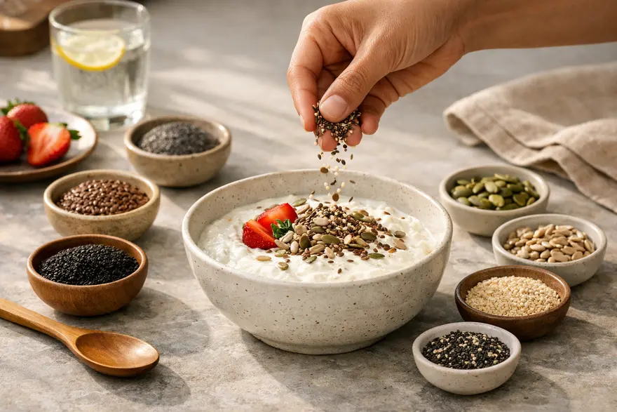
15 Healthy Seeds To Eat Daily With Benefits And Uses
Healthy seeds are small, but they can add a lot to your plate. They are rich in fibre, healthy fats, plant protein, and important vitamins and minerals. When you eat them in sensible portions, they can support heart health, digestion, satiety, and overall wellness. The key is balance. You do not need to eat all 15 seeds every day. It is better to include a few regularly, rotate them through your meals, and use them in ways that suit your body and routine. This guide will help you understand which seeds are worth adding to your diet, what they offer, and how to use them easily in everyday meals. Introduction To Healthy Seeds Healthy seeds are nutrient-dense plant foods that can fit into many eating patterns. Some, such as chia, flax, and sunflower, are commonly used in daily diets. Others, such as garden cress, basil seeds, and melon seeds, are less common but still nutritious. Most seeds offer a useful mix of: Fibre Unsaturated fats Plant protein Antioxidants Minerals such as magnesium, zinc, iron, and calcium No single seed does everything. What matters more is variety, moderation, and consistency. Why You Should Eat Seeds Regularly Eating seeds regularly can be a simple way to improve the nutritional quality of your meals. Fibre-rich seeds can help support digestion and help you feel full for longer. Seeds with healthy fats may support heart health when used as part of a balanced diet. Some seeds also add plant protein, which can help with satiety and meal balance. Others bring in minerals such as magnesium, zinc, and iron. Seeds are also versatile. You can add them to curd, smoothies, salads, soups, oats, rotis, laddoos, or trail mixes without changing your meals too much. That said, seeds are not a magic fix. They work best as part of an overall healthy eating pattern. 15 Healthy Seeds To Include In Your Diet 1. Chia Seeds Chia seeds are one of the most popular healthy seeds for eating. They are rich in fibre and omega-3 fats. When soaked, they form a gel-like texture, which can help with fullness and make them easy to add to puddings, smoothies, and overnight oats. Best use: Soak and add to smoothies, porridge, or curd. 2. Flaxseeds Flaxseeds are known for their fibre, lignans, and plant omega-3 fats. They are especially useful if you want to improve the nutritional quality of breakfast bowls, rotis, or smoothies. Ground flaxseed is usually better than whole flaxseed because it is easier for your body to digest and absorb. Best use: Use freshly ground flaxseed in atta, smoothies, or porridge. 3. Pumpkin Seeds Pumpkin Seeds are rich in magnesium, zinc, healthy fats, and plant protein. They make a satisfying snack and can add crunch to salads and soups. They are also easy to roast at home, but portion size still matters because they are energy-dense. Best use: Eat roasted as a snack or sprinkle over salads and vegetables. 4. Sunflower Seeds Sunflower seeds provide healthy fats, vitamin E, and useful minerals. They are easy to eat and work well as a topping for sandwiches, curd, salads, and homemade seed mixes. Choose unsalted or lightly salted versions if you want to keep sodium intake under control. Best use: Add to salads, curd bowls, or homemade granola. 5. Sesame Seeds Sesame Seeds are tiny but nutrient-rich. They provide healthy fats, fibre, and minerals, and they are easy to use in Indian cooking. They work well in chutneys, laddoos, stir-fries, and salad toppings. They are also one of the easiest seeds to include if you want a simple flavour and texture boost. Best use: Sprinkle over sabzi, mix into chutney, or use in tahini-style spreads. 6. Hemp Seeds Hemp seeds, often sold as hemp hearts, are a good plant-based source of protein and healthy fats. They also contain essential amino acids, which makes them especially useful if you are trying to improve protein intake in vegetarian meals. They have a mild, nutty taste and blend easily into many dishes. Best use: Add to smoothies, salads, curd, or soups. 7. Sabja Seed (Basil Seeds) Sabja Seed, also called basil seed, is commonly soaked before use. Like chia, it absorbs water and swells, which gives drinks and desserts a thicker texture. It is often used in summer beverages and light desserts. It is best to soak it properly before eating. Best use: Soak and add to drinks, falooda, or fruit bowls. 8. Garden Cress Seeds Garden cress seeds are small but nutrient-dense. They contain protein, essential fatty acids, and useful minerals. In Indian households, they are often added to laddoos or simple seed mixes. They have a slightly peppery taste, so a little goes a long way. Best use: Add to laddoos, porridges, or seed mixes in small amounts. 9. Watermelon Seeds Watermelon seeds are often thrown away, but the edible kernels can be a useful source of protein, healthy fats, iron, and zinc. Roasted watermelon seeds make a convenient snack and can also be blended into seed mixes. Best use: Roast and eat as a snack or add to trail mix. 10. Muskmelon Seeds Muskmelon seeds are commonly used in Indian gravies, sweets, and seed mixes. They contain protein, fats, and fibre, and they can add creaminess when blended into recipes. Best use: Use in gravies, kormas, sweets, or snack mixes. 11. Poppy Seeds Poppy seeds are usually used in small amounts, but they can still add flavour, healthy fats, and texture. They work well in baking, curries, and traditional sweets. Because they are usually used in small quantities, think of them as a supportive ingredient rather than a major nutrition source. Best use: Add to curries, baked dishes, and traditional desserts. 12. Nigella Seeds Nigella seeds, often called kalonji, are commonly used more for flavour than for volume. They contain healthy fats and plant compounds and can be a useful part of a varied diet. You usually do not need much, as their taste is strong and distinctive. Best use: Use in pickles, breads, or savoury dishes. 13. Lotus Seeds (Makhana) Lotus seeds, or makhana, are technically not used in the same way as tiny sprinkle seeds, but they still belong in this list because they are seed-based, widely eaten, and nutritious. They offer protein, minerals, and a light crunch, especially when roasted. They can be a better snack choice than many fried alternatives. Best use: Dry roast for snacking or add to trail mixes and kheer. 14. Quinoa Quinoa is technically a pseudocereal, but it is seed-like in its nutrition and use. It provides fibre, protein, and a useful range of micronutrients. It is easy to use in place of rice or in salads. Best use: Use in grain bowls, khichdi-style meals, or salads. 15. Amaranth Amaranth is another pseudocereal that works like a seed in many recipes. It offers protein, fibre, and minerals and can be used in porridges, laddoos, and snacks. Popped amaranth is especially easy to add to homemade mixes. Best use: Use in porridge, laddoos, or popped snack mixes. Best Way To Incorporate Seeds Into Your Diet The best way to eat seeds is the way you can follow regularly. You can start by adding one or two types to meals you already eat. Sprinkle seeds over curd, smoothie bowls, salads, soups, oats, or poha. Mix ground flaxseed into atta. Add chia or basil seeds to drinks. Use pumpkin or sunflower seeds in snack mixes. Blend melon seeds into gravies for texture. You do not need complex recipes. A simple daily habit is usually enough. Whole, Ground, Soaked, Or Roasted: What Works Best? Different seeds work better in different forms. Ground: Flaxseeds are often best eaten ground. Soaked: Chia and basil seeds are commonly soaked before use. Roasted: Pumpkin, sunflower, watermelon, and makhana can be roasted for snacking. Whole or sprinkled: Sesame, poppy, and nigella seeds are often used whole in cooking. The right method can improve texture, digestibility, and ease of use. How Many Seeds Should You Eat Daily? You do not need large amounts. A practical starting point for most adults is: 1 to 2 tablespoons of one or two seed varieties a day Smaller amounts for stronger-flavoured seeds such as nigella or poppy A small handful for snack-style seeds such as roasted pumpkin or sunflower seeds Rotation across the week rather than trying to eat everything daily Start small if your diet is currently low in fibre. Increase gradually and drink enough water. Potential Side Effects Of Eating Seeds Seeds are healthy, but more is not always better. Possible issues include: Bloating or gas if you increase fibre too quickly Digestive discomfort if you overeat them Extra calorie intake if portions are too large Allergic reactions in people with seed allergies Poor tolerance of certain seeds if you have a sensitive stomach Difficulty swallowing if dry seeds are eaten carelessly without enough fluid This does not mean you should avoid seeds. It simply means portion size and preparation matter. Who Should Be Careful With Certain Seeds? You may need extra care if: You have a known seed allergy You have digestive sensitivity and do not tolerate high-fibre foods well You are choosing seeds for a small child and need to avoid choking risk Your doctor has advised a special diet because of a health condition If you have an ongoing medical condition or symptoms such as severe bloating, abdominal pain, or food-related reactions, it is best to ask your doctor or dietitian before making major dietary changes. How To Choose And Store Seeds Choose plain, good-quality seeds with minimal added salt, sugar, or flavouring. A few simple rules help: Buy from trusted brands Check expiry dates Prefer unsalted versions where possible Store seeds in an airtight container Keep ground flaxseed in the refrigerator Keep seeds away from heat, light, and moisture Buying small amounts at a time can help you keep them fresh. Conclusion Seeds are one of the easiest ways to make your meals more nutritious. They can add fibre, healthy fats, plant protein, crunch, and flavour without much effort. You do not need to eat all 15 every day. What helps more is using a few regularly, rotating them through the week, and choosing forms that are easy for your body to digest and enjoy. If you are working on your overall health, weight management, cholesterol, or blood sugar goals, it is also important to look beyond single foods and focus on your full lifestyle. And if your doctor advises health tests as part of preventive care, Metropolis Healthcare offers a wide range of diagnostic services, convenient home sample collection, and easy booking options. Frequently Asked Questions What Are The Healthiest Seeds To Eat Daily? Some of the most popular and nutrient-dense choices are chia, flax, pumpkin, sunflower, hemp, sesame, and basil seeds. You do not need all of them every day. Pick one or two and rotate them through the week. Can Seeds Help With Weight Loss? Seeds can support weight loss as part of a balanced diet because many are rich in fibre, protein, and healthy fats, which can help with fullness. But they still contain calories, so portion control matters. How Can You Incorporate Seeds Into Daily Meals? You can sprinkle them over curd, salads, oats, and soups, blend them into smoothies, mix ground seeds into atta, or eat roasted seeds as a snack. Start with simple additions you can repeat regularly. Are There Any Health Risks With Eating Seeds? Seeds are safe for most people when eaten in moderate amounts. Problems usually happen when portions are too large, fibre is increased too quickly, or a person has an allergy or digestive sensitivity. Is It Better To Eat Seeds Raw, Roasted, Or Soaked? That depends on the seed. Flaxseeds are usually better ground. Chia and basil seeds are commonly soaked. Pumpkin and sunflower seeds can be eaten raw or roasted. Sesame seeds are often sprinkled whole into dishes. How Much Seed Mix Can You Eat In A Day? For most adults, a small serving is enough. A practical amount is around 1 to 2 tablespoons of mixed seeds a day, or a small handful if you are eating larger snack-style seeds such as pumpkin or sunflower seeds. References Mayo Clinic. Flaxseed: Is Ground Better Than Whole? Mayo Clinic. Cleveland Clinic. The 6 Best Seeds to Eat. Cleveland Clinic. Cleveland Clinic. The Many Health Benefits of Sunflower Seeds. Cleveland Clinic. Hrnčič MK, Ivanovski M, Cör D, Knez Ž. Chia Seeds (Salvia hispanica L.): An Overview—Phytochemical Profile, Isolation Methods, and Application. Molecules. 2020;25(1):11. PMID: 31861466. Bravo HC, Ccanto NRV, Zura-Bravo L, et al. Basil Seeds as a Novel Food, Source of Nutrients and Functional Ingredients with Beneficial Properties: A Review. Molecules. 2021;26(15):4597. PMID: 34202798. Melo D, Machado N, Matos C, et al. Nutritional and Chemical Characterization of Poppy Seeds, Cold-Pressed Oil, and Cake: Poppy Cake as a High-Fibre and High-Protein Ingredient for Novel Food Production. Foods. 2022;11(19). PMID: 36230103. Siddique R, Sahar A, Riaz A, et al. Lotus Seeds (Nelumbinis semen) as an Emerging Therapeutic Seed: A Comprehensive Review. Food Science & Nutrition. 2021. Tufail T, Shah MA, Arfat Y, et al. Garden Cress Seeds: A Review on Nutritional Composition, Therapeutic Potential, and Industrial Utilization. Food Science & Nutrition. 2024. Wei P, Meng Q, Liu R, et al. Sesame (Sesamum indicum L.): A Comprehensive Review of Nutritional Value, Phytochemical Composition, Health Benefits, Development of Food, and Industrial Applications. Nutrients. 2022.
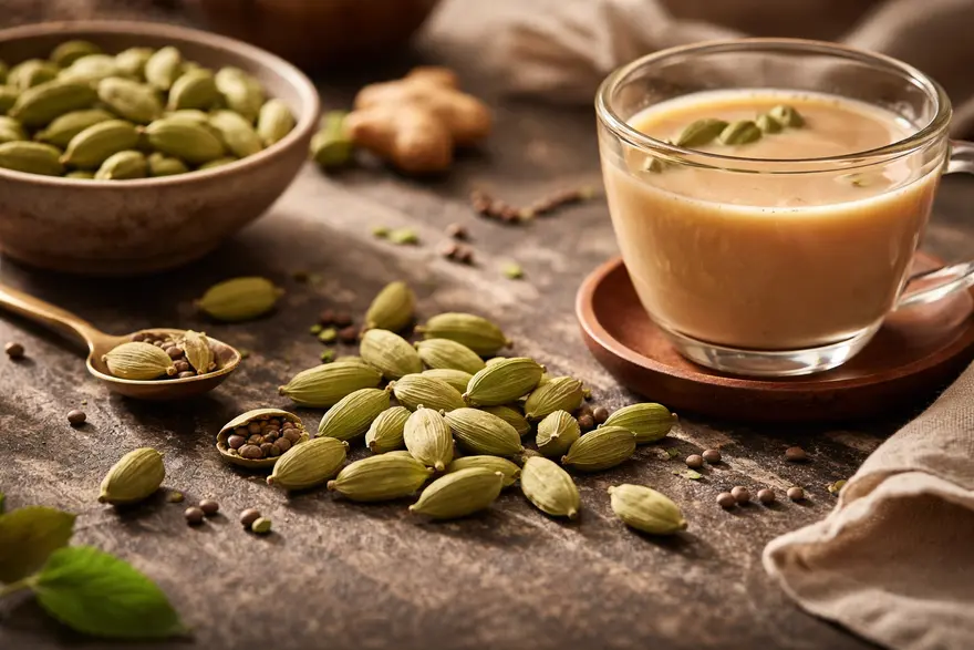
10 Elaichi (Cardamom) Benefits And Best Ways To Use It
Elaichi, also known as cardamom, is one of the most loved spices in Indian kitchens. You may add it to chai, kheer, curries, or desserts for its warm aroma and slightly sweet taste. Beyond flavour, elaichi has also been used for generations in everyday wellness routines. Modern research suggests that cardamom may support digestion, freshen breath, and help with some aspects of heart and metabolic health. At the same time, it is best to keep expectations realistic. Elaichi is a helpful spice, not a cure. The best results come when you use it as part of a balanced diet and healthy lifestyle. What Is Elaichi (Cardamom)? Elaichi is a spice made from the seeds inside the pods of plants from the ginger family. Green cardamom is the variety most people use in daily cooking, while black cardamom has a smokier flavour and is often used in savoury dishes. In this article, the focus is mainly on green elaichi. It is rich in aroma because of its natural essential oils and plant compounds. That is why even a small amount can add a lot of taste to your food and drinks. Nutritional Value Of Elaichi (Cardamom) Elaichi is usually eaten in small amounts, so you do not consume large quantities of its nutrients at one time. Even so, it contains fibre, minerals, and antioxidant plant compounds that contribute to its value as a spice. Nutritional Breakdown Of Cardamom (Per 100g) Nutrient Approximate Amount Per 100g Energy 311 kcal Carbohydrates 68.47 g Fibre 28 g Protein 10.76 g Fat 6.70 g Calcium 383 mg Iron 13.97 mg Magnesium 229 mg Potassium 1119 mg Vitamin C 21 mg Key Active Compounds In Cardamom 1,8-cineole Alpha-terpinyl acetate Linalool Other volatile oils and antioxidant compounds 10 Elaichi (Cardamom) Benefits 1. May Support Digestion Elaichi is widely used after meals because it may help ease bloating, gas, and a feeling of heaviness. Traditional use and early research suggest it may support digestive comfort and stimulate digestion. This is one reason many people like to chew a pod after food or add it to warm drinks. 2. May Freshen Breath And Support Oral Health Elaichi is often used as a natural mouth freshener. Its aromatic oils can help leave your mouth feeling cleaner and fresher after meals. Some laboratory and review-based evidence also suggests cardamom compounds may act against certain oral bacteria. This makes elaichi a simple and pleasant addition to your oral care routine, though it does not replace brushing and flossing. 3. May Help Support Healthy Blood Pressure Cardamom contains antioxidant compounds, and a small clinical study found that cardamom powder may help lower blood pressure in adults with newly diagnosed high blood pressure. This does not mean elaichi should replace your medicines. But adding it to your diet may support heart-friendly eating habits. 4. May Support Heart Health Because cardamom may help with blood pressure and oxidative stress, it may also support overall cardiovascular health. This benefit is best understood in the bigger picture. If you eat a balanced diet, stay active, sleep well, and manage stress, elaichi can be one small supportive part of that routine. 5. May Help Reduce Inflammation Cardamom is rich in antioxidant plant compounds. These compounds may help protect your cells from oxidative stress and may support the body’s response to inflammation. Human evidence is still limited, but some reviews and trials suggest that cardamom may improve certain inflammatory markers. 6. May Support Respiratory Comfort Elaichi is commonly added to warm tea and home drinks when you have throat discomfort or a heavy feeling in the chest. Traditional use suggests it may help you feel more comfortable during mild cough or congestion. Some research also points to possible airway-relaxing and antimicrobial effects, though stronger human studies are still needed. 7. May Help With Metabolic Health Some small studies suggest cardamom may help improve certain markers related to blood sugar control, triglycerides, and insulin resistance. That said, the evidence is not strong enough to treat it as a diabetes remedy. You should think of it as a helpful spice that may fit into a healthy eating pattern, not as a substitute for medical care. 8. May Support Fluid Balance Cardamom has been linked with a mild diuretic effect in some studies. This means it may help your body manage fluid balance more efficiently. This is one reason people sometimes associate it with feeling lighter or less bloated. It is better to view this as supportive, not as a detox cure. 9. May Provide Antioxidant Protection One of the clearest strengths of elaichi is its antioxidant content. Antioxidants help protect your cells from damage caused by free radicals. Over time, antioxidant-rich foods and spices may support overall wellbeing and help you build a more protective daily diet. 10. May Promote A Sense Of Everyday Wellness Elaichi does not need to be dramatic to be useful. Its aroma, taste, and warmth can make simple foods feel more satisfying. That matters because when healthy food tastes better, you are more likely to enjoy it regularly. This is one of the easiest and most practical cardamom benefits. Elaichi Benefits For Men There are no proven cardamom effects that are exclusive to men, but elaichi may still be useful in a few practical ways. It may support digestion after heavy or rich meals. It may help freshen breath naturally after tea, coffee, or meals. It may support heart-friendly and metabolic-friendly eating habits. It can add flavour without needing extra sugar in drinks or desserts. It may fit into a weight-conscious routine when used in place of sweet flavourings. If you are searching for elaichi benefits for male health, the safest answer is that elaichi may support general wellness, but there is no strong evidence that it has a unique male-specific effect. Elaichi Water Benefits Elaichi water is simply water infused with lightly crushed cardamom pods. Many people drink it for its mild aroma and soothing taste. The main elaichi water benefits are linked to hydration, digestive comfort, and a light fresh feeling after meals. It may also help you reduce sugary drinks if you enjoy flavoured water. It is important to keep this realistic. Elaichi water benefits can be a useful part of a healthy routine, but it is not a miracle drink. How To Make Elaichi Water Lightly crush 1 to 2 green cardamom pods. Add them to 1 to 2 cups of warm or boiled water. Let the water steep for 5 to 10 minutes. Strain and drink it warm or at room temperature. Best Ways To Use Elaichi (Cardamom) Elaichi is easy to include in your daily meals. You do not need supplements to enjoy it. In fact, culinary use is the simplest and safest approach for most people. How To Use Elaichi In Cooking Add crushed pods to tea or milk. Use powdered elaichi in kheer, phirni, custard, and baked desserts. Mix a pinch into porridge or oats. Add it to smoothies for aroma and warmth. Use it in pulao, biryani, and rich gravies. Sprinkle a little into fruit bowls or yoghurt. How To Make Elaichi Tea For Health Benefits Boil 1 to 2 cups of water. Lightly crush 2 to 3 cardamom pods. Add them to the water and simmer for a few minutes. Add tea leaves if you want regular chai. Strain and drink warm. Cardamom Oil For Aromatherapy Cardamom oil has a warm, spicy aroma. Some people use it in diffusers for a comforting atmosphere. It may help make your space feel calmer and fresher. Do not ingest essential oil unless a qualified clinician advises it. Always dilute it properly before applying it to the skin. Elaichi As A Spice For Weight Loss Elaichi can add flavour to food without adding sugar. It may help you enjoy simpler foods more. It can fit into a weight-conscious diet when you use it in tea, oats, or fruit. Direct evidence for major weight loss is limited, so do not rely on it alone. Elaichi Banana Benefits A simple banana and elaichi combination can work well in smoothies, porridge, or a quick mashed snack. Banana gives you carbohydrates, potassium, and fibre, while elaichi adds flavour and a lighter digestive feel. The easiest elaichi banana benefits come from making a wholesome snack taste better without needing too much added sugar. If you enjoy homemade smoothies, elaichi banana benefits can include better flavour, more satisfaction, and a more balanced snack option. Possible Side Effects And Precautions Elaichi is generally safe in normal food amounts. Still, there are a few points to keep in mind. Too much may cause stomach discomfort in some people. If you are pregnant, culinary use is usually preferred over concentrated supplements. If you have a medical condition, speak with your doctor before taking cardamom extracts or capsules. If you take regular medicines, especially for blood pressure or blood sugar, avoid self-prescribing supplements. Essential oils should not be consumed casually. How Much Elaichi Can You Have In A Day? There is no standard daily requirement for elaichi when used in food. In normal cooking amounts, it is usually well tolerated. For most people, 1 to 2 pods in tea or meals, or a small pinch of powder, is a practical amount. You do not need large quantities to enjoy the flavour or potential health value. If you want to try elaichi water benefits regularly, keep the preparation light and moderate rather than very concentrated. Conclusion Elaichi is much more than a fragrant kitchen spice. It may support digestion, freshen breath, contribute to antioxidant intake, and offer mild support for heart and metabolic health. The evidence is promising, but it is still best to think of cardamom as a supportive food ingredient rather than a treatment. The easiest way to use it is in daily cooking, tea, or light infused water. Simple habits such as adding elaichi to warm drinks, fruit bowls, or healthy snacks can make wholesome eating more enjoyable. If you want more reliable health guidance on nutrition, prevention, symptoms, and testing, you can explore more expert-led articles from Metropolis Healthcare. With advanced diagnostic technology, expert pathologists, 4,000+ tests, and convenient home sample collection, Metropolis Healthcare helps you make informed health decisions with greater confidence. FAQs What Are The Health Benefits Of Cardamom? Cardamom may support digestion, oral freshness, antioxidant intake, and some aspects of blood pressure and metabolic health. The strongest evidence is still limited, so it is best used as part of a healthy diet rather than as a remedy on its own. What Are The Health Benefits Of Elaichi For Men? Elaichi may support digestion, breath freshness, and heart-friendly eating habits in men, just as it may in women. There is no strong evidence for a unique men-only effect. Can Cardamom Help In Weight Loss? Cardamom may help indirectly by making healthier foods and drinks more flavourful without extra sugar. However, there is not enough evidence to say that cardamom alone causes meaningful weight loss. Is Cardamom Good For Your Heart? It may be. Some small studies and meta-analyses suggest cardamom may support blood pressure control and inflammation markers. Still, it should complement, not replace, medical care and healthy lifestyle habits. How Should You Use Cardamom For Skin Benefits? There is not enough strong human evidence to recommend cardamom as a skin treatment. It is safer to use elaichi as a food spice and follow a proper skincare plan for specific skin concerns. What Are Elaichi Water Benefits? Elaichi water benefits may include a mild digestive boost, better hydration support, and an easy way to replace sugary drinks with something lighter. Keep your expectations moderate and use it as part of an overall healthy routine. Can You Have Elaichi Every Day? Yes, most people can have elaichi daily in small food amounts. Moderate use in tea, meals, or desserts is generally considered safe. Is Elaichi Safe During Pregnancy? Elaichi used in normal cooking is generally preferred over concentrated supplements during pregnancy. If you are pregnant and want to use cardamom extracts, oils, or capsules, speak to your doctor first. References United States Department of Agriculture, Agricultural Research Service. FoodData Central: Spices, cardamom. Accessed April 2, 2026. Singletary K. Cardamom: Potential Health Benefits. Nutr Today. 2022;57(1):38-49. Verma SK, Jain V, Katewa SS. Blood pressure lowering, fibrinolysis enhancing and antioxidant activities of cardamom (Elettaria cardamomum). Indian J Biochem Biophys. 2009;46(6):503-506. PMID: 20361714. Izadi B, et al. The effect of green cardamom on blood pressure and inflammatory markers among patients with metabolic syndrome and related disorders: A systematic review and meta-analysis of randomized clinical trials. J Hum Hypertens. 2023. PMID: 36181264. Heydarian A, et al. Effect of cardamom consumption on inflammation and blood pressure in adults: A systematic review and meta-analysis of randomized clinical trials. Food Sci Nutr. 2024. PMID: 38268891. Aghasi M, Koohdani F, Qorbani M, et al. Beneficial effects of green cardamom on serum SIRT1, glycemic indices and triglyceride levels in patients with type 2 diabetes mellitus: a randomized double-blind placebo controlled clinical trial. J Sci Food Agric. 2019;99(8):3933-3940. PMID: 30701554. Yahyazadeh R, et al. The effect of Elettaria cardamomum (cardamom) on the metabolic syndrome: Narrative review. Iran J Basic Med Sci. 2021. PMCID: PMC8917848. Sharma R. Cardamom comfort. Dent Res J (Isfahan). 2012;9(4):523. PMCID: PMC3353705. Jesylne P, Soundarajan S, Murthykumar K, Meenakshi M. The Role of Cardamom Oil in Oral Health: A Short Review. Res J Pharm Technol. 2016;9(3):272-274. doi:10.5958/0974-360X.2016.00050.0. WebMD. Cardamom: Uses, Side Effects, and More. Accessed April 2, 2026.
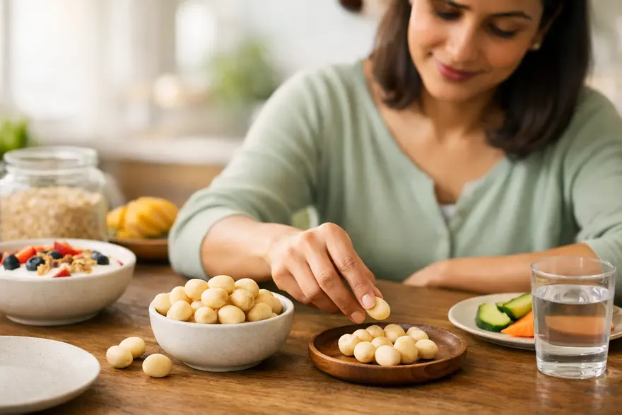
9 Macadamia Benefits And How Much To Eat
Macadamia nuts are creamy, rich, and easy to enjoy as a snack. But they offer more than taste. These nuts are packed with monounsaturated fats, fibre, and useful minerals, which is why they are often included in heart-conscious eating plans. They are also calorie-dense, so portion size matters. When you eat them in moderation, macadamia nuts can fit well into a balanced diet and may support heart health, satiety, and overall metabolic wellness. This guide explains what macadamia nuts are, their nutrition, their key benefits, how much to eat, possible side effects, and whether they fit into keto or diabetes-friendly meal plans. What Are Macadamia Nuts? Macadamia nuts are tree nuts originally native to Australia. They have a mild, buttery flavour and a soft crunch, which makes them popular in snacks, baked foods, nut butters, and premium trail mixes. Nutritionally, they are best known for being rich in monounsaturated fats. These are the same broad type of fats associated with heart-friendly eating patterns. Macadamias are also naturally low in sugar and relatively low in carbohydrate compared with many processed snack foods. Nutritional Value Of Macadamia Nuts The values below are approximate for 1 ounce, or about 28 grams, which is roughly 10 to 12 macadamia nuts. Nutrient Approximate Amount Per 28 g Calories 204 Total Fat 21.5 g Protein 2.2 g Carbohydrates 3.9 g Fibre 2.4 g Sugar 1.3 g Manganese 58% of daily value Thiamine 22% of daily value Copper 11% of daily value Magnesium 9% of daily value Macadamia nuts are high in fat, but most of that fat is monounsaturated. They also provide small amounts of vitamin E and other plant compounds with antioxidant activity. 9 Macadamia Nuts Benefits 1. Supports Heart Health Macadamia nuts are rich in monounsaturated fats, which are commonly linked with better cardiovascular health when they replace less healthy fats in your diet. This makes them a smart option if you are trying to improve snack quality. 2. May Help Improve Cholesterol Profile Some research suggests that macadamia nuts may help reduce LDL cholesterol and support a healthier blood lipid profile. This is one reason they are often discussed as part of heart-friendly eating patterns. 3. Can Support Fullness And Weight Management Macadamias are energy-dense, but they also contain fat, fibre, and a little protein, which can help you feel satisfied. That means a measured portion may help control hunger better than many refined snacks. 4. May Support Metabolic Health Macadamia nuts may fit well into eating patterns aimed at reducing cardiometabolic risk. Their fat profile and low sugar content make them a better option than many packaged snack foods. 5. Can Fit Into Blood Sugar-Friendly Eating Patterns These nuts are low in sugar and relatively low in carbohydrate. When eaten in a controlled portion, they are less likely to cause a sharp rise in blood sugar than high-carb snacks. 6. Provides Antioxidants Macadamia nuts contain antioxidant compounds, including forms of vitamin E and other plant compounds. These help protect cells from oxidative stress, although they should be seen as one part of an overall healthy diet, not a cure-all. 7. Supports Digestive Health Macadamias contain fibre, which can support digestive function and contribute to fullness. They do not provide as much fibre as some seeds or legumes, but they still add to your total intake. 8. May Support Skin Health The healthy fats and antioxidant compounds in macadamia nuts may support skin health as part of a varied diet. This does not mean they act like a treatment, but they can contribute to overall nutritional support for your skin. 9. Contributes Minerals That Support Bone Health Macadamia nuts provide minerals such as manganese, magnesium, and copper. These nutrients play roles in bone structure and general metabolic function. Macadamia Nuts Benefits For Women There are no proven benefits that apply only to women, but macadamia nuts can support health goals that many women may focus on. They can be a filling snack when you are trying to manage portion sizes Their healthy fats fit well into heart-conscious eating They provide minerals that support overall nutritional intake Their fat and antioxidant content may support skin health as part of a balanced diet If you are pregnant, breastfeeding, or following a medically advised diet, it is sensible to check portion sizes and nut tolerance with your doctor or dietitian. Macadamia Nuts Benefits For Men Macadamia nuts are not specifically a “men’s food”, but they can fit well into common health goals many men may have. They can be a better snack choice than fried or heavily processed foods Their monounsaturated fats support heart-conscious eating patterns They may help with satiety during weight-management efforts They provide minerals that support overall nutrition and daily energy metabolism The key point is the same for everyone: the benefits come from moderate, regular intake within a balanced diet. How Much Macadamia Nuts Should You Eat? A practical serving is about 1 ounce a day, which is roughly 10 to 12 nuts. That amount is usually enough to give you nutritional value without adding too many calories. Because macadamias are calorie-dense, it is easy to overeat them, especially straight from a large pack. A few simple rules can help: Measure your portion instead of eating from the packet Choose raw or dry-roasted versions where possible Prefer unsalted options if you are watching your sodium intake Use them to replace less healthy snacks, not add extra calories on top of your usual intake Macadamia Nuts Side Effects Macadamia nuts are safe for most people, but there are a few things to keep in mind. First, they are high in calories. Eating large amounts regularly can increase your daily calorie intake more than you realise. Second, they are a tree nut. If you have a tree nut allergy, you should not try them without medical guidance. Nut allergies can be serious. Third, flavoured, salted, chocolate-coated, or oil-roasted versions may come with extra sodium, sugar, or fat, which can reduce the overall nutritional value. Whole nuts may also be a choking risk for very young children. Macadamia Oil Benefits Macadamia oil is used in cooking and sometimes in skin and hair products. In food, its main value comes from its fat profile. Possible macadamia oil benefits include: It is rich in monounsaturated fats It can be used in small amounts in heart-conscious meal planning It works well in salad dressings and light cooking It may be a better choice than some heavily refined cooking fats, depending on the rest of your diet Macadamia oil is still energy-dense, so portion size matters here as well. Macadamia Nuts Vs Other Nuts: A Comparison Macadamia nuts are one healthy nut option among many. They are not automatically better than almonds, walnuts, pistachios, or peanuts. They are simply different. Compared with many other nuts, macadamias are: Higher in fat and calories Lower in protein Very low in carbohydrate Mild and buttery in flavour If your goal is higher protein, another nut may suit you better. If your goal is a lower-carb, satisfying snack, macadamias may be a good fit. Variety is usually the best approach. Are Macadamia Nuts Keto-Friendly? Yes, macadamia nuts are generally considered keto-friendly because they are low in carbohydrate and high in fat. That said, keto-friendly does not mean unlimited. They still contain a lot of calories, so portion control remains important. If you follow a keto diet, macadamias can fit in well as a measured snack or topping. Can Macadamia Nuts Help With Diabetes Management? Macadamia nuts are not a treatment for diabetes, but they may fit well into a diabetes-friendly eating pattern. They are low in sugar They are relatively low in carbohydrate They contain healthy fats They may be more satisfying than many refined snacks They can be used as part of portion-controlled meals and snacks If you have diabetes, the most important thing is your total eating pattern, medication plan, physical activity, and regular monitoring. Macadamia nuts can be one useful food within that bigger picture. Best Ways To Eat Macadamia Nuts You do not need complex recipes to use macadamias well. Try them in simple ways: Eat a small raw or dry-roasted portion as a snack Chop and add them to porridge or yoghurt Sprinkle them over salads for crunch Blend them into nut butter Add a few to homemade granola Use them in baking, but keep portions realistic Keeping them plain is often the best option if your goal is health rather than indulgence. Conclusion Macadamia nuts are rich, satisfying, and nutrient-dense. Their main strengths are their monounsaturated fat content, low sugar content, and ability to fit into heart-conscious and lower-carb eating patterns. The biggest mistake is not choosing macadamias. It is eating too many of them without noticing the calorie load. A small daily portion can work well for many people. Focus on plain, minimally processed options, and treat them as part of a balanced routine rather than a miracle food. If you are trying to improve cholesterol, blood sugar, weight, or overall preventive health, it also helps to look at your bigger health picture. Metropolis Healthcare offers a wide range of diagnostic services, convenient home sample collection, and easy booking options. FAQs What Are Macadamia Nuts Good For? Macadamia nuts are mainly valued for their healthy fats, satiety, and fit within heart-conscious meal plans. They may also support a healthier lipid profile and provide useful minerals. Are Macadamia Nuts Safe To Eat Every Day? For many people, yes. A small daily serving, around 1 ounce, can fit into a balanced diet. The main concern is portion size because they are calorie-dense. Can Macadamia Nuts Help With Weight Loss? They may support weight management when eaten in moderation because they can help you feel full. But they will not help if large portions add excess calories. Do Macadamia Nuts Help With Skin Health? They may support skin health indirectly because they contain healthy fats and antioxidant compounds. They should be seen as part of overall nutrition, not a direct skin treatment. How Do Macadamia Nuts Compare To Other Nuts? Macadamia nuts are usually higher in fat and calories and lower in protein than many other nuts. They are especially popular in lower-carb eating patterns because they are low in carbohydrate. Are Macadamia Nuts Good For Cholesterol? They may be. Their monounsaturated fat content is one reason they are often included in heart-healthy eating plans, and some studies suggest they may support lower LDL cholesterol. How Many Macadamia Nuts Should You Eat In A Day? A practical amount is about 10 to 12 nuts, or roughly 1 ounce, per day. References Jones JL, Sabaté J, Heskey C, Oda K, Miles F, Rajaram S. Macadamia nut effects on cardiometabolic risk factors: a randomised trial. J Nutr Sci. 2023;12:e55. PMID: 37180485. Guasch-Ferré M, Tessier AJ, Petersen KS, Sapp PA, Tapsell LC, Salas-Salvadó J, Ros E, Kris-Etherton PM. Effects of Nut Consumption on Blood Lipids and Lipoproteins: A Comprehensive Literature Update. Nutrients. 2023;15(3):596. PMID: 36771303. Glenn AJ, Aune D, Freisling H, Mohammadifard N, Kendall CWC, Salas-Salvadó J, Jenkins DJA, Hu FB, Sievenpiper JL. Nuts and Cardiovascular Disease Outcomes: A Review of the Evidence and Future Directions. Nutrients. 2023;15(4):911. PMID: 36839269. United States Department of Agriculture, Agricultural Research Service. FoodData Central: Nuts, Macadamia Nuts, Raw. American Academy of Allergy, Asthma & Immunology. Everything You Need to Know About Tree Nut Allergy. 2023.
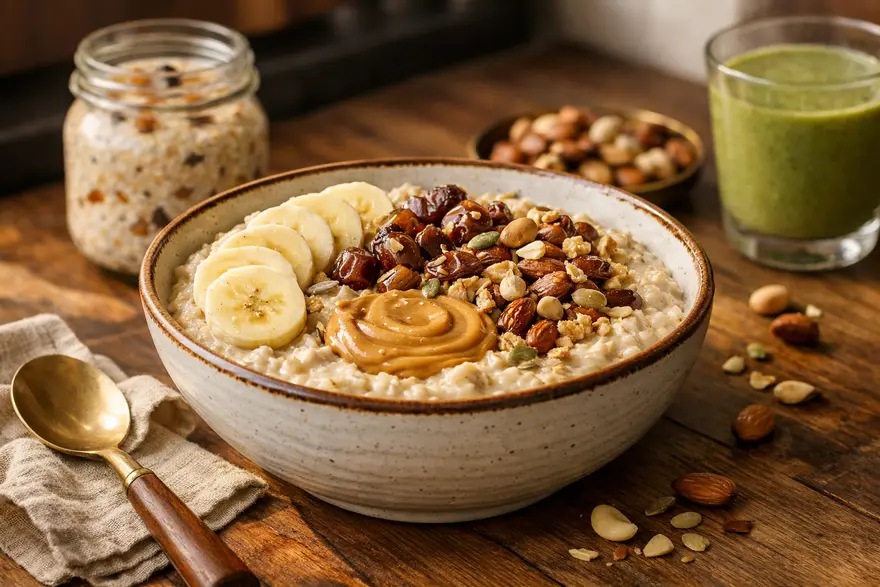
12 Oats Recipes For Weight Gain With High-Calorie Ideas
If you want to gain weight in a healthy way, oats can be a smart food to include in your meals. They are easy to prepare, affordable, filling, and easy to combine with calorie-dense ingredients such as whole milk, yoghurt, nuts, seeds, nut butters, and fruits. Oats on their own do not magically increase your weight. What helps is eating them in a way that adds more total calories and enough protein to your day. That makes oats useful for healthy weight gain, especially if you want to build strength and support muscle gain at the same time. Is Oats Good For Weight Gain? Yes, oats can support weight gain when you eat them as part of a calorie surplus. They are rich in carbohydrates, contain some protein, and pair well with higher-calorie foods. This is why many people ask, is oats good for weight gain. The answer is yes, but the way you prepare it matters. A plain bowl made with water will be much lighter than oats cooked with whole milk, peanut butter, dates, nuts, and seeds. Why Oats Are Good For Weight Gain Oats work well for weight gain for a few simple reasons. They give you steady energy from complex carbohydrates. They also provide fibre, which supports digestion, and a moderate amount of protein. Most importantly, oats are flexible. You can turn them into porridge, overnight oats, shakes, smoothies, snack bowls, or savoury meals without much effort. They are also useful if you are trying to gain muscle, because oats pair well with protein-rich foods such as milk, curd, paneer, Greek yoghurt, and whey protein. If you train regularly, this combination can support recovery and help you eat enough calories across the day. Best Oats For Weight Gain If you are choosing the best oats for weight gain, these are the main options: Rolled Oats: Usually the best oats for weight gain. They cook soft, taste good, and work well in porridge, overnight oats, and shakes. Steel-Cut Oats: Less processed and slightly chewier. They are filling and good for hearty bowls. Instant Oats: Convenient and quick. These can still work well if you add calorie-rich toppings. High-Protein Oats Blends: Useful if you want extra protein in one meal, but always check the label for added sugar and portion size. For most people, rolled oats are the easiest and most practical choice. Nutritional Value Of Oats For Weight Gain Oats are nutrient-dense, which means they provide useful nutrition along with calories. They contain carbohydrates for energy, fibre for digestive support, some protein, and minerals such as magnesium, iron, and manganese. Approximate Nutrition In Plain Oats A dry 40 g serving of rolled oats gives you roughly: 150 kcal 27 g carbohydrates 5 g protein 3 g fat 4 g fibre That is a good base, but the real weight gain value comes from what you add to it. What Makes Oats Better For Weight Gain? To make oats more useful for weight gain, pair them with foods such as: Whole milk Full-fat curd or Greek yoghurt Peanut butter or almond butter Dates, raisins, figs, and other dried fruits Almonds, walnuts, cashews, and seeds Banana, mango, chikoo, or avocado Honey or jaggery Whey protein or skimmed milk powder Coconut flakes or desiccated coconut How To Use Oats For Weight Gain: 12 High-Calorie Oats Recipes 1. Oats With Whole Milk, Banana, And Peanut Butter Ingredients 1/2 cup rolled oats 300 ml whole milk 1 banana 1 to 2 tbsp peanut butter How To Make Cook the oats in milk until soft. Slice the banana on top and stir in the peanut butter. Why It Supports Weight Gain This is a simple high-calorie bowl with carbs, fat, and protein in one meal. 2. Overnight Oats With Yoghurt, Nuts, And Honey Ingredients 1/2 cup rolled oats 1/2 cup full-fat yoghurt 1/2 cup milk 1 tbsp honey Chopped almonds and walnuts How To Make Mix everything in a jar and refrigerate overnight. Why It Supports Weight Gain It is easy to eat in the morning and gives you extra calories without feeling too heavy. 3. Mango Oats Smoothie Ingredients 1/2 cup oats 1 ripe mango 250 ml whole milk 1 tbsp chia seeds 1 tbsp nut butter How To Make Blend until smooth. Why It Supports Weight Gain This is useful if you struggle with appetite and prefer drinking your calories. 4. Chocolate Protein Oats Ingredients 1/2 cup oats 300 ml milk 1 scoop chocolate protein powder 1 tsp cocoa powder 1 tbsp peanut butter How To Make Cook the oats first. Let them cool slightly, then mix in the protein powder and peanut butter. Why It Supports Weight Gain This works well after exercise because it adds both calories and protein. 5. Dry Fruit Oats Porridge Ingredients 1/2 cup oats 300 ml whole milk Chopped dates Raisins Cashews A pinch of cardamom How To Make Cook the oats in milk and stir in the dry fruits. Why It Supports Weight Gain Dry fruits raise the calorie content quickly and make the bowl more filling. 6. Banana Dates Oats Shake Ingredients 1/2 cup oats 1 banana 3 to 4 dates 250 to 300 ml milk 1 tbsp almond butter How To Make Blend all ingredients until smooth. Why It Supports Weight Gain This is one of the easiest ways to have oats for weight gain when you need a quick snack. 7. Oats With Nut Butter And Seeds Ingredients 1/2 cup oats 300 ml milk 1 tbsp peanut or almond butter 1 tbsp chia or flax seeds 1 tsp honey How To Make Cook the oats and add the toppings before serving. Why It Supports Weight Gain Healthy fats from nut butter and seeds make the meal more calorie-dense. 8. Apple Cinnamon Oats With Raisins Ingredients 1/2 cup oats 300 ml milk 1 chopped apple 2 tbsp raisins A pinch of cinnamon 1 tbsp chopped walnuts How To Make Cook the oats and milk together. Add the apple, raisins, cinnamon, and walnuts. Why It Supports Weight Gain This gives you a good mix of carbs, fibre, and extra calories from raisins and walnuts. 9. Oats And Curd Bowl With Granola Topping Ingredients 1/2 cup soaked oats 1 cup full-fat curd 2 tbsp granola 1 tbsp mixed seeds 1 tsp honey How To Make Mix the oats and curd. Top with granola, seeds, and honey. Why It Supports Weight Gain It is quick, high in energy, and easy to carry as a snack. 10. Savoury Masala Oats With Paneer Ingredients 1/2 cup oats 1 tsp ghee Onion, tomato, and peas 50 to 75 g paneer Salt and spices How To Make Cook the vegetables in ghee, add oats and water, then stir in paneer. Why It Supports Weight Gain This is a balanced savoury option with added fat and protein. 11. Oats Khichdi With Ghee And Vegetables Ingredients 1/2 cup oats Moong dal 1 to 2 tsp ghee Mixed vegetables Mild spices How To Make Cook everything together until soft. Why It Supports Weight Gain This works well if you prefer warm savoury meals instead of sweet oats. 12. High-Calorie Oats Smoothie Bowl Ingredients 1/2 cup oats 1 frozen banana 1/2 cup yoghurt 1 tbsp peanut butter 1 tbsp pumpkin seeds Chopped dates How To Make Blend the base thick and top with dates and seeds. Why It Supports Weight Gain It is dense, satisfying, and easy to customise. Best High-Calorie Add-Ins For Oats If you want to make your oats more calorie-dense, these add-ins help the most: Whole milk Peanut butter or almond butter Chopped almonds, walnuts, or cashews Chia, flax, or pumpkin seeds Dates, raisins, figs, and apricots Banana, mango, chikoo, or avocado Greek yoghurt or hung curd Whey protein Honey or jaggery Coconut flakes These ingredients increase calories without needing a huge portion size. How To Add Oats To Your Daily Diet For Weight Gain You do not need to eat oats at every meal. One or two servings a day can fit well into your routine. You can try: Oats porridge at breakfast Overnight oats as a mid-morning meal An oats smoothie after your workout Savoury oats as an evening meal A calorie-dense shake with oats before bed if your appetite is low Consistency matters more than eating a very large amount on one day. Can Oats Help With Muscle Gain? Oats can help with muscle gain, but not on their own. They give you carbohydrates for training energy and work well with protein-rich foods that support muscle repair. If your goal is muscle gain, use oats with milk, curd, whey protein, nuts, seeds, and fruit. Pair that with regular strength training and enough total daily protein. That is a much better approach than relying on oats alone. Potential Risks Of Eating Too Many Oats For Weight Gain Oats are healthy, but eating too much too quickly can cause problems. You may feel bloated, gassy, or overly full, especially if your usual fibre intake is low. Large oat portions can also reduce your appetite for other foods, which may make it harder to eat enough calories across the day. This is why it is better to build your intake gradually and combine oats with a variety of other foods. If you have coeliac disease or gluten sensitivity, choose certified gluten-free oats when needed. Are Oats The Best Food For Weight Gain? Oats are one of the better foods for healthy weight gain, but they are not the only answer. They work best as a base food that you build on with milk, protein, nuts, seeds, and fruits. So, if you are wondering about the best oats for weight gain, the more useful question is how you prepare them. A balanced calorie surplus, enough protein, good sleep, and regular exercise matter much more than any single food. Conclusion: Final Thoughts On Oats For Weight Gain Oats can be a very practical part of a healthy weight gain plan. They are easy to prepare, easy to customise, and simple to combine with high-calorie ingredients. That makes them useful whether you prefer sweet bowls, shakes, smoothies, or savoury meals. The key is to make your oats energy-rich and balanced. Use whole milk, nut butters, fruits, nuts, seeds, curd, or protein powder to make each serving count. If you want a broader nutrition view, you can also explore our article on Oats Benefits. If you are trying to gain weight but also have poor appetite, digestive discomfort, fatigue, or unexplained weight loss, it is worth speaking with your doctor. Metropolis Healthcare offers a wide range of diagnostic tests, expert support, and convenient home sample collection to help you understand your health better and make informed choices. FAQs Is Oats Good For Weight Gain? Yes, oats can support weight gain when you eat them in a calorie surplus and combine them with calorie-dense foods like milk, nut butter, nuts, seeds, and dried fruits. Can I Add Dragon Fruit To Oats For Better Weight Gain? Yes, you can add dragon fruit to oats for taste, fibre, and variety. However, dragon fruit is not very calorie-dense, so for better weight gain, pair it with milk, yoghurt, nut butter, seeds, or nuts. How Can I Make My Oats More Calorie-Dense? You can make your oats more calorie-dense by adding: Whole milk Peanut butter or almond butter Nuts and seeds Banana, mango, or dates Full-fat yoghurt Whey protein Honey or jaggery Coconut flakes How Often Should I Eat Oats For Weight Gain? One or two servings a day can work well for many people. The right amount depends on your appetite, your total diet, and your weight gain goal. Can Oats Cause Bloating If Eaten Too Much? Yes. Oats are high in fibre, so eating too much too quickly can cause bloating, gas, or a very full feeling. Start with moderate portions and increase slowly. What Are Some Savoury Oats Recipes For Weight Gain? Some good savoury options include: Masala oats with paneer Oats khichdi with ghee Oats upma with peanuts Cheesy oats with vegetables Oats and dal porridge Are Oats The Best Food For Weight Gain? Oats are one of the better foods for healthy weight gain, but they are not the only one. They work best when you include them in an overall high-calorie, balanced diet. Can Oats Help With Muscle Gain? Yes, they can support muscle gain by giving you energy for training. They work best when you combine them with enough protein and follow a proper strength training routine. Which Are The Best Oats For Weight Gain? Rolled oats are usually the best oats for weight gain because they are easy to cook, easy to digest, and work well in both sweet and savoury recipes. References U.S. Department of Agriculture, Agricultural Research Service. FoodData Central: Oats, Whole Grain, Rolled, Old Fashioned. Accessed April 2, 2026. Paudel D, Dhungana B, Caffe M, Krishnan P. A Review of Health-Beneficial Properties of Oats. Foods. 2021;10(11):2591. PMID: 34828872. Whitehead A, Beck EJ, Tosh S, Wolever TMS. Cholesterol-Lowering Effects of Oat Beta-Glucan: A Meta-Analysis of Randomized Controlled Trials. Am J Clin Nutr. 2014;100(6):1413-1421. PMID: 25411276. Zurbau A, Noronha JC, Khan TA, Sievenpiper JL, Wolever TMS. The Effect of Oat Beta-Glucan on Postprandial Blood Glucose and Insulin Responses: A Systematic Review and Meta-Analysis. Eur J Clin Nutr. 2021;75(11):1540-1554. PMID: 33608654. Jäger R, Kerksick CM, Campbell BI, Cribb PJ, Wells SD, Skwiat TM, et al. International Society of Sports Nutrition Position Stand: Protein and Exercise. J Int Soc Sports Nutr. 2017;14:20. PMID: 28642676. NHS. Healthy Ways to Gain Weight. Accessed April 2, 2026.