


Latest Blogs
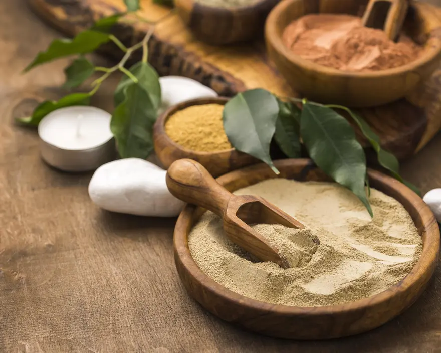
ત્રિફળા ચૂર્ણ: ફાયદા, ઉપયોગ, માત્રા અને આડઅસરો સમજાવેલી
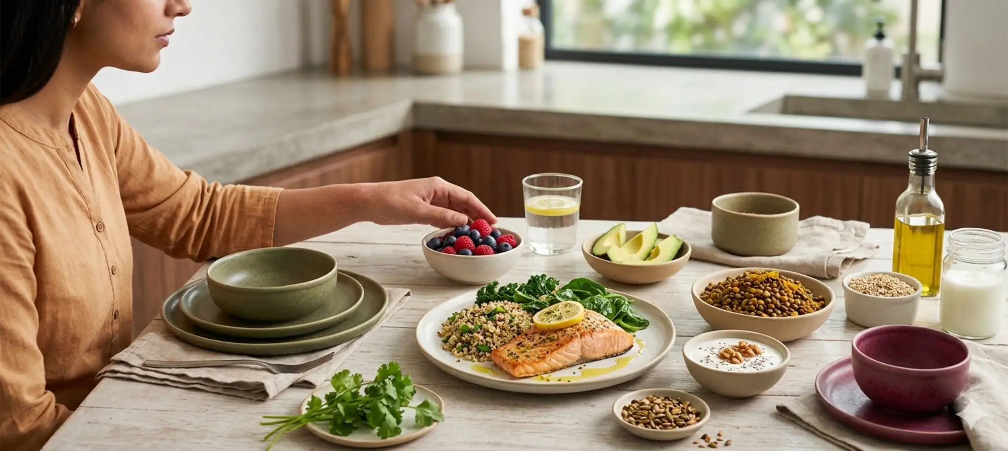
ત્રિફળા ચૂર્ણ શું છે? ત્રિફળા ચૂર્ણ, જેનો સંસ્કૃતમાં અર્થ “ત્રણ ફળો” થાય છે, એ આમળા, હરિતકી અને બિભીતકીનું પરંપરાગત આયુર્વેદિક મિશ્રણ છે, જેમાં દરેક ફળ અલગ-અલગ ઉપચારાત્મક ગુણધર્મો આપે છે એવું માનવામાં આવે છે. આમળા વિટામિન C અને એન્ટીઑક્સિડન્ટ્સ સાથે રોગપ્રતિકારક શક્તિ વધારે છે, હરિતકી પાચનમાં મદદ કરે છે અને બિભીતકી કોલેસ્ટ્રોલ તથા શ્વસન સ્વાસ્થ્યને ટેકો આપે છે. સાથે મળીને, તેનો પરંપરાગત રીતે પાચન, ડિટૉક્સિફિકેશન અને સમગ્ર જીવનશક્તિને ટેકો આપવા માટે ઉપયોગ થાય છે, જેના કારણે ત્રિફળા દીર્ઘાયુષ્ય અને જીવનશક્તિ માટે એક પરંપરાગત આયુર્વેદિક રસાયન ગણાય છે. ડિટૉક્સિફિકેશન માટે ત્રિફળા ચૂર્ણ કેવી રીતે કામ કરે છે? નેશનલ ઇન્સ્ટિટ્યુટ્સ ઓફ હેલ્થ (NIH) ડેટાબેઝમાં સંદર્ભિત અભ્યાસો અનુસાર, ત્રિફળા પાચન અને યકૃતના કાર્યને ટેકો આપનાર હળવા ડિટૉક્સિફાઇંગ અને એન્ટીઑક્સિડન્ટ એજન્ટ તરીકે કામ કરી શકે છે. તે પાચનતંત્રને લક્ષ્ય બનાવે છે, જેને આયુર્વેદમાં ઘણીવાર સમગ્ર સ્વાસ્થ્યનું મૂળ કહેવામાં આવે છે, અને આંતરડાના કુદરતી ફ્લોરાને ખલેલ પહોંચાડ્યા વગર સંગ્રહિત ઝેરી તત્ત્વો (આમ) દૂર કરે છે. તે આ રીતે કામ કરે છે: આમળા યકૃતના કાર્યને ટેકો આપે છે અને એન્ટીઑક્સિડન્ટ એન્ઝાઇમની પ્રવૃત્તિ વધારે છે, જે કુદરતી ચયાપચય પ્રક્રિયાઓમાં મદદ કરે છે. હરિતકી સ્વસ્થ મળવિસર્જનને પ્રોત્સાહન આપે છે અને કોલોનને સાફ કરે છે, જે ઝેરી તત્ત્વો દૂર કરવામાં મદદ કરે છે. બિભીતકી લિપિડ ચયાપચયને નિયંત્રિત કરે છે અને ભારે ધાતુઓ તથા ફ્રી રેડિકલ્સ બહાર કાઢવામાં ટેકો આપે છે. આયુર્વેદમાં માનવામાં આવે છે કે નિયમિત અને મધ્યમ ઉપયોગ શરીર પર ભાર મૂકતા ઑક્સિડેટિવ તણાવકારક તત્ત્વોને દૂર કરીને ત્વચાના સ્વાસ્થ્ય, ઊર્જા અને રક્તપરિભ્રમણના સંતુલનને ટેકો આપે છે. તે ત્રણ દોષો: વાત, પિત્ત અને કફ વચ્ચે સંતુલન જાળવવાની આયુર્વેદિક રીત છે. ત્રિફળા ચૂર્ણનું પોષણ મૂલ્ય ફળ વૈજ્ઞાનિક નામ મુખ્ય પોષક તત્ત્વો મુખ્ય ફાયદા આમળા એમ્બ્લિકા ઑફિસિનાલિસ વિટામિન C, પોલીફિનોલ્સ, ફ્લેવોનોઇડ્સ રોગપ્રતિકારક શક્તિને મજબૂત બનાવે છે, યકૃત અને હૃદયના સ્વાસ્થ્યને ટેકો આપે છે હરિતકી ટર્મિનાલિયા ચેબુલા ટેનિન્સ, એમિનો એસિડ્સ, ગેલિક એસિડ પાચન સુધારે છે, ચયાપચય વધારે છે અને તંતુઓને ડિટૉક્સિફાઇ કરે છે બિભીતકી ટર્મિનાલિયા બેલિરિકા લિગ્નાન્સ, એલેજિક એસિડ, એન્ટીઑક્સિડન્ટ્સ કોલેસ્ટ્રોલનું સંચાલન કરવામાં મદદ કરે છે, શ્વસન સ્વાસ્થ્ય વધારે છે પાચન સ્વાસ્થ્ય માટે ત્રિફળા પાચન સ્વાસ્થ્ય ત્રિફળાના સૌથી જાણીતા ફાયદાઓમાંનું એક છે. તે આ માટે જાણીતું છે: સ્વસ્થ આંતરડાની ગતિ અને નિયમિત મળવિસર્જનને ટેકો આપે છે પેટ ફૂલવું, ગેસ અને એસિડિટીમાંથી રાહત આપે છે ભૂખ અને પોષક તત્ત્વોના શોષણમાં સુધારો કરે છે કોલોનના કુદરતી ડિટૉક્સિફિકેશનમાં મદદ કરે છે આંતરડાના માઇક્રોબિયલ સંતુલનને ફરીથી સ્થાપિત કરે છે તમારા સ્વાસ્થ્ય માટે ત્રિફળા ચૂર્ણના 10 ફાયદા પાચન સ્વાસ્થ્ય વધારે છે: ત્રિફળા નિયમિત મળવિસર્જનને પ્રોત્સાહન આપીને અને કબજિયાત, એસિડિટી તથા પેટ ફૂલવું ઘટાડીને સ્વસ્થ પાચનને ટેકો આપે છે. તે આંતરડાના કાર્યને મજબૂત બનાવે છે અને પોષક તત્ત્વોના અસરકારક શોષણને ટેકો આપે છે. રોગપ્રતિકારક શક્તિ વધારે છે: આમળામાંથી મળતા એન્ટીઑક્સિડન્ટ્સ અને વિટામિન Cથી સમૃદ્ધ ત્રિફળા રોગપ્રતિકારક તંત્રને મજબૂત બનાવે છે, જે શરીરને ચેપ સામે લડવામાં અને ઑક્સિડેટિવ તણાવને કુદરતી રીતે ઘટાડવામાં મદદ કરે છે. વજન સંચાલનમાં મદદ કરે છે: ચયાપચય સુધારીને અને પાચનતંત્રને ડિટૉક્સિફાઇ કરીને, ત્રિફળા ભૂખ નિયંત્રિત કરવામાં મદદ કરે છે અને ધીમે-ધીમે, સ્વસ્થ રીતે વજન ઘટાડવામાં ટેકો આપે છે. યકૃતનું કાર્ય સુધારે છે: તેના ડિટૉક્સિફાઇંગ ગુણધર્મો યકૃતના એન્ઝાઇમ્સને ઉત્તેજિત કરે છે, જેનાથી ઝેરી તત્ત્વો દૂર કરવામાં અને ચરબી તથા પિત્તના ચયાપચયમાં સુધારો થાય છે. ચમકદાર ત્વચાને પ્રોત્સાહન આપે છે: નિયમિત સેવન લોહીને શુદ્ધ કરે છે, અંદરના ઝેરી તત્ત્વો દૂર કરે છે અને એક્ને અથવા પિગ્મેન્ટેશન ઘટાડે છે, જેના કારણે ત્વચાને કુદરતી અને સ્વસ્થ ચમક મળે છે. આંખોના સ્વાસ્થ્યને જાળવે છે ત્રિફળામાં રહેલા આમળાની પુનર્જીવિત કરવાની ક્રિયા દૃષ્ટિ સ્વાસ્થ્યને ટેકો આપે છે, આંખોનો થાક ઘટાડે છે અને ઉંમર સંબંધિત ક્ષયને અટકાવવામાં મદદ કરે છે. સ્વસ્થ બ્લડ શુગરને ટેકો આપે છે: પ્રારંભિક અભ્યાસો સૂચવે છે કે સંતુલિત આહાર સાથે ઉપયોગ કરવામાં આવે ત્યારે ત્રિફળા ગ્લુકોઝ ચયાપચય સુધારવામાં મદદ કરી શકે છે, અને સંતુલિત આહાર સાથે જોડાય ત્યારે સમય જતાં બ્લડ શુગરના સ્તરને સ્થિર કરવામાં મદદ કરે છે. કોલેસ્ટ્રોલનું સંચાલન કરે છે: બિભીતકી સમગ્ર જીવનશૈલી યોજનાના ભાગરૂપે સ્વસ્થ કોલેસ્ટ્રોલ સ્તરને ટેકો આપવામાં મદદ કરી શકે છે, જે વધુ સ્વસ્થ લિપિડ પ્રોફાઇલ અને હૃદય-રક્તવાહિની કાર્યને પ્રોત્સાહન આપે છે. મૌખિક સ્વાસ્થ્યને ટેકો આપે છે: તેના એન્ટિમાઇક્રોબિયલ ગુણધર્મોને કારણે, ત્રિફળા મોઢાના છાલા અટકાવવામાં મદદ કરે છે, પ્લાકનું જમાવટ ઘટાડે છે અને તાજો શ્વાસ તથા સ્વસ્થ મસૂડા પ્રોત્સાહિત કરે છે. વાળ અને માથાની ત્વચાને મજબૂત બનાવે છે: પોષક તત્ત્વોથી સમૃદ્ધ સંયોજન વાળના ફોલિકલ્સને પોષણ આપે છે, સમય પહેલાં વાળ સફેદ થવાથી બચાવે છે અને વધુ મજબૂત, ચમકદાર વાળ માટે માથાની ત્વચાનું સમગ્ર સ્વાસ્થ્ય વધારે છે. મહત્તમ ફાયદા માટે ત્રિફળા ચૂર્ણનો ઉપયોગ કેવી રીતે કરવો વ્યક્તિગત પસંદગી મુજબ ત્રિફળા ચૂર્ણ પાઉડર, કેપ્સ્યુલ અથવા ટેબ્લેટ સ્વરૂપે લઈ શકાય છે. પરંપરાગત રીતે, તેને ગરમ પાણી અથવા મધ સાથે મિક્સ કરીને વહેલી સવારે ખાલી પેટે અથવા સૂતા પહેલાં લેવામાં આવે છે. પાચનને ટેકો માટે: 1 ચમચી ગરમ પાણીમાં મિક્સ કરો, તેને 10 મિનિટ સુધી પલળવા દો અને સૂતા પહેલાં પીવો. પાચન સ્વાસ્થ્ય માટે: નાસ્તા પહેલાં હૂંફાળા પાણીમાં ઓછી માત્રા લો. ત્વચા અને રોગપ્રતિકારક શક્તિ માટે: શોષણ અને પોષક સહકાર વધારવા માટે મધ અથવા ઘી સાથે મિક્સ કરો. ત્રિફળા ચૂર્ણની માત્રા માટે ભલામણો પુખ્ત વયના લોકો: 3–5 grams (લગભગ 1 ચમચી) દિવસમાં એક કે બે વખત ગરમ પાણી સાથે. કબજિયાત માટે: સૂતા પહેલાં ગરમ પાણી અથવા દૂધ સાથે લો. વજન સંચાલન માટે: વહેલી સવારે ભોજન પહેલાં લો. બાળકો: અડધી માત્રા (ફક્ત દેખરેખ હેઠળ). કેપ્સ્યુલ સ્વરૂપે: સામાન્ય રીતે દરેક માત્રામાં 500–1000 mg. શું ત્રિફળા ચૂર્ણનો ઉપયોગ અન્ય સપ્લિમેન્ટ્સ સાથે કરી શકાય? વ્યવસાયિક માર્ગદર્શન હેઠળ ત્રિફળાને અશ્વગંધા અથવા હળદર જેવી અન્ય આયુર્વેદિક જડીબુટ્ટીઓ સાથે જોડીને લઈ શકાય છે, જે એન્ટીઑક્સિડન્ટ અને પુનર્જીવિત કરનારા ગુણધર્મો વધારે છે. જોકે, તેને અન્ય લૅક્સેટિવ્સ અથવા ડિટૉક્સ જડીબુટ્ટીઓ સાથે જોડવાનું ટાળવું જોઈએ, સિવાય કે ડૉક્ટર ભલામણ કરે, કારણ કે તે આંતરડાની પ્રવૃત્તિ વધારી શકે છે અથવા દવાઓના શોષણમાં ફેરફાર કરી શકે છે. જો તમે ડાયાબિટીસ, રક્તચાપ અથવા થાઇરૉઇડની દવા લઈ રહ્યા હો, તો ત્રિફળા સામેલ કરતાં પહેલાં તમારી નિયમિત રીત વિશે તમારા હેલ્થકેર પ્રદાતા સાથે ચર્ચા કરો, કારણ કે તે ચયાપચય દરને સૂક્ષ્મ રીતે અસર કરી શકે છે. ત્રિફળા ચૂર્ણ: આડઅસરો અને સાવચેતીઓ ભલામણ કરેલી માત્રામાં લેવાય ત્યારે મોટાભાગના પુખ્ત વયના લોકો માટે ત્રિફળા સુરક્ષિત માનવામાં આવે છે. જોકે, વધુ અથવા દેખરેખ વગરનું સેવન કારણે થઈ શકે છે: પેટમાં અસ્વસ્થતા અથવા ખેંચાણ પાતળા ઝાડા અથવા ડિહાઇડ્રેશન કેટલીક દવાઓનું શોષણ ઘટવું સાવચેતીઓ: ગર્ભવતી અથવા સ્તનપાન કરાવતી મહિલાઓએ ત્રિફળાનો ઉપયોગ કરતાં પહેલાં યોગ્ય હેલ્થકેર પ્રદાતાની સલાહ લેવી જોઈએ, કારણ કે તેના શુદ્ધિકરણ ગુણધર્મો યોગ્ય ન હોઈ શકે. દીર્ઘકાલીન ઝાડા, આઈબીએસ અથવા લો બ્લડ પ્રેશર ધરાવતા લોકોએ તેનો ઉપયોગ કરતાં પહેલાં ડૉક્ટરની સલાહ લેવી જોઈએ. અચાનક ડિટૉક્સ પ્રતિક્રિયાઓથી બચવા માટે ઓછી માત્રાથી શરૂ કરીને ધીમે-ધીમે વધારવું શ્રેષ્ઠ છે. શું ત્રિફળા ચૂર્ણ લાંબા ગાળાના ઉપયોગ માટે સુરક્ષિત છે? ભલામણ કરેલી માત્રામાં લેવાય ત્યારે, ત્રિફળા સામાન્ય રીતે લાંબા ગાળાના ઉપયોગ માટે યોગ્ય માનવામાં આવે છે. રાસાયણિક લૅક્સેટિવ્સથી વિપરીત, તે આદત પાડતું નથી અને સમય જતાં આંતરડાની અંદરની પડને પોષણ આપે છે. જોકે, આયુર્વેદિક નિષ્ણાતો સતત ઉપયોગના દર થોડા મહિનાઓ પછી શરીરની કુદરતી પ્રણાલીઓને ફરીથી સંતુલિત થવા દેવા માટે થોડો વિરામ (લગભગ 1–2 અઠવાડિયા) લેવાની ભલામણ કરે છે. શું ત્રિફળા વજન ઘટાડવામાં મદદ કરી શકે? આહાર અને વ્યાયામ સાથે જોડાય ત્યારે ત્રિફળા સ્વસ્થ ચયાપચય અને લિપિડ સંતુલનને ટેકો આપી શકે છે. તેની હળવી ડિટૉક્સિફાઇંગ અસર પાચનતંત્રને સાફ કરે છે, જે પોષક તત્ત્વોના શોષણમાં મદદ કરે છે. તૃપ્તિની લાગણી સુધારે છે અને રાત્રે મોડે થતી ખાવાની ઇચ્છાને નિયંત્રિત કરવામાં મદદ કરે છે. સંતુલિત આહાર અને નિયમિત શારીરિક પ્રવૃત્તિ સાથે જોડાય ત્યારે શ્રેષ્ઠ કામ કરે છે. તે “ઝડપી ઉપાય” નથી, પરંતુ સતત વજન સંચાલન અને ચયાપચય સ્વાસ્થ્ય માટે સહાયક જડીબુટ્ટી છે. ત્રિફળા ચૂર્ણ અને અન્ય આયુર્વેદિક ડિટૉક્સ વચ્ચે તફાવત સંતુલિત ફોર્મ્યુલા: ત્રિફળા ત્રણ જડીબુટ્ટીઓને જોડે છે: આમળા, હરિતકી અને બિભીતકી, જે ત્રણેય દોષો (વાત, પિત્ત અને કફ)નું સંતુલન કરે છે, જ્યારે એક જ જડીબુટ્ટી ધરાવતા ડિટૉક્સ માત્ર એક પાસાને લક્ષ્ય બનાવે છે. હળવું છતાં અસરકારક: કઠોર પર્ગેટિવ ક્લેન્સથી વિપરીત, તે નબળાઈ અથવા ડિહાઇડ્રેશન કર્યા વગર પાચનતંત્રને ધીમે-ધીમે સાફ કરે છે. પોષક તત્ત્વોથી ભરપૂર ડિટૉક્સ: ત્રિફળા ઝેરી તત્ત્વો દૂર કરતી વખતે તંતુઓને પોષણ આપે છે અને મહત્વપૂર્ણ પોષક તત્ત્વોને ફરીથી પૂરાં પાડે છે, જેથી પોષક તત્ત્વોની ખોટ વગર ડિટૉક્સ થાય છે. સિસ્ટેમિક ક્રિયા: યકૃત, આંતરડા અને લોહી પર સમગ્ર રીતે કામ કરે છે, જ્યારે અન્ય ડિટૉક્સ ઘણીવાર માત્ર એક અંગ પર ધ્યાન કેન્દ્રિત કરે છે. નિયમિત ઉપયોગ માટે સુરક્ષિત: આદત પાડતું નથી અને લાંબા ગાળાના સ્વાસ્થ્ય માટે યોગ્ય છે, જેના કારણે તે આક્રમક અથવા ટૂંકા ગાળાના આયુર્વેદિક ક્લેન્સ કરતાં વધુ સુરક્ષિત બને છે. પુનર્જીવિત અસર: તે ક્લેન્સર અને રસાયન (પુનર્જીવક) બંને તરીકે કામ કરે છે, જે સામાન્ય ડિટૉક્સિફિકેશનથી આગળ જઈ જીવનશક્તિ અને દીર્ઘાયુષ્યને પ્રોત્સાહન આપે છે. નિષ્કર્ષ ત્રિફળા ચૂર્ણ એ સમયની કસોટી પર ખરા ઉતરેલું આયુર્વેદિક ફોર્મ્યુલેશન છે, જેનો પરંપરાગત રીતે પાચન સ્વાસ્થ્ય, ચયાપચય અને સમગ્ર સંતુલનને ટેકો આપવા માટે ઉપયોગ થાય છે અને જે પાચન, ડિટૉક્સિફિકેશન, ચયાપચય તથા સમગ્ર પુનર્જીવનને ટેકો આપે છે. સવારે નિયમિત રીતના ભાગરૂપે લેવાય કે રાત્રિના ડિટૉક્સ તરીકે, તેના ફાયદા આંતરડાથી લઈને ત્વચા સુધી વિસ્તરે છે, જે સાચું આંતરિક સંતુલન દર્શાવે છે. મેટ્રોપોલિસ હેલ્થકેર પસંદ કરો — ભારતની વિશ્વસનીય ડાયગ્નોસ્ટિક બ્રાન્ડ, જેમાં 4,000+ ટેસ્ટ, NABL & CAP-પ્રમાણિત લેબ્સ અને ચોકસાઈ તથા વિશ્વસનીયતા સુનિશ્ચિત કરતા નિષ્ણાત પેથોલોજિસ્ટ્સ છે. 10,000+ સંપર્ક-બિંદુઓ પર ઘરેથી સેમ્પલ કલેક્શન, ઝડપી રિપોર્ટ ટર્નઅરાઉન્ડ અને વેબસાઇટ, કૉલ, ઍપ અથવા વોટ્સએપ દ્વારા સરળ બુકિંગની સુવિધાનો અનુભવ કરો. વારંવાર પૂછાતા પ્રશ્નો ત્રિફળા ચૂર્ણ શેમાંથી બને છે? ત્રિફળા ચૂર્ણ આમળા, હરિતકી અને બિભીતકીમાંથી બને છે, ત્રણ ફળોને સુકવી, પીસી અને સમાન પ્રમાણમાં મિક્સ કરવામાં આવે છે. તે 100% વનસ્પતિ આધારિત છે અને કુદરતી રીતે એન્ટીઑક્સિડન્ટ્સથી સમૃદ્ધ છે. મારે ત્રિફળા ચૂર્ણ કેટલા વખત લેવું જોઈએ? દિવસમાં એક વખત સામાન્ય સ્વાસ્થ્ય માટે દિવસમાં બે વખત ખાસ ડિટૉક્સ અથવા પાચન સંબંધિત લક્ષ્યો માટે દીર્ઘકાલીન ઉપયોગ માટે હંમેશા વ્યવસાયિક દેખરેખ હેઠળ શું ત્રિફળા ચૂર્ણ કબજિયાતમાં મદદ કરી શકે? હા. તે હળવા કુદરતી લૅક્સેટિવ તરીકે કામ કરે છે, મળવિસર્જન સુધારે છે અને કૃત્રિમ લૅક્સેટિવ્સથી વિપરીત આધારતા ઊભી કર્યા વગર ઝેરી તત્ત્વો દૂર કરે છે. શું ત્રિફળા ચૂર્ણ ગર્ભવતી મહિલાઓ માટે સુરક્ષિત છે? ના. તેના શુદ્ધિકરણ ગુણધર્મોને કારણે ગર્ભાવસ્થા દરમિયાન સામાન્ય રીતે તેની ભલામણ કરવામાં આવતી નથી, કારણ કે તે ગર્ભાશયના સંકોચનોને ઉત્તેજિત કરી શકે છે. હંમેશા પહેલા હેલ્થકેર નિષ્ણાતની સલાહ લો. શું ત્રિફળા ચૂર્ણ ત્વચાનું સ્વાસ્થ્ય સુધારી શકે? ચોક્કસ. લોહીને શુદ્ધ કરીને, પાચન સુધારીને અને ઑક્સિડેટિવ તણાવ ઘટાડીને, ત્રિફળા વધુ સ્વચ્છ, તેજસ્વી અને યુવાન દેખાતી ત્વચા મેળવવામાં મદદ કરે છે.
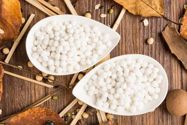
સાબુદાણાના 10 આશ્ચર્યજનક ફાયદા જે તમે જાણતા ન હતા
જો તમે ક્યારેય ઉપવાસ દરમિયાન અથવા સ્વસ્થ થતી વખતે સાબુદાણા તરફ વળ્યા છો, તો તમને જાણીને આશ્ચર્ય થઈ શકે છે કે તે માત્ર ઝડપી ઊર્જા કરતાં વધુ આપે છે. આ સરળ ખાદ્ય પદાર્થ તમારા પેટ માટે હળવો છે અને અનપેક્ષિત રીતે તમારા સ્વાસ્થ્યને ટેકો આપી શકે છે. તમે પાચન સંભાળતા હો, હળવા પોષણની શોધમાં હો અથવા ફક્ત આરોગ્યપ્રદ વિકલ્પો શોધી રહ્યા હો, સાબુદાણા ઘણું આપી શકે છે. આ બ્લોગમાં, તમે ઓછા જાણીતા ફાયદાઓ વિશે જાણશો, જે તમને તમારા શરીર અને આરોગ્યની જરૂરિયાતો માટે યોગ્ય પસંદગી કરવામાં વધુ વિશ્વાસ આપી શકે છે. સાબુદાણા (ટૅપિયોકા પર્લ્સ) શું છે? સાબુદાણા, જેને ટૅપિયોકા પર્લ્સ તરીકે પણ ઓળખવામાં આવે છે, કસાવા છોડના મૂળમાંથી બનતો સ્ટાર્ચયુક્ત ખાદ્ય પદાર્થ છે. આ નાના, સફેદ દાણા રાંધવામાં આવે ત્યારે નરમ અને પારદર્શક બને છે, જેના કારણે તેઓ પચવામાં સરળ અને ખાવામાં આરામદાયક લાગે છે. સામાન્ય રીતે ઉપવાસ દરમિયાન ઉપયોગમાં લેવાતા સાબુદાણાને પેટ માટે હળવા અને શરીરને ઊર્જા આપનાર તરીકે ઓળખ મળી છે. તેનો ઉપયોગ ઘણીવાર વિવિધ ખીરો, નાસ્તા અને મસાલેદાર વાનગીઓમાં થાય છે. તેની સાદાઈ હોવા છતાં, સાબુદાણા માત્ર ઊર્જાનો સ્રોત નથી—તેમાં સ્વાસ્થ્યને ટેકો આપતા ઘણા ગુણધર્મો છે, જેને ઘણા લોકો અવગણે છે. સાબુદાણાનું પોષણ મૂલ્ય સાબુદાણા મુખ્યત્વે કાર્બોહાઇડ્રેટનો સ્રોત છે, તેમ છતાં તેમાં અન્ય પોષક તત્ત્વો અલ્પ માત્રામાં હોય છે, જે સંતુલિત માત્રામાં લેવાય ત્યારે તમારા સ્વાસ્થ્યને ટેકો આપે છે. સાબુદાણાનું પોષણ મૂલ્ય સમજવાથી તેને તમારા ભોજનમાં સામેલ કરવા અંગે માહિતીસભર પસંદગી કરવામાં મદદ મળે છે. પોષક તત્ત્વ દર 100g (રાંધ્યા વગરના સાબુદાણા) ઊર્જા 358 kcal કાર્બોહાઇડ્રેટ્સ 88.7 g પ્રોટીન 0.2 g ચરબી 0.2 g ફાઇબર 0.9 g કેલ્શિયમ 20 mg આયર્ન 1.58 mg પોટેશિયમ 11 mg સોડિયમ 1 mg પ્રોટીન અને ચરબીમાં ઓછો હોવા છતાં, સાબુદાણા ઝડપી ઊર્જા આપે છે, જેના કારણે તે સ્વસ્થ થવા અને ઉપવાસના આહાર માટે યોગ્ય બને છે. દૂધ, શિંગદાણા અથવા શાકભાજી જેવા ઘટકો સાથે મિક્સ કરવામાં આવે ત્યારે તે વધુ પોષક બને છે. સાબુદાણાના 10 આરોગ્ય ફાયદા તમે કદાચ ઉપવાસ દરમિયાન સાબુદાણાના ઉપયોગથી પહેલેથી પરિચિત હશો, પરંતુ જાણવા જેવું ઘણું વધુ છે. અહીં દસ આશ્ચર્યજનક સાબુદાણાના ફાયદા આપેલા છે, જે તમારા સ્વાસ્થ્ય અને સુખાકારીને ટેકો આપી શકે છે: 1. તરત ઊર્જા વધારે છે સાબુદાણા કાર્બોહાઇડ્રેટ્સથી સમૃદ્ધ છે, ખાસ કરીને સ્ટાર્ચથી, જે ઊર્જાના સ્તરને ઝડપથી પુનઃસ્થાપિત કરવામાં મદદ કરે છે. જો તમે નબળાઈ અનુભવો છો અથવા બીમારીમાંથી સાજા થઈ રહ્યા છો, તો સાબુદાણા તમને ફરીથી તાકાત મેળવવામાં મદદરૂપ થતો સરળતાથી પચી શકે એવો ઊર્જા સ્રોત આપી શકે છે. 2. પાચન સ્વાસ્થ્યને ટેકો આપે છે સાબુદાણાનો હળવો અને અસ્વસ્થતા ન કરનાર સ્વભાવ તેને તમારા પાચનતંત્ર માટે નરમ બનાવે છે. સંવેદનશીલ પેટ ધરાવતા લોકો માટે તેની ઘણીવાર ભલામણ કરવામાં આવે છે, કારણ કે તે તમારા આંતરડાં પર વધુ ભાર મૂક્યા વગર પાચન સંબંધિત અસ્વસ્થતાને શાંત કરવામાં મદદ કરે છે. 3. ગ્લુટન-સંવેદનશીલ લોકો માટે મુક્ત વિકલ્પ જો તમને ગ્લુટન પ્રત્યે સંવેદનશીલતા હોય અથવા સિલિયક રોગ હોય, તો સાબુદાણા એક સુરક્ષિત વિકલ્પ છે. કુદરતી રીતે ગ્લુટન-મુક્ત હોવાથી, તેનો ઉપયોગ ઘઉં આધારિત અનાજની જગ્યાએ પાચન સંબંધિત સમસ્યા કર્યા વગર કરી શકાય છે. 4. સ્નાયુઓની મરામતમાં મદદ કરી શકે છે પોતે પ્રોટીનમાં ઓછો હોવા છતાં, દૂધ અથવા શિંગદાણા સાથે રાંધવામાં આવે ત્યારે સાબુદાણા સારો રિકવરી ખોરાક બને છે. આ સ્રોતોમાંથી મળતું વધારાનું પ્રોટીન તમારા શરીરને તંતુઓની મરામત કરવામાં અને શારીરિક પ્રવૃત્તિ અથવા બીમારી પછી પુનઃપ્રાપ્તિમાં મદદ કરે છે. 5. હાડકાંની મજબૂતીને પ્રોત્સાહન આપે છે સાબુદાણામાં કેલ્શિયમ અને આયર્ન અલ્પ માત્રામાં હોય છે, જે હાડકાંના સ્વાસ્થ્યને ટેકો આપે છે. જોકે તે તમારો એકમાત્ર સ્રોત ન હોવો જોઈએ, દૂધજન્ય પદાર્થો અથવા પાંદડાવાળા શાકભાજી સાથે તેને મિક્સ કરવાથી જરૂરી ખનિજોના સેવનમાં વધારો થઈ શકે છે. 6. ચિંતા ઘટાડવા માટે હળવી શાંત કરનારી અસર કેટલાક પરંપરાગત ઉપયોગો સૂચવે છે કે સાબુદાણાની શાંત કરનારી અસર હોય છે. વૈજ્ઞાનિક પુરાવા મર્યાદિત હોવા છતાં, તેની સરળ પાચ્યતા અને ઊર્જાનો ટેકો તમને વધુ સ્થિર અને ઓછી ચિંતા અનુભવવામાં મદદ કરી શકે છે. 7. ઓછું વજન ધરાવતા લોકોમાં વજન વધારવામાં મદદ કરે છે તેની ઊંચી કેલરી અને કાર્બોહાઇડ્રેટ સામગ્રીને કારણે, સાબુદાણા સ્વસ્થ વજન વધારવાની જરૂર હોય તેવા લોકો માટે યોગ્ય છે. પોષક તત્ત્વોથી ભરપૂર ખોરાક સાથે રાંધવામાં આવે ત્યારે તે લાંબા સમય સુધી ઊર્જા અને પોષણ આપે છે. 8. ગર્ભાવસ્થા દરમિયાન ઉપયોગી (મર્યાદિત માત્રામાં) મર્યાદિત માત્રામાં, સાબુદાણા ગર્ભાવસ્થા દરમિયાન ઊર્જા પૂરી પાડવામાં અને થાક ઘટાડવામાં મદદ કરી શકે છે. તે તમારી વ્યક્તિગત આહાર જરૂરિયાતો માટે યોગ્ય છે કે નહીં તેની ખાતરી કરવા માટે હંમેશા તમારા હેલ્થકેર પ્રદાતા સાથે વાત કરો. 9. પુરુષોના સ્વાસ્થ્ય માટે ફાયદાકારક પુરુષોના સ્વાસ્થ્ય માટે ઓછા જાણીતા સાબુદાણાના ફાયદામાં હોર્મોનલ સંતુલન અને સ્ટેમિનાને સંભવિત ટેકોનો સમાવેશ થાય છે, ખાસ કરીને જ્યારે તેને દૂધ અને નટ્સ જેવા પોષક તત્ત્વોથી ભરપૂર ખોરાક સાથે તૈયાર કરવામાં આવે. 10. ડિટૉક્સ આહાર માટે મદદરૂપ થઈ શકે છે કારણ કે તે સરળતાથી પચી શકે છે અને તીખો નથી, સાબુદાણા ઘણીવાર હળવા અથવા ડિટૉક્સિફાઇંગ આહારમાં સામેલ કરવામાં આવે છે. તેની સાદાઈ તમારા પાચનતંત્રને આરામ આપે છે અને હળવી જડીબુટ્ટીઓ અથવા કુદરતી મસાલા સાથે ઉપયોગમાં લેવાય ત્યારે સોજો ઘટાડવામાં મદદ કરી શકે છે. આ સાબુદાણાના ફાયદા તેને તમારી આહાર યોજનામાં બહુમુખી ઉમેરો બનાવે છે, ખાસ કરીને જ્યારે તેને અન્ય આરોગ્યપ્રદ ખોરાક સાથે મિક્સ કરવામાં આવે. ફક્ત યાદ રાખો—મર્યાદા જ મુખ્ય છે. સાબુદાણામાં ગ્લાયસેમિક ઇન્ડેક્સ સાબુદાણાનો ગ્લાયસેમિક ઇન્ડેક્સ (GI) ઊંચો હોય છે, એટલે કે તે ખાધા પછી બ્લડ શુગરના સ્તરમાં ઝડપથી વધારો કરે છે. જ્યારે તમારી ઊર્જા ઓછી હોય અને ઝડપી વધારો જોઈએ ત્યારે આ ઉપયોગી થઈ શકે છે. જોકે, જો તમે ડાયાબિટીસ, પ્રીડાયાબિટીસ અથવા ઇન્સ્યુલિન રેઝિસ્ટન્સ સંભાળી રહ્યા હો, તો તમારી માત્રા અને તેને કેવી રીતે તૈયાર કરો છો તેની કાળજી રાખવી મહત્વપૂર્ણ છે. સાબુદાણા એકલા ખાવાથી ગ્લુકોઝમાં અચાનક વધારો થઈ શકે છે. આ અસર ઘટાડવા માટે, તેને ફાઇબરથી ભરપૂર શાકભાજી, નટ્સ અથવા દહીં કે શિંગદાણા જેવા પ્રોટીન સ્રોતો સાથે લો. આ સંયોજન કાર્બોહાઇડ્રેટ્સના પાચનને ધીમું કરવામાં મદદ કરે છે અને તમારા રક્તપ્રવાહમાં શુગર ધીમે અને સ્થિર રીતે છોડે છે. સંતુલિત ભોજનમાં સમજદારીથી સામેલ કરવામાં આવે તો, તમે બ્લડ શુગર પર નજર રાખતા હો ત્યારે પણ સાબુદાણા તમારા આહારનો ભાગ બની શકે છે. હંમેશા તમારા શરીરની પ્રતિક્રિયા સાંભળો અને શંકા હોય ત્યારે તમારા હેલ્થકેર પ્રદાતાની સલાહ લો. શું સાબુદાણા ખાવાથી કોઈ આડઅસરો થાય છે? સાબુદાણા મર્યાદિત માત્રામાં લેવાય ત્યારે મોટાભાગના લોકો માટે સામાન્ય રીતે સુરક્ષિત છે, ખાસ કરીને જ્યારે તે સંતુલિત ભોજનનો ભાગ હોય. જોકે, સંભવિત ચિંતાઓ સમજવાથી તમને તમારા શરીર અને સ્વાસ્થ્ય માટે શ્રેષ્ઠ પસંદગી કરવામાં મદદ મળી શકે છે. 1. બ્લડ શુગરમાં અચાનક વધારો થવાની શક્યતા ઉચ્ચ કાર્બોહાઇડ્રેટ સામગ્રીને કારણે, સાબુદાણા બ્લડ શુગરના સ્તરને ઝડપથી વધારી શકે છે. જો તમે ડાયાબિટીસ અથવા પ્રીડાયાબિટીસ સંભાળી રહ્યા હો, તો તેનું સેવન મર્યાદિત રાખવું અથવા શુગરના શોષણને ધીમું કરવા માટે સાબુદાણાને પ્રોટીન અને ફાઇબર સાથે લેવો મહત્વપૂર્ણ છે. 2. એકલા ખાવામાં આવે ત્યારે પોષક તત્ત્વોમાં ઓછો એકલો સાબુદાણા પૂરતું પ્રોટીન, ફાઇબર અથવા આવશ્યક વિટામિન્સ આપતો નથી. આ કારણસર, તેની પોષક પ્રોફાઇલ વધારવા માટે તેને દૂધ, ઘી, નટ્સ અથવા શાકભાજી જેવા અન્ય ઘટકો સાથે ઉપયોગમાં લેવું શ્રેષ્ઠ છે. 3. વધારે ખાવામાં આવે તો વજન વધી શકે છે સાબુદાણા ઓછું વજન ધરાવતા લોકોને મદદ કરી શકે છે, પરંતુ જો તમે નિયમિતપણે મોટી માત્રામાં તેનો ઉપયોગ કરો, તો તે અનિચ્છનીય વજન વધારવામાં ફાળો આપી શકે છે. પૂરતા ફાઇબર અથવા પ્રોટીન વગરની ઊંચી કેલરી સામગ્રી સમય જતાં ચરબીના સંગ્રહ તરફ દોરી શકે છે. 4. કેટલાક લોકોને પેટ ફૂલવાની સમસ્યા થઈ શકે છે કેટલાક કિસ્સાઓમાં, સાબુદાણા પેટ ફૂલવું અથવા ગેસનું કારણ બની શકે છે, ખાસ કરીને જો તે સારી રીતે રાંધવામાં ન આવ્યો હોય અથવા મોટી માત્રામાં ખવાયો હોય. યોગ્ય તૈયારી અને માત્રા નિયંત્રણ આ અસ્વસ્થતા ઘટાડવામાં મદદ કરી શકે છે. 5. યોગ્ય રીતે પ્રક્રિયા ન કરવામાં આવે તો દૂષિત થવાનું જોખમ કાચા ટૅપિયોકામાંથી સાઇનાઇડ જેવા કુદરતી રીતે બનતા ઝેરી તત્ત્વો દૂર કરવા માટે યોગ્ય પ્રક્રિયા જરૂરી છે. સુરક્ષા સુનિશ્ચિત કરવા માટે હંમેશા વિશ્વસનીય સ્રોતોથી સાબુદાણા ખરીદો અને તેને સારી રીતે રાંધો. આ સાબુદાણાની આડઅસરો સમજવાથી તમે તેના ફાયદાઓનો સચેત રીતે આનંદ લઈ શકો છો. હંમેશા તમારા શરીરની પ્રતિક્રિયા સાંભળો અને ખાધા પછી તમને કેવું લાગે છે તેના આધારે માત્રા સમાયોજિત કરો. સાબુદાણાની રેસીપી જે તમારે ઘરે જરૂર અજમાવવી જોઈએ તમારા રસોડામાં સાબુદાણા ઉમેરવું બોરિંગ હોવું જરૂરી નથી. અહીં કેટલીક સ્વાદિષ્ટ, આરોગ્યપ્રદ અને સરળતાથી બની શકે તેવી રેસીપી આપવામાં આવી છે, જેના દ્વારા તમે તેના ફાયદાનો અલગ-અલગ રીતે આનંદ લઈ શકો છો. 1. સાબુદાણા ખીચડી ઉપવાસ માટેની ઉત્તમ વાનગી, સાબુદાણા ખીચડી સાગોને રાતભર પલાળીને અને તેને શિંગદાણા, જીરું અને બાફેલા બટાકા સાથે રાંધીને બનાવવામાં આવે છે. સ્વાદ અને તાજગી માટે સમારેલી કોથમીર અને થોડો લીંબુનો રસ ઉમેરો. 2. સાબુદાણા ખીર આ ક્રીમી મીઠાઈમાં સાબુદાણાને દૂધમાં એલચી અને થોડું ગોળ અથવા ખાંડ સાથે ધીમે તાપે ઉકાળવામાં આવે છે. ટેક્સચર અને વધારેલા પોષણ માટે સમારેલી બદામ અથવા કાજુ ઉમેરો. 3. સાબુદાણા વડા આ કરકરા વડામાં પલાળેલા સાબુદાણા, મસળેલા બટાકા, લીલા મરચાં અને શેકેલા શિંગદાણા મિક્સ કરવામાં આવે છે. વધુ આરોગ્યપ્રદ વિકલ્પ માટે તેને ઓછી તેલમાં તળવામાં અથવા એર-ફ્રાય કરવામાં આવે છે, જે કરકરાપણું અને નરમાશનું મનભાવન સંતુલન આપે છે. 4. સાબુદાણા થાલીપીઠ (ફ્લેટબ્રેડ) પલાળેલા સાબુદાણાને મસળેલા બટાકા, મસાલા અને રાજગરા અથવા ચોખાના લોટ જેવા લોટના વિકલ્પો સાથે મિક્સ કરો. તેને રોટલા જેમ વણીને તવા પર શેકો. પેટ ભરાય એવો નાસ્તો બનાવવા માટે દહીં સાથે પીરસો. 5. સાબુદાણા ઉપમા ખીચડી જેવી જ પરંતુ વધુ હળવી, સાબુદાણા ઉપમામાં ઓછા મસાલા વપરાય છે અને તેમાં ખમણેલું નાળિયેર અથવા ગાજર જેવી શાકભાજી ઉમેરવામાં આવે છે. દિવસના કોઈપણ સમયે હળવા અને પોષક તત્ત્વોથી ભરપૂર ભોજન માટે તે ઉત્તમ છે. આ રેસીપી માત્ર તમારી થાળીમાં વિવિધતા લાવે છે એટલું જ નહીં, પણ સંતુલિત ખાવા માટે ટેકો આપે તેવી રીતે સાબુદાણાના ફાયદા માણવા દે છે. હવે સુધીમાં, તમે જાણી લીધું છે કે સાબુદાણાના ફાયદા તમે ધાર્યા હશે તેનાથી ઘણાં આગળ જાય છે. તમે વજન ઘટાડવા માટે સાબુદાણા વિશે વિચારી રહ્યા હો અથવા ફક્ત સાગો રાંધવાની નવી રીતો શોધી રહ્યા હો, યોગ્ય અભિગમ સાથે આ સરળ ખાદ્ય પદાર્થ તમારા રસોડાનો નિયમિત ભાગ બની શકે છે. નિષ્કર્ષ સાબુદાણા, જેને ઘણીવાર ઓછું આંકવામાં આવે છે, માત્ર ઊર્જા કરતાં ઘણું વધુ આપે છે—તે સમજદારીથી ઉપયોગમાં લેવાય ત્યારે પાચન, પુનઃપ્રાપ્તિ અને પુરુષોના સ્વાસ્થ્યને પણ ટેકો આપે છે. તેને સંતુલિત ભોજનમાં સામેલ કરીને, તમે આરામ અને પોષણ બંનેનો આનંદ લઈ શકો છો. જો તમે વધુ માહિતીસભર આહાર સંબંધિત નિર્ણયો લેવા માંગતા હો અથવા આવા ખોરાક તમારા સ્વાસ્થ્યને કેવી રીતે અસર કરે છે તે અંગે સ્પષ્ટતા જોઈએ, તો વ્યાવસાયિક ડાયગ્નોસ્ટિક ચેકનો વિચાર કરો. મેટ્રોપોલિસ હેલ્થકેર તમારા સ્વાસ્થ્ય પ્રવાસને ટેકો આપવા માટે વિશ્વસનીય, ચોક્કસ ટેસ્ટિંગ અને નિષ્ણાત વેલનેસ પેકેજિસ આપે છે. વારંવાર પૂછાતા પ્રશ્નો પ્રશ્ન 1. શું દરરોજ સાબુદાણા ખાવા યોગ્ય છે? હા, તમે દરરોજ ઓછી માત્રામાં સાબુદાણા ખાઈ શકો છો, પરંતુ તેને ફાઇબર, પ્રોટીન અને અન્ય પોષક તત્ત્વો સાથે સંતુલિત કરો. પ્રશ્ન 2. શું સાબુદાણા ચોખા કરતાં વધુ આરોગ્યપ્રદ છે? સાબુદાણા ઝડપી ઊર્જા આપે છે પરંતુ તેમાં પોષક તત્ત્વો ઓછાં હોય છે. ઉપયોગમાં લેવાયેલી જાતિ પર આધાર રાખીને ચોખા વધુ ફાઇબર અને પ્રોટીન આપી શકે છે. પ્રશ્ન 3. શું સાબુદાણામાં પ્રોટીન વધુ હોય છે? ના, સાબુદાણામાં પ્રોટીન ખૂબ ઓછું હોય છે. તેની પોષક પ્રોફાઇલ સુધારવા માટે તેને દૂધ, નટ્સ અથવા દાળ સાથે લો. પ્રશ્ન 4. શું સાબુદાણા ફાઇબરથી સમૃદ્ધ છે? સાબુદાણામાં ફાઇબર ખૂબ ઓછું હોય છે, તેથી તે એકલા પાચન સુધારવા માટે યોગ્ય નથી. વધુ સારા પરિણામો માટે શાકભાજી અથવા બીજ ઉમેરો. પ્રશ્ન 5. કોને સાબુદાણા ટાળવા જોઈએ? ડાયાબિટીસ અથવા ઇન્સ્યુલિન રેઝિસ્ટન્સ ધરાવતા લોકોએ તેના ઊંચા ગ્લાયસેમિક ઇન્ડેક્સ અને ઝડપથી શુગર વધારવાના કારણે સાબુદાણા મર્યાદિત રાખવા જોઈએ. પ્રશ્ન 6. શું સાબુદાણા મેંદાથી બને છે? ના, સાબુદાણા ટૅપિયોકા (કસાવા મૂળ)માંથી બને છે, મેંદામાંથી નહીં. તે કુદરતી, ગ્લુટન-મુક્ત સ્ટાર્ચ ઉત્પાદન છે.
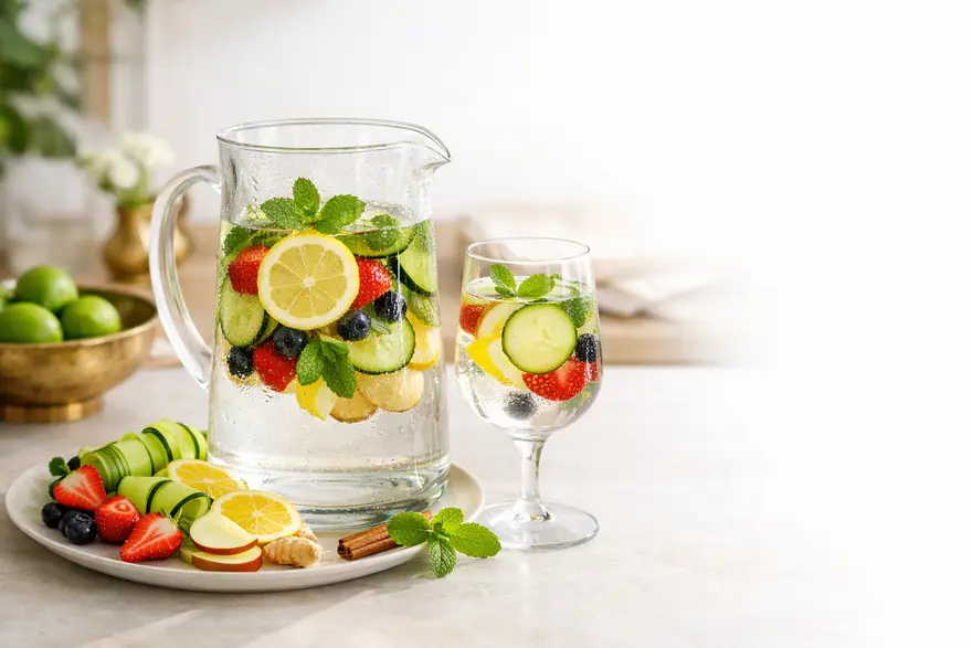
દૈનિક હાઇડ્રેશન માટે ડિટૉક્સ પાણીની 15 રેસીપી
ડિટૉક્સ પાણી એટલે ફળો, શાકભાજી, જડીબુટ્ટીઓ અથવા મસાલા સાથે ભેળવેલું પાણી. સાદા પાણીને વધુ આકર્ષક બનાવવાની આ તાજગીભરી રીત છે, ખાસ કરીને જો તમને દિવસ દરમિયાન પૂરતું પાણી પીવામાં મુશ્કેલી થતી હોય. “ડિટૉક્સ”ના વિચારને યોગ્ય રીતે સમજવું પણ મહત્વપૂર્ણ છે. ડિટૉક્સ પાણી તમારા શરીરને કોઈ ખાસ અથવા જાદુઈ રીતે શુદ્ધ કરતું નથી. તમારું યકૃત અને કિડની પહેલેથી જ આ કામ કુદરતી રીતે કરે છે. ડિટૉક્સ પાણી તમને હાઇડ્રેટેડ રહેવામાં, ખાંડવાળા પીણાં ઓછા કરવામાં અને હળવી, વધુ સ્વાદિષ્ટ દૈનિક રીત અપનાવવામાં મદદ કરી શકે છે. ડિટૉક્સ પાણી શું છે? ડિટૉક્સ પાણી એ સાદું પાણી છે, જેમાં લીંબુ, કાકડી, ફુદીનો, આદુ, બેરી અથવા સફરજનની સ્લાઇસ જેવા ઘટકો ઉમેરવામાં આવે છે. જ્યૂસથી વિપરીત, તેમાં બ્લેન્ડ કરેલો પલ્પ અથવા કુદરતી ખાંડની મોટી માત્રા હોતી નથી. ઘણા પૅકેજ્ડ પીણાંથી વિપરીત, તેમાં સિરપ અથવા ઉમેરેલા મીઠાશકારકોની પણ જરૂર પડતી નથી. તમે તેને ઇન્ફ્યુઝ્ડ વોટર તરીકે પણ જોઈ શકો છો. રોજિંદા ઉપયોગમાં, બંને શબ્દો સામાન્ય રીતે એક જ અર્થ આપે છે. ડિટૉક્સ પાણી હાઇડ્રેશનમાં કેવી રીતે મદદ કરે છે ઘણા લોકોને પાણી તાજું અને સુખદ સ્વાદવાળું લાગે ત્યારે વધુ પાણી પીવું સરળ લાગે છે. અહીં ડિટૉક્સ પાણી મદદરૂપ થઈ શકે છે. તે તમને ફિઝી પીણાં, પૅકેજ્ડ જ્યૂસ અને અન્ય મીઠાં પીણાંનો સરળ વિકલ્પ આપે છે. આ અર્થમાં, ડિટૉક્સ પાણી તમારા નિયમિત હાઇડ્રેશન પીણાંમાંના એક તરીકે સારી રીતે કામ કરી શકે છે. તે તમને દિવસભર વધુ નિયમિત રીતે પાણી પીવા માટે પણ પ્રોત્સાહિત કરી શકે છે, જે ઘણીવાર સાચો આરોગ્યલાભ હોય છે. ડિટૉક્સ પાણીમાં વપરાતા કેટલાક ઘટકો, જેમ કે સિટ્રસ ફળો, બેરી અને જડીબુટ્ટીઓમાં વનસ્પતિ સંયોજનો અને કુદરતી સ્વાદ પણ હોય છે. પરંતુ સૌથી મોટો ફાયદો હજી પણ પાણીમાંથી જ મળે છે. ડિટૉક્સ પાણીમાં સામાન્ય રીતે વપરાતા ઘટકો ડિટૉક્સ પાણીની રેસીપીમાં કેટલાક ઘટકો વારંવાર જોવા મળે છે, કારણ કે તે પીણાને ભારે અથવા વધારે મીઠું બનાવ્યા વગર સ્વાદ ઉમેરે છે. લીંબુ તેજસ્વી સ્વાદ ઉમેરે છે અને સૌથી લોકપ્રિય પસંદગીઓમાંનું એક છે. કાકડી પાણીનો સ્વાદ હળવો અને ઠંડક આપનારો બનાવે છે. ફુદીનો તાજગી ઉમેરે છે અને ઉનાળાના પીણાંમાં ઘણીવાર વપરાય છે. આદુ ગરમ, તીખો સ્વાદ આપે છે અને પાચન આરામ માટે ઘણીવાર પસંદ કરવામાં આવે છે. બેરી રંગ અને કુદરતી ફળવાળો સ્વાદ ઉમેરે છે. સફરજન હળવી મીઠાશ માટે સારી રીતે કામ કરે છે. તજ ગરમાશ અને ઊંડાણ ઉમેરે છે. બેઝિલ, તુલસી અને રોઝમેરી ડિટૉક્સ પાણીને વધુ સુગંધિત અને રસપ્રદ બનાવી શકે છે. ઘરે ડિટૉક્સ પાણી કેવી રીતે બનાવવું ડિટૉક્સ પાણી બનાવવું સરળ છે. તમારા ઘટકોને સારી રીતે ધોઈ લો. ફળો અથવા શાકભાજીને પાતળી સ્લાઇસમાં કાપો જેથી તેઓ સ્વાદ વધુ સરળતાથી છોડે. તેમને ઠંડા પાણીની બોટલ, જાર અથવા જગમાં ઉમેરો અને મિશ્રણને ઓછામાં ઓછા 1 to 2 કલાક માટે ફ્રિજમાં રહેવા દો. વધુ મજબૂત સ્વાદ માટે, તમે તેને રાતભર રાખી શકો છો. સ્વચ્છ કન્ટેનર વાપરો, પાણીને ફ્રિજમાં રાખો અને શ્રેષ્ઠ સ્વાદ તથા ખાદ્ય સુરક્ષા માટે દરરોજ તાજું બેચ બનાવો. ડિટૉક્સ પાણીની 15 સરળ રેસીપી લીંબુ અને ફુદીનાનું ડિટૉક્સ પાણી ઘટકો: 1 લીંબુ, સ્લાઇસ કરેલું ફુદીનાના પાનનો નાનો મુઠ્ઠીભર ભાગ 1 litre પાણી કેવી રીતે બનાવવું: ઠંડા પાણીમાં લીંબુ અને ફુદીનો ઉમેરો અને થોડા કલાકો માટે ફ્રિજમાં રાખો. તમને તે કેમ ગમી શકે: આ ડિટૉક્સ પાણીની સૌથી સરળ રેસીપીમાંની એક છે અને ગરમ હવામાનમાં ખાસ કરીને તાજગીભરી લાગે છે. કાકડી, લીંબુ અને ફુદીનાનું ડિટૉક્સ પાણી ઘટકો: અડધી કાકડી, સ્લાઇસ કરેલી 1 લીંબુ, સ્લાઇસ કરેલું ફુદીનાના થોડા પાન 1 litre પાણી કેવી રીતે બનાવવું: બધું એક જગમાં ઉમેરો અને 2 to 4 કલાક માટે ઠંડું કરો. તમને તે કેમ ગમી શકે: આ એક ક્લાસિક મિશ્રણ છે, જેનો સ્વાદ તાજો, ઠંડકભર્યો અને હળવો લાગે છે. દિવસભર ફ્રિજમાં રાખવા માટે આ સૌથી સરળ હાઇડ્રેશન પીણાંમાંનું એક છે. લીંબુ અને આદુનું ડિટૉક્સ પાણી ઘટકો: 1 લીંબુ, સ્લાઇસ કરેલું તાજા આદુની થોડા પાતળી સ્લાઇસ 1 litre પાણી કેવી રીતે બનાવવું: પીતા પહેલાં તેને થોડા કલાકો માટે ફ્રિજમાં ભેળવાઈ રહેવા દો. તમને તે કેમ ગમી શકે: જો તમને વધુ તીખો સ્વાદ ગમે છે, તો આ સંયોજન ગરમાશવાળું છતાં તાજું લાગી શકે છે. આદુ આધારિત કુદરતી ડિટૉક્સ પીણાં ગમતા લોકો ઘણીવાર તેને પસંદ કરે છે. સફરજન અને તજનું ડિટૉક્સ પાણી ઘટકો: અડધું સફરજન, પાતળી સ્લાઇસ કરેલું તજનો 1 નાનો ટુકડો 1 litre પાણી કેવી રીતે બનાવવું: પાણીમાં સફરજન અને તજ ઉમેરો અને ઘણા કલાકો માટે ફ્રિજમાં રાખો. તમને તે કેમ ગમી શકે: આ રેસીપીનો સ્વાદ હળવો અને આરામદાયક છે અને જો તમને સિટ્રસ પાણી કરતાં ઓછું ખાટું કંઈક ગમતું હોય તો તે સારી રીતે કામ કરે છે. સ્ટ્રોબેરી અને બેઝિલનું ડિટૉક્સ પાણી ઘટકો: 4 to 5 સ્ટ્રોબેરી, સ્લાઇસ કરેલી બેઝિલના થોડા પાન 1 litre પાણી કેવી રીતે બનાવવું: ઘટકોને ભેગા કરો અને પીરસતા પહેલાં સારી રીતે ઠંડું કરો. તમને તે કેમ ગમી શકે: આ મિશ્રણ ભારે બન્યા વગર થોડું મીઠું અને સુગંધિત લાગે છે. નારંગી અને ફુદીનાનું ડિટૉક્સ પાણી ઘટકો: 1 નારંગી, સ્લાઇસ કરેલી ફુદીનાના થોડા પાન 1 litre પાણી કેવી રીતે બનાવવું: 2 to 4 કલાક માટે ફ્રિજમાં પલળવા દો. તમને તે કેમ ગમી શકે: તેનો સ્વાદ તેજસ્વી અને તાજગીભર્યો લાગે છે અને જો સાદું પાણી બોરિંગ લાગે તો તે સારો વિકલ્પ બની શકે છે. અનાનસ અને આદુનું ડિટૉક્સ પાણી ઘટકો: અનાનસના થોડા નાના ટુકડા આદુની થોડા પાતળી સ્લાઇસ 1 litre પાણી કેવી રીતે બનાવવું: થોડા કલાકો માટે ફ્રિજમાં રાખો અને ઠંડું પીરસો. તમને તે કેમ ગમી શકે: આ રેસીપીમાં વધુ મજબૂત ટ્રોપિકલ સ્વાદ છે અને જો તમે તમારી દૈનિક રીતમાં વિવિધતા ઈચ્છતા હો તો તે સારી રીતે કામ કરે છે. તરબૂચ અને ફુદીનાનું ડિટૉક્સ પાણી ઘટકો: 1 cup તરબૂચના ક્યુબ્સ ફુદીનાના થોડા પાન 1 litre પાણી કેવી રીતે બનાવવું: ઠંડા પાણીમાં ઉમેરો અને થોડા સમય માટે ભેળવાઈ રહેવા દો. તમને તે કેમ ગમી શકે: આ ઉનાળાનો એક સુખદ વિકલ્પ છે, જેનો સ્વાદ તાજો અને કુદરતી રીતે મીઠો લાગે છે. બેરી ડિટૉક્સ પાણી ઘટકો: થોડી સ્ટ્રોબેરી થોડી બ્લૂબેરી થોડી રાસ્પબેરી 1 litre પાણી કેવી રીતે બનાવવું: પાણીમાં ઉમેરતા પહેલાં થોડા બેરીને હળવેથી કચડી લો, પછી ફ્રિજમાં રાખો. તમને તે કેમ ગમી શકે: બેરી પાણી રંગીન, હળવા ફળવાળા સ્વાદવાળું અને બપોર દરમિયાન પીવા માટે સરળ હોય છે. કાકડી અને લાઇમનું ડિટૉક્સ પાણી ઘટકો: અડધી કાકડી, સ્લાઇસ કરેલી 1 લાઇમ, સ્લાઇસ કરેલું 1 litre પાણી કેવી રીતે બનાવવું: પીતા પહેલાં 2 to 4 કલાક માટે ઠંડું કરો. તમને તે કેમ ગમી શકે: આ મિશ્રણ સ્વચ્છ અને ઠંડકભર્યું છે. બહાર સમય વિતાવ્યા પછી તે ખાસ કરીને તાજગીભર્યું લાગી શકે છે. બીટરૂટ અને લીંબુનું ડિટૉક્સ પાણી ઘટકો: બીટરૂટની થોડા પાતળી સ્લાઇસ અડધું લીંબુ, સ્લાઇસ કરેલું 1 litre પાણી કેવી રીતે બનાવવું: સ્વાદ સંતુલિત રહે તે માટે બીટરૂટની માત્રા ઓછી જ વાપરો. ઉપયોગ પહેલાં ઠંડું કરો. તમને તે કેમ ગમી શકે: આ રેસીપી તમારા પાણીને આકર્ષક રંગ અને વધુ માટી જેવી ગંધવાળો સ્વાદ આપે છે. તુલસી અને લીંબુનું ડિટૉક્સ પાણી ઘટકો: તુલસીના થોડા પાન અડધું લીંબુ, સ્લાઇસ કરેલું 1 litre પાણી કેવી રીતે બનાવવું: પાણીમાં ઉમેરો અને થોડા કલાકો માટે ફ્રિજમાં રાખો. તમને તે કેમ ગમી શકે: આ હળવું અને સુગંધિત લાગે છે અને જો તમને સરળ અને હર્બલ કંઈક જોઈએ હોય તો સારો વિકલ્પ છે. સફરજન, લીંબુ અને આદુનું ડિટૉક્સ પાણી ઘટકો: સફરજનની થોડા સ્લાઇસ અડધું લીંબુ, સ્લાઇસ કરેલું આદુની થોડા સ્લાઇસ 1 litre પાણી કેવી રીતે બનાવવું: તેને થોડા કલાકો માટે ફ્રિજમાં પલળવા દો. તમને તે કેમ ગમી શકે: આ મિશ્રણ એક જ ગ્લાસમાં મીઠાશ, તાજગી અને ગરમાશ આપે છે. ગ્રેપફ્રૂટ અને રોઝમેરીનું ડિટૉક્સ પાણી ઘટકો: ગ્રેપફ્રૂટની થોડા સ્લાઇસ રોઝમેરીની એક નાની ડાળી 1 litre પાણી કેવી રીતે બનાવવું: પીતા પહેલાં ઘણા કલાકો માટે ઠંડું કરો. તમને તે કેમ ગમી શકે: તેનો સ્વાદ મજબૂત અને થોડો કડવો હોય છે, જે ખૂબ મીઠાં પીણાં ન ગમતા લોકોને અનુકૂળ પડે છે. કિવી અને ફુદીનાનું ડિટૉક્સ પાણી ઘટકો: 1 કિવી, સ્લાઇસ કરેલું ફુદીનાના થોડા પાન 1 litre પાણી કેવી રીતે બનાવવું: 2 to 4 કલાક માટે ફ્રિજમાં રાખો. તમને તે કેમ ગમી શકે: આ રેસીપીનો સ્વાદ તાજો અને ફળવાળો લાગે છે અને વધુ સામાન્ય લીંબુ આધારિત વિકલ્પો કરતાં સારો ફેરફાર છે. ડિટૉક્સ પાણી પીવાનો શ્રેષ્ઠ સમય ડિટૉક્સ પાણી પીવાનો કોઈ એક જ પરફેક્ટ સમય નથી. શ્રેષ્ઠ સમય એ છે જ્યારે તે તમને નિયમિત રીતે વધુ પાણી પીવામાં મદદ કરે. કેટલાક લોકો તેને સવારે સૌથી પહેલા પીવાનું પસંદ કરે છે, જ્યારે કેટલાક દિવસભર પોતાની સાથે બોટલ રાખે છે. બંને રીતો સારી રીતે કામ કરી શકે છે. સવારે ડિટૉક્સ પાણી સવારનું ડિટૉક્સ પાણી તમને દિવસની હળવી શરૂઆત કરવામાં મદદ કરી શકે છે, ખાસ કરીને જો તમે સામાન્ય રીતે સૂકાપો અથવા સુસ્તી અનુભવતા જાગો છો. ઘણા લોકોને આ સમયે લીંબુ આધારિત રેસીપી ગમે છે. જો તમને લીંબુ પાણીના ફાયદામાં રસ હોય, તો સરળ લીંબુ અને ફુદીનો અથવા લીંબુ અને આદુનું મિશ્રણ શરૂ કરવા માટે સરળ આદત બની શકે છે. તેમ છતાં, જો સિટ્રસથી તમારા પેટમાં તકલીફ થાય, તો તેને જબરદસ્તી પીવાની જરૂર નથી. કાકડી અથવા બેરી આધારિત વિકલ્પો વધુ હળવા હોઈ શકે છે. દિવસભર ડિટૉક્સ પાણી ડિટૉક્સ પાણી આખો દિવસ પીવાના પીણાં તરીકે પણ સારી રીતે કામ કરે છે. તે કામ, મુસાફરી અથવા ગરમ હવામાન દરમિયાન ખાંડવાળા પીણાંની જગ્યાએ ઉપયોગી થઈ શકે છે. ચોક્કસ રેસીપી કરતાં નજીકમાં બોટલ રાખવી ઘણીવાર વધુ મહત્વપૂર્ણ હોય છે. જો તમે વ્યાયામ કરો છો, વધુ પરસેવો આવે છે અથવા લાંબા કલાકો બહાર વિતાવો છો, તો યાદ રાખો કે સાદું પાણી પણ મહત્વપૂર્ણ છે. દરરોજ કેટલું ડિટૉક્સ પાણી પીવું જોઈએ? ડિટૉક્સ પાણી તમારા કુલ પ્રવાહી સેવનમાં ગણાઈ શકે છે. મોટાભાગના પુખ્ત વયના લોકો માટે સામાન્ય પ્રવાહી માર્ગદર્શન લગભગ 6 to 8 કપ અથવા ગ્લાસ દરરોજ છે, જોકે ગરમી, બીમારી, વ્યાયામ અથવા ગર્ભાવસ્થા દરમિયાન તમારી જરૂરિયાત વધી શકે છે. તમારે ફક્ત ડિટૉક્સ પાણી જ પીવાની જરૂર નથી. તે સાદા પાણીને સંપૂર્ણપણે બદલે નહીં, પરંતુ તેને પૂરક હોવું જોઈએ. એક વ્યવહારુ અભિગમ એ છે કે તમારા સામાન્ય દૈનિક પ્રવાહીઓ સાથે ઇન્ફ્યુઝ્ડ પાણીની એક કે બે બોટલ અથવા ગ્લાસ લેવાં. શું ડિટૉક્સ પાણી વજન ઘટાડવામાં મદદ કરી શકે છે? ડિટૉક્સ પાણી સીધું ચરબી બર્ન કરતું નથી. તે વજન ઘટાડવાનો શોર્ટકટ નથી અને તેને એવી રીતે રજૂ કરવું જોઈએ નહીં. તે શું કરી શકે છે તે છે વધુ આરોગ્યપ્રદ પસંદગીઓને ટેકો આપવો. જો તમે ખાંડવાળા પીણાંની જગ્યાએ ડિટૉક્સ પાણી વાપરો છો, તો તમે વધારાની કેલરી ઘટાડીને શકો છો. તે કેટલાક લોકોને તરસને ભૂખ સમજીને થતા નાસ્તાને સંભાળવામાં પણ મદદ કરી શકે છે. તેથી હા, ડિટૉક્સ પાણી વજન સંચાલનને પરોક્ષ રીતે ટેકો આપી શકે છે, પરંતુ ફક્ત સંતુલિત આહાર અને સક્રિય જીવનશૈલીના ભાગરૂપે. શું ડિટૉક્સ પાણી ખરેખર તમારા શરીરને ડિટૉક્સ કરે છે? ઘણા ટ્રેન્ડ્સ સૂચવે છે તે રીતે નહીં. તમારા શરીરમાં પહેલેથી જ પોતાનું ડિટૉક્સ તંત્ર છે, મુખ્યત્વે યકૃત અને કિડની દ્વારા. ડિટૉક્સ પાણી તે કામ સંભાળતું નથી. તે હાઇડ્રેશનને ટેકો આપે છે, અને યોગ્ય હાઇડ્રેશન તમારા શરીરને સારી રીતે કાર્ય કરવામાં મદદ કરે છે. એટલા માટે ડિટૉક્સ પાણીને શુદ્ધિકરણ ઉપચાર કરતાં આરોગ્યપ્રદ આદત તરીકે વિચારવું વધુ સારું છે. કુદરતી ડિટૉક્સ પીણાંમાં, તેની સૌથી મોટી શક્તિ એ છે કે તે ઉમેરેલી ખાંડ વગર તમને વધુ પાણી પીવા માટે પ્રોત્સાહિત કરે છે. ડિટૉક્સ પાણી અંગે કોને સાવચેત રહેવું જોઈએ ડિટૉક્સ પાણી મોટાભાગના સ્વસ્થ પુખ્ત વયના લોકો માટે સામાન્ય રીતે સુરક્ષિત છે, પરંતુ કેટલાક લોકોએ વધુ સાવચેત રહેવું જોઈએ. જો તમને એસિડ રિફ્લક્સ હોય, તો સિટ્રસ ફળો, ફુદીનો અને તીવ્ર સ્વાદવાળું પાણી લક્ષણોને વધુ ખરાબ કરી શકે છે. જો તમને ક્રોનિક કિડની રોગ હોય અથવા તમને પ્રવાહી મર્યાદિત રાખવાનું કહેવામાં આવ્યું હોય, તો તમારું સેવન વધારતા પહેલાં તમારા ડૉક્ટર સાથે વાત કરો. જો કોઈ ચોક્કસ ફળ, જડીબુટ્ટી અથવા મસાલો તમને અનુકૂળ ન હોય, તો તેને ટાળો. બાળકો, ગર્ભવતી મહિલાઓ અને વૃદ્ધોએ રેસીપી સરળ રાખવી જોઈએ અને ડૉક્ટરે અન્યથા સલાહ ન આપી હોય ત્યાં સુધી અસામાન્ય જડીબુટ્ટીઓ અથવા ખૂબ મજબૂત મિશ્રણો ટાળવા જોઈએ. ડિટૉક્સ પાણી ક્યારે ટાળવું તમારે ડિટૉક્સ પાણી ટાળવું અથવા મર્યાદિત કરવું જોઈએ જો: સિટ્રસ અથવા ફુદીનો હાર્ટબર્ન શરૂ કરે આદુ અથવા મસાલા તમારા પેટમાં ચીડ ઉત્પન્ન કરે તમને પ્રવાહી મર્યાદિત રાખવાનું કહેવામાં આવ્યું હોય તમે એવા ઘટકો વાપરી રહ્યા હો જે એલર્જી કરી શકે પાણી લાંબા સમય સુધી બહાર રહ્યું હોય અથવા તાજી સુગંધ આવતી ન હોય જો કોઈ રેસીપીથી તમને અસ્વસ્થતા થાય, તો ઘટકો બદલો અથવા ફરીથી સાદા પાણી પર પાછા આવો. ડિટૉક્સ પાણીને દૈનિક આદત બનાવવા માટેની ટીપ્સ સરળ રીતે શરૂ કરો. એક બોટલમાં પાંચ ઘટકોની જરૂર નથી. એક ફળ અને એક જડીબુટ્ટી ઘણીવાર પૂરતી હોય છે. જો સવાર વ્યસ્ત હોય, તો તમારી બોટલ અગાઉની રાત્રે તૈયાર કરો. ઋતુ અનુસાર મળતા ઉત્પાદનો વાપરો. ફ્રિજમાં એક જગ રાખો. ખાંડ, સિરપ અથવા પૅકેજ્ડ સ્વાદવર્ધકો ઉમેરવાનું ટાળો. તમારી દૈનિક રીત જેટલી સરળ લાગે, તેને ચાલુ રાખવાની શક્યતા તેટલી વધારે હોય છે. સુરક્ષા અને સંગ્રહ માટેની ટીપ્સ ફળો, શાકભાજી અને જડીબુટ્ટીઓનો ઉપયોગ કરતાં પહેલાં તેમને સારી રીતે ધોઈ લો. કાપેલા ઉત્પાદનો પાણીમાં ઉમેર્યા પછી તેને ફ્રિજમાં રાખો. શ્રેષ્ઠ તાજગી માટે, એક દિવસમાં પૂરી કરી શકો તેટલું જ બનાવો. જો ઘટકો ભીંજાઈને ઢીલા દેખાય, પાણીમાંથી ખરાબ ગંધ આવે અથવા સ્વાદ કડવો થઈ જાય, તો તેને ફેંકી દો અને તાજું બેચ બનાવો. નિષ્કર્ષ ડિટૉક્સ પાણી દૈનિક હાઇડ્રેશનને ટેકો આપવાની સરળ અને આનંદદાયક રીત છે. તે સાદા પાણીને વધુ રસપ્રદ બનાવી શકે છે, ખાંડવાળા પીણાં ઓછા કરવામાં મદદ કરી શકે છે અને સંતુલિત વેલનેસ રૂટીનમાં સરળતાથી ફિટ થઈ શકે છે. તે શું કરી શકતું નથી તે છે તમારા શરીરના કુદરતી ડિટૉક્સ તંત્રને બદલવું અથવા આરોગ્ય સંબંધિત ચિંતાઓને એકલા હાથે ઉકેલવું. તેને ચમત્કારી ઉપચાર નહીં, પરંતુ વ્યવહારુ આદત તરીકે વિચારો. જો તમે વ્યાપક સ્વાસ્થ્ય તપાસ અથવા વેલનેસ યોજનાના ભાગરૂપે વધુ આરોગ્યપ્રદ દૈનિક રીતો બનાવી રહ્યા હો, તો મેટ્રોપોલિસ હેલ્થકેર વ્યાપક ડાયગ્નોસ્ટિક સેવાઓ, સુવિધાજનક ઘરેથી સેમ્પલ કલેક્શન અને વેબસાઇટ, ઍપ, કૉલ અથવા વોટ્સએપ દ્વારા સરળ બુકિંગ પ્રદાન કરે છે. વારંવાર પૂછાતા પ્રશ્નો શું ડિટૉક્સ પાણી ખરેખર તમારા શરીરને ડિટૉક્સ કરે છે? ના, તબીબી અર્થમાં નહીં. તમારું યકૃત અને કિડની પહેલેથી જ તમારા શરીરને ડિટૉક્સિફાઇ કરે છે. ડિટૉક્સ પાણી મુખ્યત્વે હાઇડ્રેશનને ટેકો આપીને મદદ કરે છે. શું હું દરરોજ ડિટૉક્સ પાણી પી શકું? હા, મોટાભાગના લોકો તેને દરરોજ માણી શકે છે, જો ઘટકો તેમને અનુકૂળ હોય અને તે સંતુલિત પ્રવાહી સેવનનો ભાગ હોય. ડિટૉક્સ પાણી કેટલો સમય સંગ્રહ કરી શકાય? તે તાજું બનાવવું અને ફ્રિજમાં રાખવું શ્રેષ્ઠ છે. શ્રેષ્ઠ સ્વાદ અને સુરક્ષા માટે, તે જ દિવસે પૂરું કરવાનો પ્રયત્ન કરો. શું ડિટૉક્સ પાણી સામાન્ય પાણીની જગ્યા લઈ શકે? ના. તે તમારા પ્રવાહી સેવનમાં ગણાઈ શકે છે, પરંતુ સાદું પાણી હજી પણ તમારા દિવસનો નિયમિત ભાગ રહેવું જોઈએ. શું ડિટૉક્સ પાણી બાળકો માટે સુરક્ષિત છે? મોટા બાળકો માટે, સરળ ફળ-મિશ્રિત પાણી મર્યાદિત માત્રામાં યોગ્ય હોઈ શકે છે. ઘટકો હળવા રાખો, મજબૂત જડીબુટ્ટીઓ અથવા મસાલા ટાળો અને તેને નિયમિત પાણીના બદલે ઉપયોગમાં ન લો.
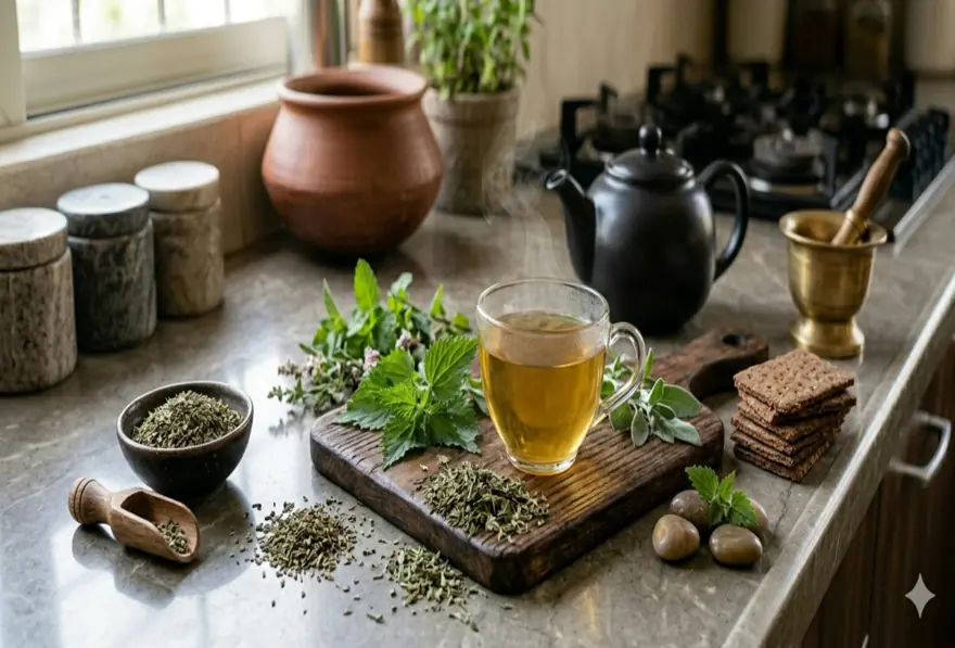
નેટલ ચાના 10 ફાયદા: સોજા ઘટાડવામાં, ડિટૉક્સ અને એલર્જી રાહતમાં મદદ
નેટલ ચા એ સ્ટિંગિંગ નેટલ છોડના પાનમાંથી બનેલું હર્બલ પીણું છે. તાજો છોડ તમારી ત્વચામાં ચુભન કરી શકે છે, પરંતુ પાનને સૂકવવાથી અથવા ઉકાળવાથી તેની અસર દૂર થાય છે અને તે ચામાં ઉપયોગ માટે સુરક્ષિત બને છે. નેટલ ચાનો પરંપરાગત હર્બલ પ્રથામાં લાંબા સમયથી ઉપયોગ થાય છે, અને હવે તેના પોષક તત્ત્વો તથા સંભવિત સ્વાસ્થ્ય ફાયદાઓને કારણે તે ધ્યાન ખેંચી રહી છે. જો તમે નેટલ ચાના ફાયદા વિશે જાણકારી મેળવી રહ્યા હો, તો અપેક્ષાઓ વાસ્તવિક રાખવી મહત્વપૂર્ણ છે. નેટલ ચા સ્વસ્થ જીવનશૈલીનો ઉપયોગી ભાગ બની શકે છે, પરંતુ તે દીર્ઘકાલીન બીમારીઓનો ઇલાજ નથી. કેટલાક ફાયદાઓ માટે વધુ સારા પુરાવા છે, જ્યારે કેટલાક દાવાઓ માટે હજુ વધુ માનવીય સંશોધનની જરૂર છે. નેટલ ચા શું છે? નેટલ ચા ઉર્ટિકા ડાયોઇકા, જેને સ્ટિંગિંગ નેટલ પણ કહેવામાં આવે છે, તેમાંથી બને છે. તે પાંદડાવાળો છોડ છે, જે યુરોપ, એશિયા અને ઉત્તર અમેરિકાના ઘણા ભાગોમાં ઉગે છે. પાનને સૂકવ્યા અથવા ઉકાળ્યા પછી તેની ચુભનવાળી અસર નિષ્ક્રિય થઈ જાય છે. આ ચાનો સ્વાદ માટી જેવો અને થોડો ઘાસ જેવો હોય છે. કેટલાક લોકોને તે પાલક અથવા અન્ય લીલી હર્બલ ચા જેવી લાગે છે. તમે તેને સૂકવેલા પાન, ટી બેગ્સ અથવા કાળજીપૂર્વક તૈયાર કરેલા તાજા પાનથી બનાવી શકો છો. આ જાણવું પણ ઉપયોગી છે કે નેટલ પાન અને નેટલ મૂળ એકસરખાં નથી. નેટલ પાનનો ઉપયોગ ચા અને સામાન્ય હર્બલ ઉપયોગ માટે વધુ થાય છે, જ્યારે નેટલ મૂળ પર પ્રોસ્ટેટ સંબંધિત ચિંતાઓ માટે વધુ અભ્યાસ કરવામાં આવ્યો છે. નેટલ ચાનું પોષણ પ્રોફાઇલ સ્ટિંગિંગ નેટલ તેના સમૃદ્ધ પોષણ પ્રોફાઇલ માટે જાણીતું છે. છોડમાં વિટામિન A, C અને K જેવા વિટામિન્સ સાથે કેલ્શિયમ, આયર્ન, મેગ્નેશિયમ અને પોટેશિયમ જેવા ખનિજો હોય છે. તેમાં પોલીફિનોલ્સ અને ફ્લેવોનોઇડ્સ જેવા વનસ્પતિ સંયોજનો પણ હોય છે. તેમ છતાં, નેટલ ચાનો એક કપ નેટલ પાનની સંપૂર્ણ સર્વિંગ ખાવાથી મળતા પોષક તત્ત્વો જેટલી માત્રા આપશે નહીં. ચા હજી પણ હળવો સ્રોત છે, પરંતુ તે કેફીન-મુક્ત હર્બલ પીણું બની શકે છે, જે તમારી દૈનિક રીતમાં વિવિધતા ઉમેરે છે. નેટલ ચાના 10 ફાયદા એન્ટીઑક્સિડન્ટ્સથી સમૃદ્ધ નેટલમાં એન્ટીઑક્સિડન્ટ સંયોજનો હોય છે, જે તમારા કોષોને ઑક્સિડેટિવ તણાવથી બચાવવામાં મદદ કરે છે. આ એક કારણ છે કે તેને સામાન્ય સુખાકારી માટે સહાયક જડીબુટ્ટી તરીકે ઘણીવાર ચર્ચવામાં આવે છે. શરીરની સોજા સંબંધિત પ્રતિક્રિયાને ટેકો આપવામાં મદદ કરી શકે છે નેટલનો ઉપયોગ ઘણીવાર તેની સોજા ઘટાડવાની સંભાવના માટે થાય છે. નેટલ ચા પીવાના આ સૌથી વધુ ચર્ચાતા ફાયદાઓમાંનો એક છે, ખાસ કરીને રોજિંદા સ્વાસ્થ્ય માટે વધુ વનસ્પતિ આધારિત ટેકો ઈચ્છતા લોકોમાં. કેટલીક એલર્જી રાહત આપી શકે છે મોસમી એલર્જી માટે નેટલનો હર્બલ પ્રથામાં લાંબા સમયથી ઉપયોગ થાય છે. કેટલાક પ્રારંભિક માનવીય સંશોધન સૂચવે છે કે તે કેટલાક લોકોમાં છીંક અને નાકમાં ચીડ જેવા લક્ષણો ઘટાડવામાં મદદ કરી શકે છે. સાંધાના આરામને ટેકો આપી શકે છે નેટલ સોજા ઘટાડવાની પ્રવૃત્તિ સાથે જોડાયેલી હોવાથી, સાંધાના આરામ માટે ટેકો ઈચ્છતા લોકો પણ ક્યારેક તેનો ઉપયોગ કરે છે. તે સારવારની જગ્યા લેવું જોઈએ નહીં, પરંતુ તે વધુ વ્યાપક વેલનેસ અભિગમમાં ફિટ થઈ શકે છે. મૂત્ર સ્વાસ્થ્યને ટેકો આપી શકે છે નેટલનો ક્યારેક તેના હળવા ડાય્યુરેટિક અસરને કારણે મૂત્ર સહાય માટે ઉપયોગ થાય છે. આ સામાન્ય પ્રવાહી સંતુલન અને મૂત્ર પ્રવાહને ટેકો આપવા મદદ કરી શકે છે. પુરુષોમાં પ્રોસ્ટેટ સ્વાસ્થ્યને ટેકો આપી શકે છે આ ફાયદો નેટલ ચા કરતાં નેટલ મૂળ સાથે વધુ મજબૂત રીતે જોડાયેલો છે. તેમ છતાં, મોટું થયેલું પ્રોસ્ટેટ સંબંધિત મૂત્ર લક્ષણો સાથે નેટલની ઘણીવાર ચર્ચા થાય છે. ચાને યોગ્ય તબીબી મૂલ્યાંકનનો વિકલ્પ માનવો જોઈએ નહીં. પ્રવાહી સંતુલનમાં મદદ કરી શકે છે લોકો ઘણીવાર નેટલ ચાને “ડિટૉક્સ” ચા તરીકે વર્ણવે છે, પરંતુ આ શબ્દ ગેરમાર્ગે દોરી શકે છે. તમારા શરીરમાં પહેલેથી જ ડિટૉક્સ સિસ્ટમ્સ હોય છે. નેટલ ચા શું કરી શકે છે તે છે તમારી સામાન્ય દૈનિક રીતના ભાગરૂપે હાઇડ્રેશન અને હળવા પ્રવાહી સંતુલનને ટેકો આપવો. સ્વસ્થ બ્લડ શુગર સ્તરને ટેકો આપી શકે છે નેટલ તૈયારીઓ પરના કેટલાક અભ્યાસો બ્લડ શુગર નિયંત્રણ માટે સંભવિત ટેકો સૂચવે છે. આ આશાસ્પદ છે, પરંતુ પુરાવા એટલા મજબૂત નથી કે નેટલ ચાને ડાયાબિટીસના ઉપાય તરીકે જોવામાં આવે. સ્વસ્થ બ્લડ પ્રેશરને ટેકો આપી શકે છે બ્લડ પ્રેશર પર સંભવિત અસર માટે પણ નેટલનો અભ્યાસ કરવામાં આવ્યો છે. આ ક્ષેત્રમાં હજી વધુ સંશોધનની જરૂર છે, તેથી તેને સાબિત સારવાર નહીં પરંતુ સંભવિત સહાયક ફાયદા તરીકે જોવું શ્રેષ્ઠ છે. કુદરતી રીતે કેફીન-મુક્ત હર્બલ પીણું જો તમને કેફીન વગરનું ગરમ પીણું જોઈએ હોય, તો નેટલ ચા વ્યવહારુ વિકલ્પ બની શકે છે. તે નિયમિત ચા અથવા કોફીનો હર્બલ વિકલ્પ આપે છે અને વધુ હળવું દૈનિક પીણું ઈચ્છતા લોકોને અનુકૂળ પડી શકે છે. નેટલ ચા સોજામાં કેવી રીતે મદદ કરી શકે છે સોજો તમારા શરીરની તણાવ, બીમારી અને ઇજાઓ સામેની કુદરતી પ્રતિક્રિયાનો ભાગ છે. પરંતુ સતત સોજો સમય જતાં સમસ્યા બની શકે છે. નેટલમાં એવા સંયોજનો હોય છે, જેની સોજા ઘટાડતી અસર માટે અભ્યાસ કરવામાં આવી રહ્યો છે. આ કારણે નેટલ ચાને ઘણીવાર સાંધાના આરામ અને મોસમી ચીડ માટેના ટેકા સાથે જોડવામાં આવે છે. તેમ છતાં, વધુ મજબૂત દાવાઓ લેબ અભ્યાસો, પ્રાણી સંશોધન અથવા માત્ર ચા કરતાં નેટલના અન્ય સ્વરૂપો પરના અભ્યાસોમાંથી આવે છે. તેથી નેટલ ચાને સારવાર નહીં પરંતુ સહાયક પીણું તરીકે વર્ણવવું વધુ સારું છે. શું નેટલ ચા તમારા શરીરને ડિટૉક્સ કરે છે? ઘણા લોકો નેટલ ચા પીવાના ફાયદા શોધે છે કારણ કે તેઓ કુદરતી ડિટૉક્સ પીણું ઈચ્છે છે. પરંતુ “ડિટૉક્સ” કોઈ ચોક્કસ તબીબી શબ્દ નથી. નેટલ ચા કોઈ જાદુઈ રીતે ઝેરી તત્ત્વો બહાર કાઢતી નથી. તે શું કરી શકે છે તે છે હાઇડ્રેશનને ટેકો આપવો અને હળવા ડાય્યુરેટિક તરીકે કામ કરવું, જે શરીરમાં સામાન્ય પ્રવાહી ગતિમાં મદદ કરી શકે છે. આ ફાયદાને સમજવાની આ વધુ ચોક્કસ અને ઉપયોગી રીત છે. એલર્જી રાહત માટે નેટલ ચાના ફાયદા નેટલને ક્યારેક કુદરતી એન્ટિહિસ્ટામાઇન તરીકે વર્ણવવામાં આવે છે. કેટલાક નાના અભ્યાસો સૂચવે છે કે તે મોસમી એલર્જીના લક્ષણો જેમ કે છીંક, ખંજવાળ અને વહેતું નાકમાં મદદ કરી શકે છે. જોકે, પુરાવા મિશ્ર છે. કેટલાક લોકોને તે મદદરૂપ લાગે શકે છે, જ્યારે અન્ય લોકોને બહુ ફરક ન જણાય. જો તમારી એલર્જી ગંભીર, સતત હોય અથવા શ્વાસ લેવામાં અસર કરતી હોય, તો ફક્ત હર્બલ ચા પર આધાર રાખવાને બદલે ડૉક્ટર સાથે વાત કરવી શ્રેષ્ઠ છે. નેટલ ચા અને હોર્મોનલ સંતુલન તમે હોર્મોન્સ માટે નેટલ ચાના ફાયદા વિશેની સામગ્રી જોઈ શકો છો, પરંતુ આ વિષયને કાળજીપૂર્વક સંભાળવાની જરૂર છે. નેટલ ચા વ્યાપક અથવા વિશ્વસનીય રીતે હોર્મોન્સનું સંતુલન કરે છે એવું કહેવા માટે પૂરતા મજબૂત પુરાવા નથી. વધુ જાણીતો હોર્મોનલ સંબંધ પરોક્ષ છે. નેટલ, ખાસ કરીને મૂળ, ક્યારેક પુરુષોના પ્રોસ્ટેટ સ્વાસ્થ્ય અને મૂત્ર લક્ષણો સાથે જોડાઈને ચર્ચાય છે. પરંતુ તે દરેક માટે નેટલ ચા હોર્મોનલ સંતુલનને ટેકો આપે છે એવું કહેવા જેવું નથી. અહીં વધુ દાવા ટાળવા શ્રેષ્ઠ છે. નેટલ ચા હૃદયના સ્વાસ્થ્યને કેવી રીતે ટેકો આપી શકે છે જો નેટલ ચા તમને ખાંડવાળા પીણાં ઘટાડવામાં મદદ કરે અને સ્વસ્થ જીવનશૈલીમાં ફિટ થાય, તો તે સામાન્ય અર્થમાં હૃદયના સ્વાસ્થ્યને ટેકો આપી શકે છે. બ્લડ પ્રેશર માટે નેટલની સંભવિત ભૂમિકા અંગે પણ થોડો સંશોધન રસ છે. તેમ છતાં, હાઇપરટેન્શન, ઊંચા કોલેસ્ટ્રોલ અથવા હૃદયરોગની યોગ્ય સારવારના બદલે નેટલ ચાનો ઉપયોગ કરવો જોઈએ નહીં. તેને વધુ વ્યાપક સ્વસ્થ દૈનિક રીતનો એક નાનો ભાગ માનો. નેટલ ચા સાથે રોગપ્રતિકારક કાર્યને ટેકો આપવો નેટલ વનસ્પતિ સંયોજનોથી સમૃદ્ધ છે અને સામાન્ય શરીર કાર્યને ટેકો આપતા પોષક તત્ત્વો ધરાવે છે. તેથી તેને ઘણીવાર રોગપ્રતિકારક ટેકા અંગેની ચર્ચામાં સામેલ કરવામાં આવે છે. પરંતુ નેટલ ચા નાટ્યાત્મક રીતે રોગપ્રતિકારક શક્તિ “વધારે” છે એમ કહેવા કરતાં તે સમગ્ર સુખાકારીને ટેકો આપે છે એમ કહેવું વધુ ચોક્કસ છે. સારી ઊંઘ, સંતુલિત પોષણ, વ્યાયામ અને તણાવ સંચાલન હજી પણ ઘણાં વધુ મહત્વપૂર્ણ છે. ત્વચાના સ્વાસ્થ્ય માટે નેટલ ચા નેટલમાં એન્ટીઑક્સિડન્ટ્સ હોય છે અને તે સોજા ઘટાડતા ગુણધર્મો સાથે જોડાયેલી હોવાથી, કેટલાક લોકો તેનો ઉપયોગ ત્વચા માટે અનુકૂળ દૈનિક રીતના ભાગરૂપે કરે છે. મોસમી ત્વચા ચીડ સાથે પણ ક્યારેક તેની ચર્ચા થાય છે. તેમ છતાં, નેટલ ચા ત્વચા રોગની સીધી સારવાર નથી. જો તમને સતત એક્ઝિમા, હાઇવ્સ, રેશ અથવા એક્ને હોય અને તેમાં સુધારો ન થતો હોય, તો તમારે તબીબી સલાહ લેવી જોઈએ. પાચન સ્વાસ્થ્ય: નેટલ ચાની ભૂમિકા કેટલાક લોકો સામાન્ય પાચન આરામ માટે નેટલ ચા પીવે છે. ગરમ હર્બલ પીણું શાંત કરનારું લાગી શકે છે, અને નેટલ સરળ કેફીન-મુક્ત વિકલ્પ ઈચ્છતા લોકોને અનુકૂળ પડી શકે છે. સાથે સાથે, નેટલ દરેકને અનુકૂળ નથી આવતું. કેટલાક લોકોને પેટમાં તકલીફ, પાતળા ઝાડા અથવા પેટમાં અસ્વસ્થતા અનુભવાય શકે છે. તેથી ઓછી માત્રાથી શરૂ કરીને તમારું શરીર કેવી પ્રતિક્રિયા આપે છે તે જોવું શ્રેષ્ઠ છે. શું નેટલ ચા દરેક માટે સુરક્ષિત છે? નેટલ ચા દરેક માટે યોગ્ય નથી. મધ્યમ માત્રામાં, ઘણા પુખ્ત વયના લોકો તેને સામાન્ય રીતે સારી રીતે સહન કરે છે. પરંતુ તે હજી પણ આડઅસરો આપી શકે છે અથવા દવાઓ સાથે ક્રિયા કરી શકે છે. સંભવિત આડઅસરોમાં શામેલ છે: પેટમાં તકલીફ ઝાડા અથવા કબજિયાત સંવેદનશીલ લોકોમાં ત્વચા પ્રતિક્રિયાઓ કેટલાક વપરાશકર્તાઓમાં એલર્જીના લક્ષણો ગર્ભાવસ્થામાં ખાસ સાવચેતી જરૂરી છે. ગર્ભાશયને ઉત્તેજિત કરવાની ચિંતાઓને કારણે ગર્ભાવસ્થામાં નેટલની સામાન્ય રીતે ભલામણ કરવામાં આવતી નથી. જો તમે ગર્ભવતી હો, સ્તનપાન કરાવતા હો અથવા ગર્ભધારણ કરવાનો પ્રયત્ન કરી રહ્યા હો, તો નેટલ ચાનો નિયમિત ઉપયોગ કરતાં પહેલાં ડૉક્ટરને પૂછવું શ્રેષ્ઠ છે. નેટલ ચા અંગે કોને સાવચેત રહેવું જોઈએ? તમારે નેટલ ચા અંગે ખાસ સાવચેત રહેવું જોઈએ જો તમે: ગર્ભવતી હો બ્લડ થિનર્સ લેતા હો ડાયાબિટીસની દવાઓ લેતા હો બ્લડ પ્રેશરની દવાઓ લેતા હો ડાય્યુરેટિક્સ અથવા લિથિયમ વાપરતા હો કિડની રોગ ધરાવતા હો નેટલ અથવા સમાન છોડ પ્રત્યે જાણીતી એલર્જી ધરાવતા હો કારણ કે નેટલ બ્લડ શુગર, બ્લડ પ્રેશર, પ્રવાહી સંતુલન અથવા વિટામિન Kના સેવનને અસર કરી શકે છે, તે કેટલાક લોકોના સારવાર આયોજન સાથે ક્રિયા કરી શકે છે. નેટલ ચા કેવી રીતે બનાવવી એક કપ અથવા ટીપોટમાં સૂકવેલા નેટલ પાનના 1 to 2 teaspoons ઉમેરો. તે પર તાજું ઉકાળેલું પાણી રેડો. ઢાંકી દો અને 5 to 10 minutes સુધી પલળવા દો. જરૂર હોય તો પાન ગાળી લો. ગરમ પીવો. ઈચ્છો તો લીંબુ ઉમેરી શકો છો. જો તમે તાજા નેટલ પાન વાપરી રહ્યા હો, તો ઉકાળતા પહેલાં તેને હાથમોજાં પહેરીને હેન્ડલ કરો. નેટલ ચાની માત્રા: તમારે કેટલું પીવું જોઈએ? નેટલ ચા માટે કોઈ એક જ પ્રમાણભૂત માત્રા નથી. મોટાભાગના પુખ્ત વયના લોકો માટે દિવસમાં 1 to 2 cups સમજદારીભરી શરૂઆત છે. થોડા વ્યવહારુ સૂચનો: તમને કેવું લાગે છે તે જોવા માટે એક કપથી શરૂ કરો શરૂઆતમાં ખૂબ મજબૂત ચા ટાળો હેલ્થકેર નિષ્ણાતની સલાહ વગર ચાને દવા જેવી ન માનો જો તમે નિયમિત દવાઓ લેતા હો, તો વધુ સાવચેત રહો નિષ્કર્ષ જો તમને પરંપરાગત ઉપયોગનો લાંબો ઇતિહાસ ધરાવતો કેફીન-મુક્ત વિકલ્પ જોઈએ હોય, તો નેટલ ચા એક ઉપયોગી હર્બલ પીણું બની શકે છે. તેની મુખ્ય શક્તિઓમાં એન્ટીઑક્સિડન્ટ સામગ્રી, સંભવિત સોજા ઘટાડતો ટેકો અને મોસમી એલર્જી રાહત તથા સામાન્ય સુખાકારીમાં સંભવિત ભૂમિકાનો સમાવેશ થાય છે. પરંતુ કેટલાક ઉપયોગો માટે પુરાવા અન્ય કરતાં વધુ મજબૂત છે, અને તેને દરેક સમસ્યાનો ઉપાય માનવો જોઈએ નહીં. જો તમને નિવારણ અને રોજિંદા આરોગ્ય મોનિટરિંગમાં રસ હોય, તો સ્વસ્થ આદતો યોગ્ય આરોગ્ય તપાસ સાથે જોડાય ત્યારે શ્રેષ્ઠ કામ કરે છે. સંતુલિત આહાર, ઊંઘ, વ્યાયામ અને હાઇડ્રેશન સાથે, નિયમિત બ્લડ ટેસ્ટ અને ફુલ બોડી ચેકઅપ્સ તમને તમારા સમગ્ર સ્વાસ્થ્ય વિશે માહિતગાર રહેવામાં મદદ કરી શકે છે. મેટ્રોપોલિસ હેલ્થકેર સાથે, તમે 4,000+ ટેસ્ટ, NABL અને CAP-પ્રમાણિત લેબ્સમાંથી વિશ્વસનીય રિપોર્ટ્સ અને સુવિધાજનક ઘરેથી સેમ્પલ કલેક્શન દ્વારા સક્રિય અભિગમ અપનાવી શકો છો. વારંવાર પૂછાતા પ્રશ્નો નેટલ ચા પીવાના મુખ્ય ફાયદા શું છે? નેટલ ચાના મુખ્ય ફાયદાઓમાં એન્ટીઑક્સિડન્ટ ટેકો, સંભવિત સોજા ઘટાડતી અસર, કેટલાક લોકો માટે હળવી એલર્જી રાહત અને કેફીન-મુક્ત હર્બલ પીણાં તરીકે સામાન્ય વેલનેસ ટેકોનો સમાવેશ થાય છે. શું નેટલ ચા એલર્જીમાં મદદ કરી શકે છે? તે કેટલાક લોકોને મોસમી એલર્જીના લક્ષણોમાં મદદ કરી શકે છે, પરંતુ પુરાવા મિશ્ર છે. જો તમારા લક્ષણો મહત્વપૂર્ણ હોય, તો તે યોગ્ય સારવારની જગ્યા લેવું જોઈએ નહીં. શું નેટલ ચા સોજા માટે સારી છે? નેટલનો ઉપયોગ ઘણીવાર તેની સોજા ઘટાડવાની સંભાવના માટે થાય છે. આ વધુ આશાસ્પદ ક્ષેત્રોમાંનું એક છે, પરંતુ ચાને હજી પણ ઉપચારાત્મક નહીં પરંતુ સહાયક તરીકે જોવી જોઈએ. શું તમે દરરોજ નેટલ ચા પી શકો છો? કેટલાક પુખ્ત વયના લોકો મધ્યમ માત્રામાં દરરોજ નેટલ ચા પી શકે છે. ખાસ કરીને જો તમે પહેલી વાર અજમાવી રહ્યા હો, તો દિવસમાં 1 કપથી શરૂ કરવું સમજદારીભર્યું છે. શું ગર્ભાવસ્થા દરમિયાન નેટલ ચા સુરક્ષિત છે? ડૉક્ટર અન્યથા સલાહ ન આપે ત્યાં સુધી ગર્ભાવસ્થા દરમિયાન નેટલ ચા ટાળવી સામાન્ય રીતે શ્રેષ્ઠ છે. શું નેટલ ચા દવાઓ સાથે ક્રિયા કરી શકે છે? હા. તે બ્લડ થિનર્સ, ડાયાબિટીસની દવાઓ, બ્લડ પ્રેશરની દવાઓ, લિથિયમ અને ડાય્યુરેટિક્સ સાથે ક્રિયા કરી શકે છે. શું નેટલ ચા ખરેખર શરીરને ડિટૉક્સ કરે છે? ઘણા વેલનેસ દાવાઓ સૂચવે છે તે નાટ્યાત્મક રીતે નહીં. તે હાઇડ્રેશન અને હળવા પ્રવાહી સંતુલનને ટેકો આપી શકે છે, પરંતુ તે તબીબી ડિટૉક્સ સારવાર નથી. સંદર્ભો ભુસાલ KK, મગર SK, થાપા R, et al. સ્ટિંગિંગ નેટલ (ઉર્ટિકા ડાયોઇકા L.)નું પોષણ અને ઔષધીય મહત્વ: એક સમીક્ષા. હેલિયોન. 2022;8(6):e09717. PMID: 35800714. મિટમેન P. એલર્જિક રાઇનાઇટિસની સારવારમાં ફ્રીઝ-ડ્રાઇડ ઉર્ટિકા ડાયોઇકાનો રૅન્ડમાઇઝ્ડ, ડબલ-બ્લાઇન્ડ અભ્યાસ. પ્લાન્ટા મેડ. 1990;56(1):44-47. PMID: 2192379. ક્રુબાસિક JE, રૂફોગાલિસ BD, વાગ્નર H, ક્રુબાસિક S. સ્ટિંગિંગ નેટલ અસર અને કાર્યક્ષમતા પ્રોફાઇલ્સ પર વ્યાપક સમીક્ષા. ભાગ II: ઉર્ટિકા રેડિક્સ. ફાઇટોમેડિસિન. 2007;14(7-8):568-579. PMID: 17509841. સફારીનેજાદ MR. બિનાઇન પ્રોસ્ટેટિક હાઇપરપ્લેશિયાની સારવાર માટે ઉર્ટિકા ડાયોઇકા: પ્રોસ્પેક્ટિવ, રૅન્ડમાઇઝ્ડ, ડબલ-બ્લાઇન્ડ, પ્લેસીબો-કન્ટ્રોલ્ડ, ક્રોસઓવર અભ્યાસ. જર્નલ ઓફ હર્બલ ફાર્માકોથેરપી. 2005;5(4):1-11. PMID: 16635963. ઝિયાઈ R, ફોશાટી S, હાદી A, et al. ટાઇપ 2 ડાયાબિટીસ મેલિટસ ધરાવતા દર્દીઓમાં ગ્લાયસેમિક નિયંત્રણ પર નેટલ (ઉર્ટિકા ડાયોઇકા) સપ્લિમેન્ટેશનની અસર: સિસ્ટમેટિક રિવ્યુ અને મેટા-એનાલિસિસ. ફાઇટોથેરપી રિસર્ચ. 2020;34(2):282-294. PMID: 31802554. વેબએમડી. સ્ટિંગિંગ નેટલ – ઉપયોગો, આડઅસરો અને વધુ. ઍક્સેસ કરાયું એપ્રિલ 3, 2026.
એક્ઝિમા આહાર: વધુ સારા ત્વચા સ્વાસ્થ્ય માટે ખાવા અને ટાળવાના ખોરાક
એક્ઝિમા એક સામાન્ય સોજા સંબંધિત ત્વચા સ્થિતિ છે, જે તમામ વય જૂથોના લાખો લોકોને અસર કરે છે. તે સૂકી, ખંજવાળવાળી અને ચીડાયેલી ત્વચાનું કારણ બની શકે છે, જે અચાનક ભભૂકી શકે છે અને તેના સાથે જીવતા લોકો માટે રોજિંદું જીવન અસ્વસ્થ બનાવી શકે છે. ખોરાક સીધો એક્ઝિમાનું કારણ બનતો નથી, પરંતુ કેટલાક ખોરાક કેટલાક લોકોમાં લક્ષણોને વધુ ખરાબ કરી શકે છે અથવા ફ્લેર-અપ્સ શરૂ કરી શકે છે. જેમને ખોરાકની એલર્જી અથવા સંવેદનશીલતા પણ હોય, તેમના માટે તમે શું ખાઓ છો તે તમારી ત્વચા કેટલી વાર અને કેટલી ગંભીર રીતે પ્રતિક્રિયા આપે છે તેમાં અર્થપૂર્ણ ભૂમિકા ભજવી શકે છે. એક્ઝિમા આહાર યોજના કડક નિયમો અનુસરવાની અથવા કારણ વગર આખા ખોરાક જૂથો દૂર કરવાની બાબત નથી. તે તમારા પોતાના શરીરને સમજવા, સંભવિત ટ્રિગર્સ ઓળખવા અને સંતુલિત, પોષક આહાર દ્વારા તમારી ત્વચાના સ્વાસ્થ્યને ટેકો આપવાની બાબત છે. આ માર્ગદર્શિકા તમને મદદરૂપ થઈ શકે તેવા ખોરાક, લક્ષણોને વધુ ખરાબ કરી શકે તેવા ખોરાક અને આહાર દ્વારા એક્ઝિમાને વધુ આત્મવિશ્વાસથી સંભાળવા માટે લઈ શકાય તેવા વ્યવહારુ પગલાં વિશે સમજાવે છે. એક્ઝિમા શું છે? એક્ઝિમા, જેને એટોપિક ડર્મેટાઇટિસ તરીકે પણ ઓળખવામાં આવે છે, એક દીર્ઘકાલીન સોજા સંબંધિત ત્વચા સ્થિતિ છે, જેમાં સૂકી, લાલ અને અત્યંત ખંજવાળવાળી ત્વચાના પેચિસ જોવા મળે છે. તે સૌથી સામાન્ય ત્વચા સ્થિતિઓમાંની એક છે, ખાસ કરીને બાળકોમાં, જોકે તે કોઈપણ વયના લોકોને અસર કરી શકે છે. સામાન્ય લક્ષણોમાં સતત ખંજવાળ, સૂકી અથવા પડવાળી ત્વચા, લાલાશ અને સોજો, રેશ જેવા પેચિસ જેમાંથી પ્રવાહી નીકળી શકે અથવા પોપડા પડી શકે, અને વારંવાર ખંજવાળવાથી જાડી અથવા ચામડાની જેવી બનેલી ત્વચાનો સમાવેશ થાય છે. લક્ષણો હળવાથી ગંભીર સુધી હોઈ શકે છે અને ઘણીવાર ફ્લેર-અપ્સ તરીકે ઓળખાતા ચક્રોમાં આવે-જાય છે. એક્ઝિમાનું કોઈ એક જાણીતું કારણ નથી. તેમાં આનુવંશિક પરિબળો, રોગપ્રતિકારક તંત્રની અતિપ્રવૃત્તિ, ત્વચા અવરોધનું બગડેલું કાર્ય અને પર્યાવરણીય ટ્રિગર્સનું સંયોજન સામેલ હોવાનું માનવામાં આવે છે. અસરકારક સંચાલન માટે સામાન્ય રીતે બહુદિશાત્મક અભિગમ જરૂરી હોય છે, જેમાં યોગ્ય ત્વચા સંભાળ, જાણીતા ટ્રિગર્સથી બચવું, તણાવ સંચાલન અને કેટલાક કિસ્સાઓમાં આહાર પર વધુ ધ્યાન આપવું શામેલ છે. આહાર અને એક્ઝિમા વચ્ચે કેવી રીતે સંબંધ હોઈ શકે સંશોધન સૂચવે છે કે એક્ઝિમા ધરાવતા ઘણા લોકોને ખોરાકની એલર્જી અથવા સંવેદનશીલતા પણ હોય છે. આવા લોકોમાં, કેટલાક ખોરાક ખાવાથી રોગપ્રતિકારક પ્રતિક્રિયા શરૂ થઈ શકે છે, જે સોજો વધારે છે અને ત્વચાના ફ્લેર-અપ્સ તરફ દોરી જાય છે. જોકે, ખોરાક અને એક્ઝિમા વચ્ચેનો સંબંધ ખૂબ વ્યક્તિગત હોય છે. જે ખોરાક એક વ્યક્તિમાં પ્રતિક્રિયા કરાવે છે, તે બીજી વ્યક્તિ માટે સંપૂર્ણપણે હાનિરહિત હોઈ શકે છે. આ કારણે દરેક માટે સમાન રીતે કામ કરે એવો એક જ એક્ઝિમા આહાર નથી. આહાર એક્ઝિમાનો ઇલાજ પણ નથી. આ સ્થિતિ સંભાળવા માટે વ્યાપક કાળજીની જરૂર પડે છે, જેમાં ત્વચા સંભાળ, પર્યાવરણીય ટ્રિગર્સ અને જરૂર પડે ત્યાં તબીબી સારવારનો સમાવેશ થાય છે. તેમ છતાં, તમે શું ખાઓ છો તેના પર ધ્યાન આપવું, શક્ય હોય ત્યાં સોજો ઘટાડતા ખોરાક પસંદ કરવો અને તમારા વ્યક્તિગત ટ્રિગર્સ ઓળખવા તમારી ત્વચાના સ્વાસ્થ્યને અર્થપૂર્ણ રીતે ટેકો આપી શકે છે અને ફ્લેર-અપ્સની આવર્તનતા ઘટાડી શકે છે. એક્ઝિમા આહારમાં સામેલ કરવા જેવા ખોરાક કેટલાક ખોરાકમાં એવા ગુણધર્મો હોય છે, જે સોજો ઘટાડવામાં અને સમગ્ર ત્વચા સ્વાસ્થ્યને ટેકો આપવામાં મદદ કરી શકે છે. આ સામાન્ય રીતે મોટાભાગના લોકો દ્વારા સારી રીતે સહન થાય છે, જોકે વ્યક્તિગત ખોરાકની એલર્જી અને સંવેદનશીલતા હંમેશા ધ્યાનમાં લેવી જોઈએ. સોજો ઘટાડતા ખોરાક ચરબીયુક્ત માછલી: સૅલ્મન, સાર્ડિન, મેકરેલ અને હેરિંગ ઓમેગા-3 ફેટી એસિડ્સથી સમૃદ્ધ છે, જેમાં સારી રીતે સ્થાપિત સોજો ઘટાડતા ગુણધર્મો હોય છે અને તે ત્વચાની સોજા સંબંધિત પ્રતિક્રિયાને શાંત કરવામાં મદદ કરી શકે છે. ઑલિવ તેલ: રસોઈ અને ડ્રેસિંગમાં વપરાતી સ્વસ્થ ચરબી, ઑલિવ તેલમાં સોજો ઘટાડતા સંયોજનો હોય છે અને તે ઘણી ત્વચા-અનુકૂળ આહાર પદ્ધતિઓનો મહત્વપૂર્ણ ભાગ છે. એવોકાડો: સ્વસ્થ મોનોઅનસેચ્યુરેટેડ ચરબી અને એન્ટીઑક્સિડન્ટ્સથી સમૃદ્ધ એવોકાડો ત્વચાના હાઇડ્રેશનને ટેકો આપી શકે છે અને સોજો ઘટાડવામાં મદદ કરી શકે છે. નટ્સ અને બીજ: અખરોટ, ફ્લેક્સસીડ્સ અને ચિયા સીડ્સ વનસ્પતિ આધારિત ઓમેગા-3 ફેટી એસિડ્સ આપે છે અને મોટાભાગના લોકો માટે ફાયદાકારક છે, જો તેમને ચોક્કસ નટ એલર્જી ન હોય. એન્ટીઑક્સિડન્ટ્સથી સમૃદ્ધ ફળો અને શાકભાજી બ્લૂબેરી, સ્ટ્રોબેરી અને રાસ્પબેરી જેવી બેરી સફરજન બ્રોકોલી પાલક અને અન્ય પાંદડાવાળા લીલા શાકભાજી ચેરી ટામેટાં અને કેપ્સિકમ આ ખોરાકમાં વિટામિન્સ, ખનિજો અને એન્ટીઑક્સિડન્ટ્સ વધુ હોય છે, જે કોષોને ઑક્સિડેટિવ તણાવથી બચાવવામાં મદદ કરે છે અને શરીરની કુદરતી સોજો ઘટાડતી પ્રક્રિયાઓને ટેકો આપે છે. ક્વેરસેટિન ધરાવતા ખોરાક ક્વેરસેટિન કુદરતી રીતે મળતું વનસ્પતિ સંયોજન છે, જેમાં એન્ટીઑક્સિડન્ટ અને એન્ટિહિસ્ટામાઇન ગુણધર્મો હોય છે. તે સોજો ઘટાડવામાં અને કેટલાક લોકોમાં એક્ઝિમાને વધુ ખરાબ કરી શકે તેવી હિસ્ટામાઇન પ્રતિક્રિયા ઘટાડવામાં મદદ કરી શકે છે. સારા સ્રોતોમાં શામેલ છે: સફરજન ડુંગળી કેલ ડાર્ક બેરી લાલ દ્રાક્ષ ગ્રીન ટી પ્રોબાયોટિક ખોરાક પ્રોબાયોટિક ખોરાકમાં જીવંત કલ્ચર્સ હોય છે, જે આંતરડાના સ્વાસ્થ્યને ટેકો આપે છે, અને આંતરડાનું સ્વાસ્થ્ય વધતા પ્રમાણમાં રોગપ્રતિકારક કાર્ય અને ત્વચા સ્વાસ્થ્ય સાથે જોડાય છે. તેને તમારા આહારમાં સામેલ કરવાથી એલર્જિક પ્રતિક્રિયાઓ ઘટાડવામાં અને વધુ સંતુલિત રોગપ્રતિકારક પ્રતિક્રિયાને ટેકો આપવામાં મદદ મળી શકે છે. દહીં, જો તમે ડેરી સહન કરી શકો કેફિર મિસો અનપેસ્ટરાઇઝ્ડ સાવરક્રાઉટ કુદરતી રીતે ફર્મેન્ટેડ અથાણાં ટેમ્પે ધ્યાનમાં રાખો કે ડેરી આધારિત દહીં જેવા કેટલાક પ્રોબાયોટિક ખોરાક ડેરી સંવેદનશીલતા ધરાવતા લોકો માટે યોગ્ય ન હોઈ શકે. તમારી વ્યક્તિગત સહનશક્તિ પ્રમાણે વિકલ્પો પસંદ કરો. ફાઇબરથી સમૃદ્ધ સંપૂર્ણ ખોરાક ઓટ્સ, બ્રાઉન રાઇસ અને ક્વિનોઆ જેવા સંપૂર્ણ અનાજ મસૂર અને ચણા જેવી દાળ-કઠોળ, જો સહન થાય વિવિધ રંગોના તાજા શાકભાજી જ્યૂસ કરતાં સંપૂર્ણ ફળો ફાઇબર આંતરડાના સ્વાસ્થ્યને ટેકો આપે છે અને વધુ સ્વસ્થ રોગપ્રતિકારક પ્રતિક્રિયામાં ફાળો આપી શકે છે, જે એક્ઝિમા જેવી સોજા સંબંધિત ત્વચા સ્થિતિઓમાં કેન્દ્રિય ભૂમિકા ભજવે છે. એક્ઝિમા આહારમાં ટાળવા અથવા મર્યાદિત કરવા જેવા ખોરાક જેમ કેટલાક ખોરાક ત્વચાના સ્વાસ્થ્યને ટેકો આપી શકે છે, તેમ અન્ય ખોરાક સોજો વધારી શકે છે અથવા ફ્લેર-અપ્સ શરૂ કરી શકે છે, ખાસ કરીને જો તમે તેમને પ્રત્યે સંવેદનશીલ અથવા એલર્જિક હો. નીચેના ખોરાક સામાન્ય ટ્રિગર્સ છે, પરંતુ યાદ રાખવું મહત્વપૂર્ણ છે કે એક્ઝિમા ધરાવતા દરેક વ્યક્તિ આ બધા પર પ્રતિક્રિયા આપશે જ એવું નથી. સામાન્ય ખોરાક ટ્રિગર્સ ડેરી ઉત્પાદનો: દૂધ, ચીઝ અને માખણ એક્ઝિમા માટે સૌથી સામાન્ય રીતે જણાવાતા ખોરાક ટ્રિગર્સમાંના છે, ખાસ કરીને બાળકોમાં. ઇંડા: સંવેદનશીલ લોકોમાં એક્ઝિમા ફ્લેર-અપ્સ સાથે જોડાયેલું વારંવાર જોવા મળતું એલર્જન. સોયા: ટોફુ, સોયા દૂધ અને એડામામે સહિતના સોયા ઉત્પાદનો સોયા સંવેદનશીલતા ધરાવતા લોકોમાં પ્રતિક્રિયા શરૂ કરી શકે છે. નટ્સ: કાજુ, અખરોટ અને બદામ જેવા ટ્રી નટ્સ નટ એલર્જી ધરાવતા લોકો માટે સમસ્યાજનક હોઈ શકે છે. ઘઉં અથવા ગ્લુટન: સિલિયક રોગ અથવા ગ્લુટન સંવેદનશીલતા ધરાવતા લોકોમાં ગ્લુટન એક્ઝિમાના લક્ષણોને વધુ ખરાબ કરી શકે છે, જોકે તે દરેકને અસર કરતું નથી. માછલી અથવા શેલફિશ: ચરબીયુક્ત માછલી મોટાભાગના લોકો માટે ફાયદાકારક હોઈ શકે છે, પરંતુ ચોક્કસ માછલી અથવા શેલફિશ એલર્જી ધરાવતા લોકોએ તેને સંપૂર્ણપણે ટાળવી જોઈએ. અતિ-પ્રક્રિયિત ખોરાક પૅકેજ્ડ નાસ્તા અને તૈયાર ખાવાના ભોજન ફાસ્ટ ફૂડ ટ્રાન્સ ફેટ્સથી વધુ ખોરાક જેમ કે કેટલાક માર્જરીન અને તળેલા ખોરાક રિફાઇન્ડ લોટમાંથી બનેલા સફેદ બ્રેડ અને પાસ્તા જેવા રિફાઇન્ડ કાર્બોહાઇડ્રેટ્સ આ ખોરાક શરીરમાં વધેલા સોજા સાથે જોડાયેલા છે, જે સોજા સંબંધિત ત્વચા સ્થિતિઓને વધુ ખરાબ કરી શકે છે. વધુ ખાંડવાળા ખોરાક અને પીણાં કેક, બિસ્કિટ અને પેસ્ટ્રીઝ મીઠાઈઓ અને કન્ફેક્શનરી સોફ્ટ ડ્રિંક્સ અને સોડા ખાંડવાળા કોફી પીણાં અને સ્વાદવાળા પીણાં વધુ ખાંડનું સેવન સોજાને પ્રોત્સાહન આપી શકે છે, જે સમય જતાં એક્ઝિમાને વધુ ખરાબ કરી શકે છે. જે ખોરાક તમને વ્યક્તિગત રીતે ફ્લેર શરૂ કરતા જણાય તમે શું ખાઓ છો તેનો ટ્રેક રાખવો અને તમારા લક્ષણો ક્યારે વધુ ખરાબ થાય છે તે નોંધવું ઉપયોગી છે. તમારા વ્યક્તિગત ટ્રિગર્સ સામાન્ય યાદી સાથે મેળ ખાતા ન હોઈ શકે. અન્ય લોકો શું કહે છે તેના આધારે ખોરાક દૂર કરવાને બદલે, તમારા પોતાના પેટર્ન પર ધ્યાન આપો અને કોઈપણ મોટો ફેરફાર કરતાં પહેલાં તમારી શોધ વિશે તમારા ડૉક્ટર અથવા ડાયેટિશિયન સાથે ચર્ચા કરો. સૌથી સારી એક્ઝિમા આહાર યોજના કઈ છે? કડક ખોરાક નિયમો અનુસરવાને બદલે, મોટાભાગના નિષ્ણાતો એક લવચીક, સંતુલિત આહાર પદ્ધતિ અપનાવવાની સલાહ આપે છે, જે સોજો ઘટાડે અને સમગ્ર સ્વાસ્થ્યને ટેકો આપે. એક્ઝિમા સંચાલન માટે વધુ સારા પરિણામો સાથે જોડાયેલા મુખ્ય અભિગમો અહીં આપેલા છે: મેડિટેરેનિયન-શૈલીનો આહાર મેડિટેરેનિયન આહાર પદ્ધતિ તેના સોજો ઘટાડતા ગુણધર્મો માટે વ્યાપક રીતે ભલામણ કરવામાં આવે છે. તેમાં ભાર મૂકાય છે: વિવિધ પ્રકારના ફળો અને શાકભાજી નિયમિતપણે લેવાતી ચરબીયુક્ત માછલી ઑલિવ તેલ જેવી સ્વસ્થ ચરબી સંપૂર્ણ અનાજ અને દાળ-કઠોળ ઓછા પ્રક્રિયિત અને પૅકેજ્ડ ખોરાક લીન પ્રોટીનની મધ્યમ માત્રા આ રીતે ખાવાથી કુદરતી રીતે ઓમેગા-3 ફેટી એસિડ્સ અને ક્વેરસેટિન સહિત ઘણા પોષક તત્ત્વો મળે છે, જે એક્ઝિમા સંબંધિત સોજો ઘટાડવામાં મદદ કરી શકે છે. સોજો ઘટાડતી આહાર પદ્ધતિ સોજો ઘટાડતો આહાર મેડિટેરેનિયન અભિગમ સાથે ઘણો મળતો આવે છે અને આ બાબતો પર ધ્યાન કેન્દ્રિત કરે છે: સંપૂર્ણ, ઓછા પ્રક્રિયિત ખોરાક આંતરડાના સ્વાસ્થ્યને ટેકો આપતા ફાઇબરથી સમૃદ્ધ ભોજન શાકભાજી અને ફળોના પૂરતા ભાગ ઓછી ઉમેરેલી ખાંડ અને ઓછા અતિ-પ્રક્રિયિત ખાદ્ય ઉત્પાદનો સેચ્યુરેટેડ અથવા ટ્રાન્સ ફેટ્સ કરતાં સ્વસ્થ ચરબી આ અભિગમ શરીરમાં સમગ્ર સોજો ઘટાડવામાં મદદ કરી શકે છે, જે એક્ઝિમા સહિત વિવિધ સોજા સંબંધિત સ્થિતિઓ માટે ફાયદાકારક થઈ શકે છે. એલિમિનેશન આહાર એલિમિનેશન આહારમાં નિશ્ચિત સમયગાળા માટે તમારા ભોજનમાંથી સંભવિત ખોરાક ટ્રિગર્સ દૂર કરવામાં આવે છે, પછી પ્રતિક્રિયા કરાવે છે તે ઓળખવા માટે તેમને એક પછી એક ફરીથી સામેલ કરવામાં આવે છે. પોતાના ચોક્કસ ટ્રિગર્સ વિશે અસ્પષ્ટતા ધરાવતા લોકો માટે આ અભિગમ મદદરૂપ થઈ શકે છે, પરંતુ ખાસ કરીને બાળકો માટે તે ક્યારેય સ્વતંત્ર રીતે કરવો જોઈએ નહીં. માર્ગદર્શન વગર ખોરાક જૂથો દૂર કરવાથી પોષણની ખામીઓ થઈ શકે છે અને ખરેખર કયો ખોરાક ટ્રિગર છે તે ઓળખવું વધુ મુશ્કેલ બની શકે છે. જો તમે એલિમિનેશન આહાર વિશે વિચારતા હો, તો હંમેશા ડૉક્ટર અથવા રજિસ્ટર્ડ ડાયેટિશિયન સાથે કામ કરો. ડિશાઇડ્રોટિક એક્ઝિમા અંગે ખાસ નોંધ ડિશાઇડ્રોટિક એક્ઝિમા એક ખાસ પ્રકાર છે, જેમાં સામાન્ય રીતે હાથ અને પગ પર નાના, અત્યંત ખંજવાળવાળા ફોલ્લા થાય છે. કેટલાક લોકોમાં, સંપૂર્ણ અનાજ, કેટલાક દાળ-કઠોળ, નટ્સ, ચોકલેટ અને ચા સહિતના કેટલાક ખોરાકમાં ઓછી માત્રામાં મળતા ચોક્કસ ધાતુઓ, ખાસ કરીને નિકલ અને કોબાલ્ટ પ્રત્યે સંવેદનશીલતા તેને શરૂ કરી શકે છે અથવા વધુ ખરાબ કરી શકે છે. જો તમને ડિશાઇડ્રોટિક એક્ઝિમા હોવાનું નિદાન થયું હોય, તો તમારા કેસમાં લો-નિકલ અભિગમ અજમાવવા યોગ્ય હોઈ શકે કે નહીં તે અંગે તમારા ડર્મેટોલોજિસ્ટ સાથે વાત કરો. 7-દિવસની એક્ઝિમા આહાર યોજના શરૂઆત કરવામાં મદદ માટે આ એક લવચીક, સામાન્ય માર્ગદર્શિકા છે. તે તબીબી પ્રિસ્ક્રિપ્શન નથી, અને તમારી વ્યક્તિગત સહનશક્તિઓ હંમેશા પ્રથમ આવવી જોઈએ. જાણીતી ખોરાક એલર્જી અથવા સંવેદનશીલતા આધારે ભોજનમાં ફેરફાર કરો. દિવસ નાસ્તો બપોરનું ભોજન નાસ્તો રાત્રિભોજન દિવસ 1 બ્લૂબેરી અને થોડું મધ સાથે ઓટ પોરિજ બ્રાઉન રાઇસ અને વરાળમાં બાફેલી પાલક સાથે ગ્રિલ્ડ સૅલ્મન અખરોટની નાની મુઠ્ઠી સાથે સફરજનની સ્લાઇસ રોટલી અને સાઇડ સલાડ સાથે શાકભાજી દાળ દિવસ 2 કેળું, કેફિર અને થોડા બેરી સાથે સ્મૂધી સંપૂર્ણ અનાજની બ્રેડ અને લીલા સલાડ સાથે મસૂર સૂપ હમસ સાથે સ્લાઇસ કરેલી કાકડી રોસ્ટેડ બ્રોકોલી અને ક્વિનોઆ સાથે ગ્રિલ્ડ ચિકન દિવસ 3 હળવેથી શેકેલા પાંદડાવાળા લીલા શાક સાથે સ્ક્રેમ્બલ્ડ ઇંડા (જો સહન થાય) ગ્રિલ્ડ શાકભાજી અને ઑલિવ તેલના ડ્રેસિંગ સાથે બ્રાઉન રાઇસ ચેરી સાથે સાદા દહીંનો નાનો વાટકો શક્કરિયા અને વરાળમાં બાફેલા લીલા શાક સાથે બેક કરેલી મેકરેલ દિવસ 4 ચિયા સીડ્સ, સફરજન અને બદામ દૂધ સાથે ઓવરનાઇટ ઓટ્સ ટામેટાં, કાકડી, ઑલિવ તેલ અને જડીબુટ્ટીઓ સાથે ચણાનો સલાડ કોળાના બીજની નાની મુઠ્ઠી અને ડાર્ક બેરી મિશ્ર શાકભાજી અને બ્રાઉન રાઇસ સાથે ટોફુ સ્ટિર-ફ્રાય દિવસ 5 નારિયેળની ચટણી અને સાંભાર સાથે ઇડલી તાજા સલાડ અને ઑલિવ તેલના ડ્રેસિંગ સાથે ગ્રિલ્ડ માછલી એક ચમચી બદામ બટર સાથે સ્લાઇસ કરેલું સફરજન વરાળમાં બાફેલા શાકભાજી અને સાઇડમાં સાવરક્રાઉટ સાથે મગની દાળની ખીચડી દિવસ 6 શાકભાજી સાથે બનાવેલો અને તાજી કોથમીરથી સજાવેલો પોહા બ્રાઉન રાઇસ અને કાકડીના સલાડ સાથે રાજમા હમસ સાથે ગાજર અને સેલરી સ્ટિક્સ રોસ્ટેડ શક્કરિયા અને પાલક સાથે ગ્રિલ્ડ ચિકન દિવસ 7 કેફિરના ગ્લાસ સાથે મિશ્ર બેરી, પપૈયું અને કિવીનો ફળ બાઉલ શાકભાજી સબ્જી અને મસૂર સૂપ સાથે સંપૂર્ણ અનાજની રોટલી થોડા અખરોટ અને સફરજન સાથે ગ્રીન ટી ક્વિનોઆ અને લીલા સલાડ સાથે બેક કરેલું સૅલ્મન આ યોજના તમારા પોતાના ખોરાકની પસંદગીઓ, સહનશક્તિઓ અને તમારા ડૉક્ટર અથવા ડાયેટિશિયનની કોઈપણ માર્ગદર્શિકા આધારે અનુકૂલિત કરી શકાય છે. સંતુલિત એક્ઝિમા આહારના ફાયદા સંતુલિત, સોજો ઘટાડતો આહાર અનુસરવાથી એક્ઝિમા ધરાવતા લોકોને અનેક અર્થપૂર્ણ ફાયદા મળી શકે છે: સોજો ઘટાડવામાં મદદ કરી શકે છે: ઓમેગા-3 ફેટી એસિડ્સ, એન્ટીઑક્સિડન્ટ્સ અને ફાઇબરથી સમૃદ્ધ આહાર શરીરની કુદરતી સોજો ઘટાડતી પ્રક્રિયાઓને ટેકો આપી શકે છે. આંતરડાના સ્વાસ્થ્યને ટેકો આપી શકે છે: પ્રોબાયોટિક અને ફાઇબરથી સમૃદ્ધ ખોરાક સ્વસ્થ આંતરડાના માઇક્રોબાયોમ જાળવવામાં મદદ કરી શકે છે, જે વધતા પ્રમાણમાં રોગપ્રતિકારક કાર્ય અને ત્વચા સ્વાસ્થ્ય સાથે જોડાય છે. વ્યક્તિગત ટ્રિગર્સ ઓળખવામાં મદદ કરી શકે છે: તમારા આહાર પર ધ્યાન આપવાથી તમારા ખાવાના પેટર્ન અંગે કુદરતી રીતે વધુ જાગૃતિ આવે છે, જેના કારણે ખોરાક અને ફ્લેર-અપ્સ વચ્ચેના સંબંધો ઓળખવા સરળ બને છે. સમગ્ર ત્વચા સ્વાસ્થ્યને ટેકો આપી શકે છે: ઘણા સંપૂર્ણ ખોરાકમાં મળતા વિટામિન C, વિટામિન E, સેલેનિયમ અને ઝિંક જેવા પોષક તત્ત્વો ત્વચાની મરામત અને અવરોધ કાર્યને ટેકો આપે છે. સામાન્ય સુખાકારીને ટેકો આપે છે: સંતુલિત આહાર માત્ર તમારી ત્વચા નહીં, પરંતુ તમારા ઊર્જા સ્તર, રોગપ્રતિકારક સ્વાસ્થ્ય અને લાંબા ગાળાની સુખાકારીને પણ ફાયદો આપે છે. વાસ્તવિક અપેક્ષાઓ રાખવી મહત્વપૂર્ણ છે. માત્ર આહાર એક્ઝિમાનો ઇલાજ કરી શકતો નથી, પરંતુ તે વ્યાપક સંચાલન યોજનાનો એક મદદરૂપ ભાગ બની શકે છે. વધુ સારા ત્વચા સ્વાસ્થ્યને ટેકો આપતી જીવનશૈલી ટીપ્સ ખોરાકની પસંદગી ઉપરાંત, કેટલીક જીવનશૈલી આદતો એક્ઝિમા સંચાલનને ટેકો આપી શકે છે અને ફ્લેર-અપ્સ ઘટાડવામાં મદદ કરી શકે છે: સારી રીતે હાઇડ્રેટેડ રહો: દિવસભર પૂરતું પાણી પીવું ત્વચાના હાઇડ્રેશન અને અવરોધ કાર્યને ટેકો આપે છે. દરરોજ ઓછામાં ઓછા આઠ ગ્લાસનું લક્ષ્ય રાખો. તણાવ સંભાળો: તણાવ એક જાણીતો એક્ઝિમા ટ્રિગર છે. ઊંડો શ્વાસ, યોગ, માઇન્ડફુલનેસ અથવા હળવી ચાલ જેવી પ્રથાઓ તણાવનું સ્તર નિયંત્રણમાં રાખવામાં મદદ કરી શકે છે. સારી ઊંઘને પ્રાથમિકતા આપો: ઊંઘ દરમિયાન શરીર મરામત અને પુનર્જનન કરે છે. ખરાબ ઊંઘ સોજો વધારી શકે છે અને ત્વચાની સ્થિતિઓનું સંચાલન વધુ મુશ્કેલ બનાવી શકે છે. હળવા ત્વચા સંભાળ ઉત્પાદનો વાપરો: કડક સાબુ, સુગંધવાળા ઉત્પાદનો અને કેટલાક ફેબ્રિક્સ એક્ઝિમા-પ્રવણ ત્વચામાં ચીડ પેદા કરી શકે છે. હળવા, સુગંધ-મુક્ત મોઇશ્ચરાઇઝર્સ અને ક્લેન્ઝર્સ પસંદ કરો. જાણીતા ઇરિટન્ટ્સથી બચો: તેમાં સિગારેટનો ધુમાડો, કેટલાક સફાઈ ઉત્પાદનો અને કૃત્રિમ ફેબ્રિક્સ શામેલ છે, જે સીધા ફ્લેર-અપ્સ શરૂ કરી શકે છે. નિયમિત વ્યાયામ કરો: શારીરિક પ્રવૃત્તિ સમગ્ર સ્વાસ્થ્યને ટેકો આપે છે અને તણાવ સંભાળવામાં મદદ કરે છે, જે બંને સોજા સંબંધિત સ્થિતિઓ માટે ફાયદાકારક હોઈ શકે છે. વ્યાપક વજન ઘટાડવા માટેના ડાયેટ ચાર્ટની જેમ, નિયમિત હલનચલન અને સંતુલિત ખોરાક સાથે મળીને લાંબા ગાળાના ત્વચા અને સામાન્ય સ્વાસ્થ્ય માટે વધુ મજબૂત આધાર બનાવે છે. તમારા વ્યક્તિગત એક્ઝિમા ટ્રિગર્સ કેવી રીતે શોધવા કારણ કે એક્ઝિમા ખૂબ વ્યક્તિગત છે, તમારા પોતાના ટ્રિગર્સ શોધવા માટે થોડું અવલોકન અને ધીરજ જરૂરી છે: ખોરાક અને લક્ષણોની ડાયરી રાખો: તમે દરરોજ શું ખાઓ છો તે નોંધો અને તમારી ત્વચામાં થતા કોઈપણ ફેરફારો, જેમાં ફ્લેર-અપ્સ ક્યારે થાય છે અને કેટલા ગંભીર છે તે શામેલ છે, તે લખો. વારંવાર દેખાતા પેટર્ન જુઓ: જો તમારી ડાયરીમાં ફ્લેર-અપ્સના સમયની આસપાસ સતત એક જ ખોરાક દેખાય, તો તે વિશે તમારા ડૉક્ટર સાથે ચર્ચા કરવી યોગ્ય હોઈ શકે છે. એક સાથે ઘણાં ખોરાક દૂર કરવાનું ટાળો: એકસાથે ઘણા ખોરાક જૂથો દૂર કરવાથી ચોક્કસ કઈ વસ્તુ પ્રતિક્રિયા કરાવી રહી છે તે ઓળખવું લગભગ અશક્ય બની જાય છે. લક્ષણો વારંવાર કે ગંભીર હોય તો ડૉક્ટર સાથે વાત કરો: જો ફ્લેર-અપ્સ નિયમિત થઈ રહ્યા હોય અથવા સંભાળવા મુશ્કેલ હોય, તો ડૉક્ટર અથવા એલર્જિસ્ટ યોગ્ય એલર્જી ટેસ્ટિંગ અને ક્લિનિકલ મૂલ્યાંકન દ્વારા ટ્રિગર્સ ઓળખવામાં મદદ કરી શકે છે. તમારે ડૉક્ટર અથવા ડાયેટિશિયન સાથે ક્યારે વાત કરવી જોઈએ? કેટલીક પરિસ્થિતિઓમાં વ્યાવસાયિક માર્ગદર્શન ખાસ મહત્વનું હોય છે: જો તમને શંકા હોય કે તમને અથવા તમારા બાળકને એક્ઝિમા સાથે જોડાયેલી ખોરાકની એલર્જી છે જો તમારા બાળકને એક્ઝિમા છે અને તેને ખવડાવવામાં મુશ્કેલી અથવા મર્યાદિત ખાવાની સમસ્યા થઈ રહી છે જો તમે એલિમિનેશન આહાર વિશે વિચારતા હો, ખાસ કરીને બાળક માટે જો લક્ષણો ગંભીર, વારંવાર હોય અથવા આહાર બદલ્યા છતાં સુધરતા ન હોય જો તમે ડેરી, ગ્લુટન અથવા ઇંડા જેવા મોટા ખોરાક જૂથો દૂર કરવાનો વિચાર કરતા હો જો તમે ગર્ભવતી હો અથવા સ્તનપાન કરાવતા હો અને એક્ઝિમા સંભાળી રહ્યા હો ડૉક્ટર અથવા રજિસ્ટર્ડ ડાયેટિશિયન તમારા પોષક સેવન સાથે સમાધાન કર્યા વગર સલામત, પુરાવા આધારિત પસંદગીઓ કરવામાં મદદ કરી શકે છે. મેટ્રોપોલિસ હેલ્થકેર તમને કેવી રીતે ટેકો આપી શકે તમારા શરીરને વધુ સારી રીતે સમજવું એક્ઝિમા સંભાળવાના સૌથી મહત્વપૂર્ણ પગલાંમાંનું એક છે. કેટલાક લોકો માટે, ચોક્કસ ખોરાક એલર્જી અથવા પોષણ અસંતુલન ફ્લેર-અપ્સમાં ફાળો આપે છે કે નહીં તે જાણવાથી તેમની સંભાળ યોજનામાં અર્થપૂર્ણ ફેરફાર આવી શકે છે. મેટ્રોપોલિસ હેલ્થકેરમાં, તમને એલર્જી પેનલ્સ, પોષક ખામીઓના મૂલ્યાંકન અને ફુલ બોડી ચેકઅપ્સ સહિત ડાયગ્નોસ્ટિક ટેસ્ટની વ્યાપક શ્રેણી ઉપલબ્ધ છે, જેને NABL અને CAP-પ્રમાણિત લેબોરેટરીઝ અને અનુભવી પેથોલોજિસ્ટ્સનો ટેકો છે. તમે ચોક્કસ ખોરાક એલર્જન ચકાસવા માંગતા હો, તમારું સમગ્ર પોષણ સ્થિતિનું મૂલ્યાંકન કરવા માંગતા હો અથવા ફક્ત તમારા સ્વાસ્થ્ય વિશે માહિતગાર રહેવા માંગતા હો, મેટ્રોપોલિસ તેને સરળ બનાવે છે. વેબસાઇટ, ઍપ, ફોન અથવા વોટ્સએપ દ્વારા સરળતાથી બુક કરો, અથવા 10,000થી વધુ ટચપોઇન્ટ્સના વિશ્વસનીય નેટવર્કમાં ઘરેથી સેમ્પલ કલેક્શનની સુવિધા પસંદ કરો. ઝડપી, વિશ્વસનીય રિપોર્ટ્સનો અર્થ છે કે તમને હંમેશા આત્મવિશ્વાસથી આગળનું પગલું લેવા માટે સ્પષ્ટતા મળે છે. નિષ્કર્ષ એક્ઝિમા એક જટિલ સ્થિતિ છે અને દરેક માટે તેને દૂર કરી દે એવો કોઈ એક આહાર નથી. તમે શું ખાઓ છો તે ફ્લેર-અપ્સ સંભાળવામાં સહાયક ભૂમિકા ભજવી શકે છે, ખાસ કરીને જો ખોરાક સંવેદનશીલતા અથવા એલર્જી સામેલ હોય, પરંતુ આહાર યોગ્ય ત્વચા સંભાળ, તણાવ સંચાલન અને તબીબી માર્ગદર્શન ધરાવતી વ્યાપક સંભાળ યોજનાનો ભાગ હોય ત્યારે શ્રેષ્ઠ કામ કરે છે. કઠોર ખોરાક નિયમો અનુસરવાને બદલે, સંપૂર્ણ ખોરાક, ફળો અને શાકભાજી, સ્વસ્થ ચરબી અને ફાઇબરથી સમૃદ્ધ સંતુલિત, સોજો ઘટાડતી આહાર પદ્ધતિ પર ધ્યાન આપો. સાથે સાથે, તમારા વ્યક્તિગત ટ્રિગર્સ પર ધ્યાન આપો અને તમારી ચોક્કસ પરિસ્થિતિ માટે સલામત અને અસરકારક ફેરફારો કરવા માટે હેલ્થકેર નિષ્ણાત સાથે કામ કરો. પોષક આહાર તરફના નાના, સતત પગલાં તમારી ત્વચા અને સમગ્ર સુખાકારીમાં વાસ્તવિક ફરક પાડી શકે છે. વારંવાર પૂછાતા પ્રશ્નો એક્ઝિમા માટે કયા ખોરાક સારા છે? એક્ઝિમામાં ત્વચાના સ્વાસ્થ્યને ટેકો આપવા અને સોજો ઘટાડવામાં મદદરૂપ થઈ શકે તેવા ખોરાકમાં સૅલ્મન, સાર્ડિન અને મેકરેલ જેવી ચરબીયુક્ત માછલીનો સમાવેશ થાય છે, જે ઓમેગા-3 ફેટી એસિડ્સથી સમૃદ્ધ છે. બેરી, સફરજન, બ્રોકોલી અને પાલક જેવા એન્ટીઑક્સિડન્ટ્સથી ભરપૂર ફળો અને શાકભાજી પણ ફાયદાકારક છે. ડુંગળી અને ડાર્ક બેરી જેવા ક્વેરસેટિન ધરાવતા ખોરાક, અને દહીં, કેફિર અને મિસો જેવા પ્રોબાયોટિક ખોરાક પણ સહાયક ભૂમિકા ભજવી શકે છે. સંપૂર્ણ અનાજ, દાળ-કઠોળ અને ઑલિવ તેલ તથા એવોકાડો જેવી સ્વસ્થ ચરબી એક્ઝિમા-અનુકૂળ આહાર પદ્ધતિને પૂર્ણ કરે છે. શું આહારથી એક્ઝિમા સાજું થઈ શકે? ના, માત્ર આહારથી એક્ઝિમા સાજું થઈ શકતું નથી. ચોક્કસ ખોરાક ફ્લેર-અપ્સની આવર્તનતા અથવા ગંભીરતા ઘટાડવામાં મદદ કરી શકે છે, ખાસ કરીને ખોરાકની એલર્જી અથવા સંવેદનશીલતા ધરાવતા લોકોમાં, પરંતુ એક્ઝિમા એક દીર્ઘકાલીન સ્થિતિ છે જેને વ્યાપક સંચાલનની જરૂર પડે છે. તેમાં સામાન્ય રીતે યોગ્ય ત્વચા સંભાળ, પર્યાવરણીય ટ્રિગર્સથી બચવું, તણાવ સંચાલન અને કેટલાક કિસ્સાઓમાં તબીબી સારવારનો સમાવેશ થાય છે. આહાર વ્યાપક સંભાળ યોજનામાં એક મદદરૂપ સાધન છે, સ્વતંત્ર ઇલાજ નથી. શું ગ્લુટન એક્ઝિમાને અસર કરે છે? સિલિયક રોગ અથવા નોન-સિલિયક ગ્લુટન સંવેદનશીલતા ધરાવતા લોકોમાં ગ્લુટન એક્ઝિમાના લક્ષણોને વધુ ખરાબ કરી શકે છે. આવા લોકોમાં, આહારમાંથી ગ્લુટન દૂર કરવાથી પાચન અને ત્વચા બંનેના લક્ષણોમાં સુધારો થઈ શકે છે. જોકે, ગ્લુટન એક્ઝિમા ધરાવતા દરેક વ્યક્તિને અસર કરતું નથી, અને તેને અનાવશ્યક રીતે દૂર કરવાથી પોષણ ખાડા ઊભા થઈ શકે છે. જો તમને લાગે કે ગ્લુટન તમારા માટે ટ્રિગર છે, તો તમારા આહારમાં ફેરફાર કરતાં પહેલાં ડૉક્ટર સાથે વાત કરો. યોગ્ય ટેસ્ટિંગથી સિલિયક રોગ અથવા ગ્લુટન સંવેદનશીલતા સામેલ છે કે નહીં તેની પુષ્ટિ થઈ શકે છે. શું પ્રોબાયોટિક્સ એક્ઝિમા ફ્લેર-અપ્સમાં મદદ કરી શકે? કેટલાક સંશોધન સૂચવે છે કે પ્રોબાયોટિક ખોરાક અને સપ્લિમેન્ટ્સ રોગપ્રતિકારક કાર્યને ટેકો આપવા અને એલર્જિક પ્રતિક્રિયાઓ ઘટાડવામાં મદદ કરી શકે છે, જે એક્ઝિમા ધરાવતા લોકોને ફાયદાકારક હોઈ શકે છે. દહીં, કેફિર, મિસો અને સાવરક્રાઉટ જેવા પ્રોબાયોટિકથી સમૃદ્ધ ખોરાક સ્વસ્થ આંતરડાના માઇક્રોબાયોમને ટેકો આપે છે, જે વધતા પ્રમાણમાં ત્વચા સ્વાસ્થ્ય સાથે જોડાય છે. જોકે, પુરાવા હજી વિકસતી સ્થિતિમાં છે અને પરિણામો વ્યક્તિગત રીતે બદલાય છે. સંતુલિત આહારના ભાગરૂપે પ્રોબાયોટિક ખોરાક સામેલ કરવો મોટાભાગના લોકો માટે સામાન્ય રીતે સલામત અને સમજદારીભર્યો અભિગમ છે. શું દરેક માટે શ્રેષ્ઠ એક્ઝિમા આહાર યોજના છે? ના. એક્ઝિમા ટ્રિગર્સ અને ખોરાક સંવેદનશીલતા વ્યક્તિએ વ્યક્તિએ બદલાય છે, તેથી દરેક માટે કામ કરે એવી એક જ એક્ઝિમા આહાર યોજના નથી. જોકે, મેડિટેરેનિયન આહાર અને સોજો ઘટાડતો આહાર જેવી ખાવાની પદ્ધતિઓ વ્યાપક રીતે ફાયદાકારક છે, કારણ કે તે સંપૂર્ણ ખોરાક, સ્વસ્થ ચરબી અને ફાઇબર પર ભાર મૂકે છે અને પ્રક્રિયિત તથા વધારે ખાંડવાળા ખોરાકને મર્યાદિત કરે છે. આ અભિગમો મદદરૂપ આધાર બની શકે છે, જેને પછી તમે તમારી પોતાની સહનશક્તિ અને ટ્રિગર્સ આધારે અનુકૂલિત કરી શકો છો. શું એક્ઝિમા માટે એલિમિનેશન આહાર અજમાવવો જોઈએ? ચોક્કસ ખોરાક ટ્રિગર્સ ઓળખવા માટે એલિમિનેશન આહાર ઉપયોગી હોઈ શકે છે, પરંતુ તે હંમેશા ડૉક્ટર અથવા રજિસ્ટર્ડ ડાયેટિશિયનની દેખરેખ હેઠળ જ કરવો જોઈએ. માર્ગદર્શન વગર અનેક ખોરાક જૂથો દૂર કરવાથી પોષણની ખામીઓ થઈ શકે છે અને ફ્લેર-અપ્સનું સાચું કારણ શોધવું વધુ મુશ્કેલ બની શકે છે. ખાસ કરીને બાળકો માટે આ અત્યંત મહત્વપૂર્ણ છે, જ્યાં દેખરેખ વગર ખોરાક મર્યાદિત કરવાથી વૃદ્ધિ અને વિકાસને અસર થઈ શકે છે. જો તમને ખોરાક ટ્રિગર્સની શંકા હોય, તો મોટા આહાર ફેરફારો કરતાં પહેલાં હેલ્થકેર નિષ્ણાત સાથે વાત કરો. શું એક્ઝિમા ધરાવતા બાળકો મોટા લોકો જેવો જ આહાર અનુસરી શકે? એક્ઝિમાને ટેકો આપતા આહારના સામાન્ય સિદ્ધાંતો, જેમ કે સંપૂર્ણ ખોરાક, ફળો, શાકભાજી અને સોજો ઘટાડતા વિકલ્પો પર ભાર મૂકવો, મોટા લોકો અને બાળકો બંને માટે વ્યાપક રીતે લાગુ પડે છે. જોકે, બાળકોની પોષણ જરૂરિયાતો અલગ હોય છે, અને બાળકના આહારમાંથી ખોરાક દૂર કરવા માટે ખામીઓ ટાળવા કાળજીપૂર્વક આયોજન જરૂરી છે. જો તમારા બાળકને એક્ઝિમા હોય અને તમને ખોરાક એલર્જીની શંકા હોય, તો તેમના આહારમાં કોઈ મહત્વપૂર્ણ ફેરફાર કરતાં પહેલાં પીડિયાટ્રિશિયન અથવા પીડિયાટ્રિક ડાયેટિશિયનની સલાહ લેવી મહત્વપૂર્ણ છે.
Best Fat Loss Exercises To Burn Belly Fat Fast
Belly fat is one of the most common fitness concerns. It can affect how you feel, how your clothes fit, and sometimes, how confident you feel in your body. But it is important to know one thing clearly: you cannot reduce belly fat only by doing stomach exercises. The best fat loss exercise plan is one that helps you burn overall body fat. This includes cardio, strength training, HIIT, full body movements, a balanced fat loss diet, good sleep, stress control, and regular consistency. You do not need extreme workouts or expensive equipment to start. Many effective fat loss exercises at home can help you build strength, burn calories, and improve your overall fitness when done correctly. What Are The Best Fat Loss Exercises To Burn Belly Fat Fast? The best exercises for fat loss are full body movements that increase your heart rate, use large muscle groups, and help you burn calories. These include brisk walking, jumping jacks, squats, lunges, mountain climbers, burpees, push ups, planks, cycling, swimming, and strength training exercises. HIIT workouts and compound strength movements are especially useful because they work many muscles at once. However, no exercise can directly melt fat from your belly alone. Belly fat reduces when your overall body fat reduces. For the best results, combine exercise with healthy eating, proper hydration, enough protein, and regular sleep. How Does Fat Loss Exercise Help Reduce Belly Fat? Fat loss exercise helps your body use more energy. When you burn more calories than you consume over time, your body starts using stored fat for fuel. This can include fat around your belly, hips, thighs, arms, and other areas. Exercise also helps improve insulin sensitivity, heart health, muscle strength, stamina, and mood. Strength training is especially helpful because muscle tissue uses more energy than fat tissue, even at rest. Core exercises like planks and bicycle crunches are useful too. They strengthen your abdominal muscles, improve posture, and support your back. But they should be part of a full body fat loss workout, not the only exercise you do. Types Of Fat Loss Exercises You Should Include A balanced fat loss workout should include different types of movement. This helps you burn calories, build muscle, protect your joints, and avoid boredom. Cardio-Based Fat Loss Exercises Cardio exercises increase your heart rate and help your body burn calories. These are often considered the best exercise for fat loss when combined with diet and strength training. Good cardio exercises include: Brisk walking Jogging Cycling Swimming Dancing Stair climbing Skipping Low impact aerobics Marching in place Fast walking on an incline Start with a pace that feels manageable. You should feel slightly breathless, but still able to speak in short sentences. Strength Training For Full Body Fat Loss Exercise Strength training helps you build lean muscle and improve body shape. It also supports long term fat loss by improving your resting energy use. Useful strength exercises include: Squats Lunges Push ups Glute bridges Dumbbell rows Shoulder presses Deadlifts Step ups Planks Resistance band exercises You can use body weight, dumbbells, resistance bands, or gym machines. If you are a beginner, focus on correct form before increasing intensity. HIIT Workouts For Faster Fat Burn HIIT stands for High-Intensity Interval Training. It includes short bursts of hard exercise followed by short recovery periods. Examples include: 30 seconds jumping jacks, 30 seconds rest 30 seconds mountain climbers, 30 seconds rest 20 seconds burpees, 40 seconds rest 30 seconds squat jumps, 30 seconds rest 30 seconds high knees, 30 seconds rest HIIT can be effective, but it is intense. If you have heart disease, joint pain, uncontrolled diabetes, high blood pressure, or are new to exercise, speak to your doctor before starting high intensity workouts. 10 Best Fat Loss Exercise At Home You can do these fat loss exercises at home without equipment. Start slowly and increase the number of rounds as your stamina improves. Jumping Jacks Stand tall, jump your feet out, raise your arms, and return to the starting position. This improves heart rate and warms up your full body. High Knees Run in place while lifting your knees towards your chest. Keep your core tight and move at a steady pace. Bodyweight Squats Stand with feet shoulder width apart. Bend your knees and push your hips back as if sitting on a chair. Squats work your thighs, hips, glutes, and core. Reverse Lunges Step one leg back and lower your body gently. Return to standing and repeat on the other side. Lunges help strengthen your lower body. Mountain Climbers Start in a plank position and bring one knee at a time towards your chest. This works your core, shoulders, and legs while raising your heart rate. Modified Burpees Stand tall, place your hands on the floor, step your feet back into a plank, step forward again, and stand up. Beginners can avoid the jump. Push Ups Push ups work your chest, shoulders, arms, and core. You can start with knee push ups if regular push ups feel difficult. Plank Hold your body in a straight line on your forearms or hands. Keep your stomach firm and avoid dropping your hips. Bicycle Crunches Lie on your back and move opposite elbow towards opposite knee. This strengthens the abdominal and side waist muscles. Glute Bridges Lie on your back with knees bent. Lift your hips, squeeze your glutes, and lower slowly. This supports your hips, lower back, and core. Full Body Fat Loss Workout Plan Before every workout, warm up for 5 to 10 minutes. You can march in place, do arm circles, walk slowly, or perform light mobility movements. After the workout, cool down and stretch gently. Beginner Fat Loss Workout Routine Do this 3 to 4 days a week. March in place: 1 minute Bodyweight squats: 10 reps Wall push ups: 10 reps Glute bridges: 12 reps Step ups or stair walking: 1 minute Plank: 15 to 20 seconds Rest: 60 seconds Repeat: 2 to 3 rounds Intermediate Fat Loss Workout Split Do this 4 to 5 days a week. Jumping jacks: 45 seconds Squats: 15 reps Reverse lunges: 10 reps each side Push ups: 10 to 12 reps Mountain climbers: 30 seconds Plank: 30 to 45 seconds Rest: 45 seconds Repeat: 3 to 4 rounds Advanced Fat Loss Workout Plan Do this 4 to 5 days a week, only if your fitness level allows it. Burpees: 10 reps Jump squats: 12 reps Push ups: 15 reps Mountain climbers: 45 seconds Walking lunges: 12 reps each side Plank shoulder taps: 30 seconds Bicycle crunches: 20 reps Rest: 30 to 45 seconds Repeat: 4 to 5 rounds Weekly Fat Loss Workout Schedule Day Workout Focus Monday Cardio and core Tuesday Strength training Wednesday Brisk walking or low intensity cardio Thursday HIIT or full body circuit Friday Strength training Saturday Long walk, cycling, swimming, or dance Sunday Rest, stretching, or yoga Common Mistakes That Slow Down Belly Fat Loss Small mistakes can slow your progress even when you exercise regularly. Ignoring Diet And Nutrition Exercise works best when your diet supports your goal. If you eat more calories than your body needs, belly fat loss becomes slower. Avoid these common diet mistakes: Skipping protein Drinking too many sugary drinks Eating large portions of fried snacks Depending only on a fat loss drink Using fat loss tablets without medical advice Expecting a fat loss belt to burn belly fat Eating very little and then overeating later A fat loss diet should include balanced meals with protein, vegetables, whole grains, fruits, healthy fats, and enough water. Overtraining Without Recovery More exercise is not always better. Your body needs rest to repair muscles, balance hormones, and maintain energy. Overtraining can lead to: Tiredness Poor sleep Body pain Low motivation Increased hunger Higher injury risk Take at least 1 to 2 lighter days each week. Sleep well and stretch after workouts. Only Doing Crunches For Belly Fat Crunches can strengthen your abdominal muscles, but they do not directly burn belly fat. If your routine includes only sit ups, crunches, or planks, your calorie burn may be limited. For better results, combine: Cardio Strength training HIIT Core work Healthy eating Daily movement How To Combine Diet And Fat Loss Exercise For Faster Results Exercise and diet work together. Exercise helps you burn calories and build strength. Food gives your body the nutrients it needs to recover and function well. To support fat loss: Eat protein with every main meal Add vegetables to lunch and dinner Choose whole grains over refined grains Limit sugary drinks and sweets Use healthy fats in small portions Avoid extreme crash diets Keep a regular meal pattern Track your progress through waist size, energy, strength, and clothes fit Protein Intake And Fat Loss Protein helps you feel full and supports muscle repair after exercise. Good protein choices include: Dal Lentils Beans Curd Paneer Eggs Fish Chicken Soy Tofu Nuts and seeds in small portions If you exercise regularly, your protein needs may be higher. A nutrition expert can help you choose the right amount for your body weight and health goals. Importance Of Sleep And Stress Management Poor sleep and high stress can make fat loss harder. They may increase hunger, cravings, tiredness, and emotional eating. Helpful habits include: Sleeping at a regular time Reducing screen use before bed Practising deep breathing Walking outdoors Doing yoga or stretching Taking rest days Avoiding late night heavy meals Stress control is not just good for your mind. It also supports better food choices and regular exercise. Hydration And Metabolism Water helps your body function well during exercise. Dehydration can make you feel tired, dizzy, or less focused. Simple hydration tips include: Drink water through the day Carry a bottle when you exercise Drink more in hot weather Limit sugary drinks Choose water instead of high calorie beverages You do not need a special fat loss drink to reduce belly fat. Plain water, balanced meals, and consistent movement are more useful. How Long Does It Take To Lose Belly Fat With Exercise? The time needed to lose belly fat depends on your starting weight, diet, age, hormones, sleep, stress, activity level, and medical history. Some people notice better stamina within 2 to 3 weeks. Visible waist changes often take longer. A steady approach is safer and more sustainable than rapid weight loss. Focus on progress signs such as: Better energy Improved sleep Looser clothes Better strength Lower waist measurement Improved blood test markers Better consistency with meals and workouts If you have sudden weight gain, unexplained weight loss, tiredness, irregular periods, diabetes, thyroid concerns, or other symptoms, speak to your doctor. Best Time To Do Fat Loss Exercise For Maximum Results The best time to exercise is the time you can follow consistently. Morning workouts may help some people stay disciplined. Evening workouts may suit others better because the body feels warmer and more active. Choose a time based on: Your work schedule Your energy level Meal timing Sleep routine Family responsibilities Safety and comfort Avoid intense exercise immediately after a heavy meal. Keep a small gap and listen to your body. Conclusion The best way to burn belly fat is to focus on overall fat loss, not only abdominal exercises. A complete plan should include cardio, strength training, HIIT when suitable, a balanced fat loss diet, good sleep, stress control, and regular health monitoring. Metropolis Healthcare can support your wellness journey with preventive health checkups, full body checkups, speciality testing, and reliable reports. With 4,000+ tests, expert pathologists, NABL and CAP accredited labs, easy booking through the website, call, app, and WhatsApp, and home sample collection through a strong network of 10,000 touchpoints, Metropolis makes proactive health monitoring simple and accessible. If you are working on weight management, routine blood tests can help you understand important health markers such as blood sugar, lipid profile, liver function, thyroid function, and vitamin levels. People exploring GLP-1 Agonists, a GLP-1 Diet, the GLP 1 Test Package, or the GLP-1 Monitor Package can also use structured health tracking to stay informed and make safer decisions with medical guidance. FAQ What Is The Most Effective Fat Loss Exercise? The most effective fat loss exercise is usually a combination of cardio, strength training, and full body movements. Brisk walking, squats, lunges, burpees, mountain climbers, push ups, and cycling are useful options. The best exercise is one you can do safely and regularly. Can I Do Fat Loss Exercise At Home Without Equipment? Yes, you can do fat loss exercise at home without equipment. Bodyweight exercises such as squats, lunges, push ups, planks, jumping jacks, high knees, mountain climbers, and glute bridges can help you burn calories and build strength. How Many Days A Week Should I Exercise For Fat Loss? Most adults can aim for at least 150 minutes of moderate intensity aerobic activity per week, along with muscle strengthening exercises on 2 or more days a week. If you are new to exercise, start with 3 days a week and build gradually. Do Ab Workouts Burn Belly Fat? Ab workouts strengthen your core, but they do not directly burn belly fat. Belly fat reduces when your total body fat reduces through calorie control, cardio, strength training, and consistent healthy habits. What Is GLP-1 (Glucagon-Like Peptide-1) And How Does It Support Fat Loss? GLP-1 is a natural hormone that helps regulate blood sugar, appetite, and fullness. GLP-1 Agonists are medicines that act like this hormone and may support weight management in selected people under medical supervision. They are not a replacement for healthy eating, exercise, or regular health monitoring. Can Rapid Weight Loss Cause Dumping Syndrome? Rapid weight loss itself does not usually cause dumping syndrome. Dumping syndrome is more commonly linked with rapid emptying of food from the stomach into the small intestine, often after stomach or weight loss surgery. Symptoms may include nausea, diarrhoea, bloating, cramps, dizziness, or tiredness after meals. If you have these symptoms, speak to your doctor. References World Health Organization. WHO guidelines on physical activity and sedentary behaviour. Geneva: World Health Organization; 2020. Parker T. What’s the best exercise to lose fat around your belly? British Heart Foundation. Updated 7 January 2026. Donnelly JE, Blair SN, Jakicic JM, Manore MM, Rankin JW, Smith BK. Appropriate physical activity intervention strategies for weight loss and prevention of weight regain for adults. Med Sci Sports Exerc. 2009;41(2):459-471. PMID: 19127177. Vispute SS, Smith JD, LeCheminant JD, Hurley KS. The effect of abdominal exercise on abdominal fat. J Strength Cond Res. 2011;25(9):2559-2564. PMID: 21804427. Ard J, Fitch A, Fruh S, Herman L. Weight loss and maintenance related to the mechanism of action of glucagon-like peptide 1 receptor agonists. Adv Ther. 2021;38(6):2821-2839. PMID: 33977495. National Institute of Diabetes and Digestive and Kidney Diseases. Definition and facts for dumping syndrome. Last reviewed January 2019.

Liraglutide For Weight Loss: Uses, Benefits, Dose And Side Effects
Liraglutide is a prescription injectable medicine used for weight management and type 2 diabetes care. For weight loss, it is usually used along with a reduced calorie diet, regular physical activity, and medical supervision. It belongs to a group of medicines called GLP-1 agonists. These medicines work on appetite, digestion, and blood sugar control. Liraglutide can support weight loss, but it is not a quick fix or a cosmetic slimming injection. It should only be used when prescribed by a qualified doctor. What Is Liraglutide? Liraglutide is a GLP-1 receptor agonist. GLP-1 is a natural hormone in your body that helps regulate appetite, insulin release, and how quickly food leaves your stomach. Liraglutide is given as a once daily injection under the skin. It is available in different strengths and brand forms depending on its use. For weight management, liraglutide is commonly associated with Saxenda, which contains liraglutide 3.0 mg as the target maintenance dose. Doctors may also use liraglutide for type 2 diabetes management at lower doses. The dose, purpose, and monitoring plan depend on your health needs. How Does Liraglutide Work For Weight Loss? Liraglutide helps with weight loss mainly by reducing appetite and increasing fullness. It slows stomach emptying, so you may feel satisfied for longer after meals. This can make it easier to eat smaller portions and follow a calorie controlled diet. It may also help improve some metabolic markers, especially in people with type 2 diabetes or weight related health risks. However, your results depend on your food habits, activity level, sleep, medical conditions, and how consistently you follow your doctor’s plan. Liraglutide For Weight Loss: Who Should Use It? Liraglutide for weight loss may be considered for: Adults with obesity Adults with excess weight and at least one weight related condition, such as high blood pressure, type 2 diabetes, or abnormal cholesterol Selected adolescents with obesity, only under specialist supervision People who have not achieved enough weight loss with lifestyle changes alone People who can follow regular medical review and dose monitoring People who understand that medicine must be combined with diet and exercise It is not suitable for everyone. Your doctor will check your weight history, BMI, waist measurement, medicines, diabetes status, kidney function, digestive history, and risk of side effects before prescribing it. Liraglutide Injection Uses: Medical And Weight Management Benefits Liraglutide injection may be used for: Supporting chronic weight management in suitable people Helping reduce appetite and food cravings Helping you feel full with smaller meals Improving blood sugar control in people with type 2 diabetes, when prescribed for that purpose Supporting cardiometabolic health in selected people with type 2 diabetes and heart disease risk Helping maintain weight loss when used with lifestyle changes Supporting better long term eating routines when combined with a GLP-1 diet plan Do not use liraglutide injection for casual weight loss, bodybuilding, or short term event based weight reduction. Liraglutide Benefits For Weight Loss Possible benefits of liraglutide include: Reduced appetite Better portion control Lower calorie intake Gradual weight loss when combined with diet and exercise Improved fullness after meals Possible improvement in waist measurement Better blood sugar control in suitable people Possible improvement in some cholesterol and blood pressure markers Support for long term obesity management when continued under supervision The benefits are usually gradual. Some people respond well, while others may not lose enough weight to continue treatment. Liraglutide Saxenda: What Is It And How Is It Different? Saxenda is a brand name for liraglutide used for chronic weight management. It contains liraglutide and is usually titrated up to 3.0 mg once daily if tolerated. Other liraglutide products may be used for type 2 diabetes at lower maximum doses. This is why you should not switch between liraglutide products on your own. You should also not combine Saxenda with another liraglutide medicine or another GLP-1 receptor agonist unless your doctor has clearly advised it. Liraglutide Injection: How To Use It Safely Use liraglutide only as prescribed. Your healthcare provider should teach you the correct injection method before your first dose. Check The Pen Make sure the medicine is clear and colourless. Do not use it if it looks cloudy, frozen, damaged, or expired. Choose The Injection Site Liraglutide is injected under the skin of your abdomen, thigh, or upper arm. Do not inject it into a vein or muscle. Rotate The Site Change the injection spot each day to reduce irritation, lumps, and soreness. Inject At The Same Time Daily You can take liraglutide with or without food. Choose a time that is easy to follow daily. Use A New Needle Each Time Do not reuse needles. Do not share your pen with anyone else. Dispose Of Needles Safely Use a proper sharps container. Do not throw loose needles into regular household waste. Know What To Do If You Miss A Dose Follow your doctor’s instructions. Do not double the next dose unless specifically advised. Track Symptoms Note nausea, vomiting, diarrhoea, abdominal pain, appetite changes, mood changes, and signs of dehydration. Liraglutide Dose For Weight Loss The usual liraglutide dose for weight loss starts low and increases gradually. This helps your body adjust and may reduce stomach related side effects. Never start directly at the full dose. Never increase the dose faster than prescribed. Recommended Liraglutide Dose Schedule (Weekly Breakdown) Week Usual Daily Dose Week 1 0.6 mg once daily Week 2 1.2 mg once daily Week 3 1.8 mg once daily Week 4 2.4 mg once daily Week 5 onwards 3.0 mg once daily If you cannot tolerate a higher dose, your doctor may delay the increase or review whether liraglutide is right for you. The 3.0 mg dose is the usual maintenance dose for weight management, but not everyone can tolerate it. Liraglutide Side Effects: Common vs Serious Like all medicines, liraglutide can cause side effects. Many are mild and improve as your body adjusts, especially during dose increases. Some side effects need urgent medical attention. Common Side Effects Of Liraglutide Common side effects may include: Nausea Vomiting Diarrhoea or diarrhea Constipation Indigestion Stomach pain Bloating Gas Reduced appetite Headache Dizziness Tiredness Injection site reaction Acid reflux You may reduce nausea by eating smaller meals, avoiding very oily foods, eating slowly, and stopping when you feel full. Serious Side Effects And When To See A Doctor Seek medical help quickly if you notice: Severe stomach pain, especially if it spreads to the back Persistent vomiting Symptoms of acute pancreatitis Signs of gallbladder disease, such as severe upper abdominal pain, fever, nausea, or yellowing of the skin Symptoms of dehydration, especially after vomiting or diarrhoea Reduced urination or swelling, which may suggest kidney impairment Fast or irregular heartbeat Severe allergic reaction, such as swelling of the face, lips, tongue, or throat A lump in the neck, hoarseness, trouble swallowing, or trouble breathing New or worsening depression, unusual mood changes, or suicidal thoughts Low blood sugar symptoms if you also take insulin or sulfonylureas Do not ignore severe or persistent symptoms. Your doctor may need to stop liraglutide or adjust your treatment plan. Who Should Avoid Liraglutide? Liraglutide may not be suitable if you: Are pregnant or planning pregnancy Are breastfeeding, unless your doctor advises otherwise Have a personal or family history of medullary thyroid cancer Have Multiple Endocrine Neoplasia syndrome type 2 Have had a serious allergic reaction to liraglutide Have a history of pancreatitis Have severe stomach emptying problems or gastroparesis Have gallbladder disease that needs active management Have kidney impairment or repeated dehydration Have serious liver disease Take insulin or sulfonylureas without close sugar monitoring Are already using another GLP-1 agonist Have a history of suicidal thoughts or severe depression Your doctor may still consider liraglutide in some situations with extra caution. Do not self decide. Liraglutide vs Other GLP-1 Drugs (Semaglutide, etc.) Medicine Type Dosing Pattern Common Use Key Point Liraglutide Once daily injection Weight management and type 2 diabetes, depending on dose and product Daily dosing and gradual dose increase Semaglutide Usually once weekly injection for weight management Weight management and type 2 diabetes, depending on product Longer acting than liraglutide Dulaglutide Once weekly injection Mainly type 2 diabetes Not primarily used as a weight loss medicine Tirzepatide Once weekly injection Type 2 diabetes and weight management, depending on approval and product Acts on GIP and GLP-1 pathways Exenatide Daily or weekly forms, depending on product Type 2 diabetes Older GLP-1 option The best medicine depends on your health profile, availability, cost, expected benefit, side effect risk, and doctor’s assessment. How Long Does Liraglutide Take To Show Weight Loss Results? Some people notice appetite changes within the first few weeks. Visible weight loss usually takes longer. Doctors often review progress after you have reached the maintenance dose and continued treatment for several weeks. If you do not lose enough weight after a defined trial period, your doctor may stop the medicine because continuing it may not give meaningful benefit. Your progress should be measured with more than the weighing scale. Waist size, blood sugar, cholesterol, blood pressure, energy, sleep, and eating behaviour also matter. Lifestyle Tips To Maximize Liraglutide Results To get better results with liraglutide, focus on habits you can sustain. Eat protein with each main meal Include vegetables, pulses, curd, eggs, fish, lean meat, paneer, tofu, or other suitable protein sources Reduce sugary drinks, sweets, fried snacks, and refined carbs Eat slowly and stop when comfortably full Drink enough water, especially if you have vomiting or diarrhoea Avoid heavy, greasy meals if nausea is present Follow a balanced GLP-1 diet instead of crash dieting Walk daily or follow your doctor’s exercise plan Add strength training if medically suitable Sleep 7 to 8 hours when possible Track your weight weekly, not several times a day Monitor glucose, cholesterol, kidney, and liver markers when advised Keep all follow up appointments Liraglutide supports weight management. Your routine makes the results more sustainable. Is Liraglutide Safe For Long-Term Use? Liraglutide can be used for long term weight management in suitable people under medical supervision. Regular review is important because your doctor must check whether the medicine is helping, whether side effects are manageable, and whether your health markers remain safe. Long term use may require monitoring for digestive symptoms, gallbladder disease, kidney function, blood sugar, heart rate, mood changes, and medicine interactions. If side effects become serious or weight loss is not meaningful, your doctor may change the plan. Conclusion Liraglutide can be a useful option for weight loss when obesity or excess weight is affecting your health. It works by reducing appetite, increasing fullness, and supporting better eating control. However, it must be used carefully, at the right dose, and only under medical guidance. Healthy weight management also depends on regular health monitoring. Metropolis Healthcare supports preventive healthcare with full body checkups, speciality testing, home sample collection, quick turnaround time, accurate results, and easy booking through the website, call, app, and WhatsApp. With 4,000 tests and a strong home collection network across 10,000 touchpoints, Metropolis can help you stay informed about key markers such as blood sugar, cholesterol, kidney function, liver function, and overall wellness. If you are starting or already using a GLP-1 based treatment, options such as the GLP 1 Test Package and GLP-1 Monitor Package can support proactive monitoring as part of your health journey. FAQ How Does Liraglutide Injection Help Reduce Appetite? Liraglutide acts like the natural GLP-1 hormone. It works on appetite centres in the brain, slows stomach emptying, and helps you feel full for longer. This may reduce hunger and make smaller meals feel more satisfying. Is Liraglutide The Same As Saxenda? Saxenda is a brand name for liraglutide used for weight management. Liraglutide is the active ingredient. Other liraglutide products may be used for type 2 diabetes at different doses, so you should not switch products without your doctor’s advice. Can Liraglutide Cause Dumping Syndrome? Liraglutide is not usually described as a direct cause of dumping syndrome. Dumping syndrome happens when food moves from the stomach to the small intestine too quickly, often after stomach or weight loss surgery. Liraglutide usually slows stomach emptying. However, its side effects, such as nausea, cramps, diarrhoea, and fullness, may feel similar in some people. Speak to your doctor if symptoms happen after meals or if you have had bariatric surgery. Is Liraglutide Safe Without Diabetes? Liraglutide can be prescribed for weight management in suitable people without diabetes. Your doctor will decide based on your BMI, health risks, medical history, current medicines, and side effect risk. You should not use it without medical supervision. What Is Dumping Syndrome And How Is It Related To GLP-1 Drugs? Dumping syndrome is a group of symptoms caused by rapid gastric emptying. Symptoms may include nausea, diarrhoea, stomach cramps, dizziness, sweating, and weakness after meals. GLP-1 drugs usually slow stomach emptying, so they work differently from the process that causes dumping syndrome. Still, digestive symptoms can overlap, which is why a doctor should review persistent post meal symptoms.


