


Latest Blogs
एक्झिमा आहार: त्वचेच्या चांगल्या आरोग्यासाठी खावेत आणि टाळावेत असे अन्नपदार्थ
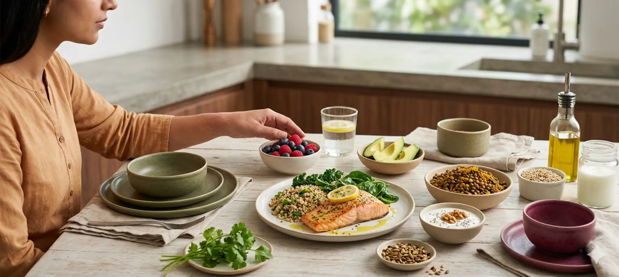
एक्झिमा ही एक सामान्य दाहक त्वचा स्थिती आहे, जी सर्व वयोगटांतील लाखो लोकांना प्रभावित करते. यामुळे त्वचा कोरडी, खाज सुटणारी आणि चिडचिडलेली होऊ शकते आणि अचानक तीव्रता वाढू शकते, ज्यामुळे यासह जगणाऱ्या लोकांचे दैनंदिन जीवन अस्वस्थ होते. अन्नामुळे थेट एक्झिमा होत नाही, पण काही अन्नपदार्थ काही व्यक्तींमध्ये लक्षणे वाढवू शकतात किंवा फ्लेअर-अप्स सुरू करू शकतात. ज्यांना अन्नाची अॅलर्जी किंवा संवेदनशीलता देखील आहे, अशा लोकांमध्ये तुम्ही काय खाता याची भूमिका तुमची त्वचा किती वेळा आणि किती तीव्रतेने प्रतिक्रिया देते यामध्ये महत्त्वाची असू शकते. एक्झिमा आहार योजना म्हणजे कोणतेही कठोर नियम पाळणे किंवा कारणाशिवाय संपूर्ण अन्नगट काढून टाकणे नाही. ती तुमचे स्वतःचे शरीर समजून घेणे, संभाव्य ट्रिगर्स ओळखणे आणि संतुलित, पोषणदायी आहार पद्धतीद्वारे त्वचेच्या आरोग्यास आधार देणे याबद्दल आहे. हा मार्गदर्शक तुम्हाला मदत करू शकणारे अन्नपदार्थ, लक्षणे वाढवू शकणारे अन्नपदार्थ आणि आहाराद्वारे एक्झिमा अधिक आत्मविश्वासाने व्यवस्थापित करण्यासाठी घेता येणारी व्यावहारिक पावले समजावतो. एक्झिमा म्हणजे काय? एक्झिमा, ज्याला अॅटॉपिक डर्माटायटिस असेही म्हणतात, ही एक दीर्घकालीन दाहक त्वचा स्थिती आहे, ज्यामध्ये त्वचेवर कोरडे, लाल आणि तीव्र खाज सुटणारे ठिपके दिसतात. हा सर्वात सामान्य त्वचा विकारांपैकी एक आहे, विशेषतः मुलांमध्ये, तरीही तो कोणत्याही वयातील लोकांना प्रभावित करू शकतो. सामान्य लक्षणांमध्ये सतत खाज येणे, कोरडी किंवा खवलेदार त्वचा, लालसरपणा आणि दाह, पुरळासारखे ठिपके ज्यातून स्त्राव होऊ शकतो किंवा पापुद्रा तयार होऊ शकतो, आणि वारंवार खाजवल्या जाणाऱ्या भागात त्वचा जाड किंवा चामड्यासारखी होणे यांचा समावेश होतो. लक्षणे सौम्य ते तीव्र असू शकतात आणि ती अनेकदा फ्लेअर-अप्स म्हणून ओळखल्या जाणाऱ्या चक्रांमध्ये येत-जात राहतात. एक्झिमाचे एकच ज्ञात कारण नाही. त्यामध्ये आनुवंशिक घटक, रोगप्रतिकारक प्रणालीची अतिसक्रियता, त्वचेच्या संरक्षणात्मक आवरणातील बिघाड आणि पर्यावरणीय ट्रिगर्स यांचे मिश्रण सहभागी असते असे मानले जाते. प्रभावी व्यवस्थापनासाठी सहसा योग्य त्वचा काळजी, ज्ञात ट्रिगर्स टाळणे, ताण व्यवस्थापन आणि काही प्रकरणांमध्ये आहाराकडे अधिक लक्ष देणे अशा अनेक बाजूंनी दृष्टिकोन आवश्यक असतो. आहार आणि एक्झिमा यांचा संबंध कसा असू शकतो? संशोधनानुसार, एक्झिमा असलेल्या अनेक लोकांना अन्नाची अॅलर्जी किंवा संवेदनशीलता देखील असते. अशा व्यक्तींमध्ये काही अन्नपदार्थ खाल्ल्याने रोगप्रतिकारक प्रतिक्रिया सुरू होऊ शकते, ज्यामुळे दाह वाढतो आणि त्वचेचे फ्लेअर-अप्स होतात. मात्र, अन्न आणि एक्झिमा यांचे नाते अत्यंत वैयक्तिक असते. एका व्यक्तीमध्ये प्रतिक्रिया निर्माण करणारा अन्नपदार्थ दुसऱ्या व्यक्तीसाठी पूर्णपणे निरुपद्रवी असू शकतो. म्हणूनच सर्वांसाठी समान परिणाम करणारा एकच एक्झिमा आहार नाही. आहार हा एक्झिमावर इलाज देखील नाही. या स्थितीचे व्यवस्थापन करण्यासाठी त्वचा काळजी, पर्यावरणीय ट्रिगर्स आणि गरजेनुसार वैद्यकीय उपचार यांचा समावेश असलेली व्यापक काळजी आवश्यक असते. तरीही, तुम्ही काय खाता याकडे लक्ष देणे, शक्य असेल तेथे दाहरोधी अन्नपदार्थ निवडणे आणि तुमचे वैयक्तिक ट्रिगर्स ओळखणे हे तुमच्या त्वचेच्या आरोग्यास अर्थपूर्ण आधार देऊ शकते आणि फ्लेअर-अप्सची वारंवारता कमी करू शकते. एक्झिमा आहारात समाविष्ट करावेत असे अन्नपदार्थ काही अन्नपदार्थांमध्ये दाह कमी करण्यास आणि एकूण त्वचेच्या आरोग्यास आधार देण्यास मदत करणारे गुणधर्म असू शकतात. बहुतेक लोकांना हे सामान्यतः चांगले सहन होतात, तरी वैयक्तिक अन्न अॅलर्जी आणि संवेदनशीलता नेहमी विचारात घेतली पाहिजे. दाहरोधी अन्नपदार्थ चरबीयुक्त मासे: सॅल्मन, सार्डिन, मॅकरेल आणि हेरिंग हे ओमेगा-3 फॅटी अॅसिड्सने समृद्ध असतात, ज्यांचे दाहरोधी गुणधर्म चांगले स्थापित आहेत आणि त्वचेची दाहक प्रतिक्रिया शांत करण्यास मदत करू शकतात. ऑलिव्ह तेल: स्वयंपाक आणि ड्रेसिंगमध्ये वापरले जाणारे आरोग्यदायी तेल, ऑलिव्ह तेलात दाहरोधी संयुगे असतात आणि त्वचेसाठी अनुकूल अनेक आहार पद्धतींचा तो महत्त्वाचा भाग आहे. अॅव्होकॅडो: आरोग्यदायी मोनोअनसॅच्युरेटेड फॅट्स आणि अँटिऑक्सिडंट्सने समृद्ध अॅव्होकॅडो त्वचेतील ओलावा टिकवण्यास आणि दाह कमी करण्यास मदत करू शकते. सुका मेवा आणि बिया: अक्रोड, जवस आणि चिया बिया वनस्पती-आधारित ओमेगा-3 फॅटी अॅसिड्स देतात आणि विशिष्ट सुका मेव्याची अॅलर्जी नसल्यास बहुतेक लोकांसाठी फायदेशीर असतात. अँटिऑक्सिडंट्सने समृद्ध फळे आणि भाज्या ब्लूबेरी, स्ट्रॉबेरी आणि रास्पबेरी यांसारख्या बेरी सफरचंद ब्रोकोली पालक आणि इतर पालेभाज्या चेरी टोमॅटो आणि बेल पेपर या अन्नपदार्थांमध्ये जीवनसत्त्वे, खनिजे आणि अँटिऑक्सिडंट्स मुबलक असतात, जे पेशींना ऑक्सिडेटिव्ह ताणापासून संरक्षण देण्यास आणि शरीराच्या नैसर्गिक दाहरोधी प्रक्रियांना आधार देण्यास मदत करतात. क्वेर्सेटिन असलेले अन्नपदार्थ क्वेर्सेटिन हे नैसर्गिकरीत्या आढळणारे वनस्पतीजन्य संयुग आहे, ज्यामध्ये अँटिऑक्सिडंट आणि अँटीहिस्टामाइन गुणधर्म असतात. काही व्यक्तींमध्ये एक्झिमा वाढवू शकणारी दाहकता आणि हिस्टामाइन प्रतिक्रिया कमी करण्यास ते मदत करू शकते. चांगले स्रोत खालीलप्रमाणे आहेत: सफरचंद कांदे केल गडद रंगाच्या बेरी लाल द्राक्षे ग्रीन टी प्रोबायोटिक अन्नपदार्थ प्रोबायोटिक अन्नपदार्थांमध्ये जिवंत कल्चर्स असतात, जे आतड्यांच्या आरोग्यास आधार देतात. आतड्यांचे आरोग्य रोगप्रतिकारक कार्य आणि त्वचेच्या आरोग्याशी वाढत्या प्रमाणात जोडले जात आहे. आहारात त्यांचा समावेश केल्याने अॅलर्जिक प्रतिक्रिया कमी करण्यास आणि अधिक संतुलित रोगप्रतिकारक प्रतिसादास आधार देण्यास मदत होऊ शकते. दही, जर तुम्हाला दुग्धजन्य पदार्थ सहन होत असतील केफिर मिसो अनपाश्चराइज्ड सॉअरक्राऊट नैसर्गिकरीत्या आंबवलेले लोणचे टेम्पेह लक्षात ठेवा की काही प्रोबायोटिक अन्नपदार्थ, जसे की दुग्धजन्य दही, दुग्धजन्य पदार्थांची संवेदनशीलता असलेल्या लोकांसाठी योग्य नसू शकतात. तुमच्या वैयक्तिक सहनशीलतेनुसार पर्याय निवडा. तंतुमय पदार्थांनी समृद्ध संपूर्ण अन्नपदार्थ ओट्स, ब्राऊन राईस आणि क्विनोआ यांसारखी संपूर्ण धान्ये सहन होत असल्यास मसूर आणि चणे यांसारखी कडधान्ये विविध रंगांतील ताज्या भाज्या ज्यूसऐवजी संपूर्ण फळे तंतुमय पदार्थ आतड्यांच्या आरोग्यास आधार देतात आणि निरोगी रोगप्रतिकारक प्रतिसादात योगदान देऊ शकतात, जो एक्झिमासारख्या दाहक त्वचा स्थितींमध्ये मध्यवर्ती भूमिका बजावतो. एक्झिमा आहारात टाळावेत किंवा मर्यादित करावेत असे अन्नपदार्थ काही अन्नपदार्थ त्वचेच्या आरोग्यास आधार देऊ शकतात, तसेच काही इतर अन्नपदार्थ दाह वाढवू शकतात किंवा फ्लेअर-अप्स सुरू करू शकतात, विशेषतः तुम्ही त्यांच्याबद्दल संवेदनशील असाल किंवा अॅलर्जिक असाल तर. खालील अन्नपदार्थ सामान्य ट्रिगर्स आहेत, पण एक्झिमा असलेल्या प्रत्येक व्यक्तीला या सर्वांवर प्रतिक्रिया येईलच असे नाही हे लक्षात ठेवणे महत्त्वाचे आहे. सामान्य अन्न ट्रिगर्स दुग्धजन्य पदार्थ: दूध, चीज आणि लोणी हे एक्झिमासाठी सर्वाधिक नोंदवले जाणारे अन्न ट्रिगर्स आहेत, विशेषतः मुलांमध्ये. अंडी: संवेदनशील व्यक्तींमध्ये एक्झिमा फ्लेअर-अप्सशी जोडलेला वारंवार आढळणारा अॅलर्जन. सोया: टोफू, सोया मिल्क आणि एडामामे यांसारखी सोया उत्पादने सोया संवेदनशीलता असलेल्या लोकांमध्ये प्रतिक्रिया निर्माण करू शकतात. सुका मेवा: काजू, अक्रोड आणि बदाम यांसारखा वृक्षजन्य सुका मेवा सुका मेव्याची अॅलर्जी असलेल्या लोकांसाठी समस्या निर्माण करू शकतो. गहू किंवा ग्लूटेन: सेलिएक आजार किंवा ग्लूटेन संवेदनशीलता असलेल्या लोकांमध्ये ग्लूटेन एक्झिमाची लक्षणे वाढवू शकते, तरी ते प्रत्येकावर परिणाम करत नाही. मासे किंवा शेलफिश: चरबीयुक्त मासे बहुतेकांसाठी फायदेशीर असू शकतात, पण विशिष्ट मासे किंवा शेलफिश अॅलर्जी असलेल्या लोकांनी ते पूर्णपणे टाळावेत. अतिप्रक्रियायुक्त अन्नपदार्थ पॅकेज्ड स्नॅक्स आणि तयार खाण्याचे पदार्थ फास्ट फूड काही मार्जरीन आणि तळलेले पदार्थ यांसारखे ट्रान्स फॅट्स जास्त असलेले अन्नपदार्थ रिफाइंड पिठापासून बनवलेला पांढरा ब्रेड आणि पास्ता यांसारखे रिफाइंड कार्बोहायड्रेट्स हे अन्नपदार्थ शरीरातील वाढलेल्या दाहाशी संबंधित असतात, ज्यामुळे दाहक त्वचा स्थिती अधिक बिघडू शकते. जास्त साखर असलेले अन्नपदार्थ आणि पेये केक, बिस्किटे आणि पेस्ट्री गोड पदार्थ आणि कन्फेक्शनरी सॉफ्ट ड्रिंक्स आणि सोडा साखरयुक्त कॉफी पेये आणि फ्लेवर्ड पेये साखरेचे जास्त सेवन दाह वाढवू शकते, ज्यामुळे कालांतराने एक्झिमा वाढू शकतो. तुम्हाला वैयक्तिकरित्या फ्लेअर्स सुरू करतात असे जाणवणारे अन्नपदार्थ तुम्ही काय खाता याची नोंद ठेवणे आणि लक्षणे कधी वाढतात ते लक्षात घेणे उपयुक्त ठरते. तुमचे वैयक्तिक ट्रिगर्स सामान्य यादीशी जुळतीलच असे नाही. इतर लोक काय सांगतात त्यावरून अन्नपदार्थ काढून टाकण्याऐवजी, तुमच्या स्वतःच्या नमुन्यांकडे लक्ष द्या आणि कोणतेही महत्त्वाचे बदल करण्यापूर्वी तुमचे निष्कर्ष डॉक्टर किंवा आहारतज्ज्ञांशी चर्चा करा. सर्वोत्तम एक्झिमा आहार योजना कोणती? कठोर अन्न नियम पाळण्याऐवजी, बहुतेक तज्ज्ञ दाह कमी करणारी आणि एकूण आरोग्यास आधार देणारी लवचिक, संतुलित आहार पद्धती स्वीकारण्याचा सल्ला देतात. एक्झिमा व्यवस्थापन सुधारण्याशी जोडल्या गेलेल्या मुख्य पद्धती येथे दिल्या आहेत: मेडिटेरेनियन-शैलीतील आहार मेडिटेरेनियन आहार पद्धतीची दाहरोधी गुणधर्मांमुळे मोठ्या प्रमाणावर शिफारस केली जाते. त्यामध्ये खालील गोष्टींवर भर दिला जातो: विविध प्रकारची फळे आणि भाज्या नियमितपणे खाल्ले जाणारे चरबीयुक्त मासे ऑलिव्ह तेलासारखे आरोग्यदायी फॅट्स संपूर्ण धान्ये आणि कडधान्ये प्रक्रियायुक्त आणि पॅकेज्ड अन्नपदार्थ कमी प्रमाणात लीन प्रथिनांचे मध्यम प्रमाण या प्रकारच्या आहारात ओमेगा-3 फॅटी अॅसिड्स आणि क्वेर्सेटिनसह अनेक पोषक घटक नैसर्गिकरीत्या समाविष्ट असतात, जे एक्झिमाशी संबंधित दाह कमी करण्यास मदत करू शकतात. दाहरोधी आहार पद्धत दाहरोधी आहार मेडिटेरेनियन पद्धतीशी मोठ्या प्रमाणात जुळतो आणि खालील गोष्टींवर लक्ष केंद्रित करतो: संपूर्ण, कमीत कमी प्रक्रिया केलेले अन्नपदार्थ आतड्यांच्या आरोग्यास आधार देणारी तंतुमय पदार्थांनी समृद्ध जेवणे भाज्या आणि फळांचे भरपूर प्रमाण कमी अतिरिक्त साखर आणि कमी अतिप्रक्रियायुक्त अन्न उत्पादने सॅच्युरेटेड किंवा ट्रान्स फॅट्सऐवजी आरोग्यदायी फॅट्स ही पद्धत शरीरातील एकूण दाह कमी करण्यास मदत करू शकते, ज्याचा एक्झिमासह विविध दाहक स्थितींना फायदा होऊ शकतो. एलिमिनेशन डाएट्स एलिमिनेशन डाएटमध्ये संशयित अन्न ट्रिगर्स ठराविक काळासाठी जेवणातून काढून टाकले जातात आणि नंतर त्यांना एकेक करून पुन्हा समाविष्ट केले जाते, जेणेकरून कोणत्या अन्नामुळे प्रतिक्रिया होते हे ओळखता येते. विशिष्ट ट्रिगर्सबद्दल खात्री नसलेल्या लोकांसाठी ही पद्धत उपयुक्त ठरू शकते, पण ती स्वतंत्रपणे कधीही करू नये, विशेषतः मुलांसाठी. मार्गदर्शनाशिवाय अन्नगट काढून टाकल्यास पोषणातील कमतरता निर्माण होऊ शकते आणि खरे ट्रिगर्स ओळखणे अधिक कठीण होऊ शकते. तुम्ही एलिमिनेशन डाएटचा विचार करत असाल, तर नेहमी डॉक्टर किंवा नोंदणीकृत आहारतज्ज्ञांसोबत काम करा. डायशिड्रॉटिक एक्झिमाबद्दल विशेष नोंद डायशिड्रॉटिक एक्झिमा हा एक विशिष्ट प्रकार आहे, ज्यामध्ये सामान्यतः हात आणि पायांवर लहान, तीव्र खाज सुटणारे फोड येतात. काही व्यक्तींमध्ये तो काही धातूंविषयीच्या संवेदनशीलतेमुळे सुरू होऊ शकतो किंवा वाढू शकतो, विशेषतः निकेल आणि कोबाल्ट, जे संपूर्ण धान्ये, काही कडधान्ये, सुका मेवा, चॉकलेट आणि चहा यांसारख्या काही अन्नपदार्थांमध्ये कमी प्रमाणात आढळतात. तुम्हाला डायशिड्रॉटिक एक्झिमाचे निदान झाले असेल, तर तुमच्या बाबतीत लो-निकेल पद्धत तपासून पाहणे योग्य ठरेल का याबद्दल तुमच्या त्वचारोगतज्ज्ञांशी बोला. 7-दिवसांची एक्झिमा आहार योजना सुरुवात करण्यासाठी ही एक लवचिक, सामान्य मार्गदर्शक योजना आहे. ही वैद्यकीय प्रिस्क्रिप्शन नाही आणि तुमची वैयक्तिक सहनशीलता नेहमी प्रथम मानली पाहिजे. ज्ञात अन्न अॅलर्जी किंवा संवेदनशीलतेनुसार जेवणात बदल करा. दिवस नाश्ता दुपारचे जेवण मधला नाश्ता रात्रीचे जेवण दिवस 1 ब्लूबेरी आणि थोड्या मधासह ओट्सची पेज ब्राऊन राईस आणि वाफवलेल्या पालकासह ग्रिल्ड सॅल्मन सफरचंदाचे काप आणि थोडे अक्रोड भाजी डाळ, रोटी आणि बाजूला सॅलड दिवस 2 केळी, केफिर आणि थोड्या बेरीसह स्मूदी संपूर्ण धान्याच्या ब्रेड आणि हिरव्या सॅलडसह मसूर सूप हुमससोबत कापलेली काकडी भाजलेल्या ब्रोकोली आणि क्विनोआसह ग्रिल्ड चिकन दिवस 3 परतलेल्या पालेभाज्यांसोबत स्क्रॅम्बल्ड अंडी (सहन होत असल्यास) ग्रिल्ड भाज्या आणि ऑलिव्ह तेल ड्रेसिंगसह ब्राऊन राईस चेरीसह साध्या दह्याची एक छोटी वाटी गोड बटाटा आणि वाफवलेल्या पालेभाज्यांसह बेक्ड मॅकरेल दिवस 4 चिया बिया, सफरचंद आणि बदाम दुधासह ओव्हरनाइट ओट्स टोमॅटो, काकडी, ऑलिव्ह तेल आणि औषधी वनस्पतींसह चण्याचे सॅलड थोड्या भोपळ्याच्या बिया आणि गडद रंगाच्या बेरी मिश्र भाज्या आणि ब्राऊन राईससह टोफू स्टर-फ्राय दिवस 5 नारळाच्या चटणी आणि सांबारासह इडली ताज्या सॅलड आणि ऑलिव्ह तेल ड्रेसिंगसह ग्रिल्ड मासे एक चमचा बदाम बटरसोबत कापलेले सफरचंद वाफवलेल्या भाज्या आणि बाजूला सॉअरक्राऊटसह मूग डाळ खिचडी दिवस 6 भाज्यांसह बनवलेला आणि ताजी कोथिंबीर घातलेला पोहा ब्राऊन राईस आणि काकडी सॅलडसह राजमा हुमससोबत गाजर आणि सेलरीच्या काड्या भाजलेल्या गोड बटाटे आणि पालकासह ग्रिल्ड चिकन दिवस 7 मिश्र बेरी, पपई आणि किवीसह फळांची वाटी आणि केफिरचा एक ग्लास भाजी, पालेभाजी आणि मसूर सूपसह संपूर्ण धान्याची रोटी काही अक्रोड आणि सफरचंदासह ग्रीन टी क्विनोआ आणि हिरव्या सॅलडसह बेक्ड सॅल्मन ही योजना तुमच्या स्वतःच्या अन्न पसंती, सहनशीलता आणि डॉक्टर किंवा आहारतज्ज्ञांच्या कोणत्याही मार्गदर्शनानुसार बदलता येते. संतुलित एक्झिमा आहाराचे फायदे संतुलित, दाहरोधी आहार पाळल्याने एक्झिमा असलेल्या लोकांना अनेक अर्थपूर्ण फायदे मिळू शकतात: दाह कमी करण्यास मदत होऊ शकते: ओमेगा-3 फॅटी अॅसिड्स, अँटिऑक्सिडंट्स आणि तंतुमय पदार्थांनी समृद्ध आहार शरीराच्या नैसर्गिक दाहरोधी प्रक्रियांना आधार देऊ शकतो. आतड्यांच्या आरोग्यास आधार देऊ शकतो: प्रोबायोटिक आणि तंतुमय पदार्थांनी समृद्ध अन्नपदार्थ निरोगी आतड्यांचा मायक्रोबायोम राखण्यास मदत करू शकतात, ज्याचा रोगप्रतिकारक कार्य आणि त्वचेच्या आरोग्याशी वाढता संबंध दिसतो. वैयक्तिक ट्रिगर्स ओळखण्यास मदत होऊ शकते: आहाराकडे लक्ष दिल्याने तुमच्या खाण्याच्या पद्धतींबद्दल नैसर्गिकरित्या अधिक जागरूकता वाढते, ज्यामुळे अन्न आणि फ्लेअर-अप्समधील संबंध ओळखणे सोपे होते. एकूण त्वचेच्या आरोग्यास आधार देऊ शकतो: अनेक संपूर्ण अन्नपदार्थांमध्ये आढळणारे व्हिटॅमिन C, व्हिटॅमिन E, सेलेनियम आणि झिंक यांसारखे पोषक घटक त्वचेची दुरुस्ती आणि संरक्षणात्मक आवरणाचे कार्य यांना आधार देतात. सर्वसाधारण आरोग्यास आधार देतो: संतुलित आहार फक्त त्वचेलाच नव्हे, तर तुमची ऊर्जा पातळी, रोगप्रतिकारक आरोग्य आणि दीर्घकालीन आरोग्यालाही फायदा करतो. वास्तववादी अपेक्षा ठेवणे महत्त्वाचे आहे. फक्त आहाराने एक्झिमा बरा होऊ शकत नाही, पण व्यापक व्यवस्थापन योजनेचा तो एक उपयुक्त भाग असू शकतो. त्वचेचे चांगले आरोग्य समर्थित करणाऱ्या जीवनशैली टिप्स अन्न निवडींपलीकडे, जीवनशैलीतील काही सवयी एक्झिमा व्यवस्थापनाला आधार देऊ शकतात आणि फ्लेअर-अप्स कमी करू शकतात: पुरेसे पाणी प्या: दिवसभर पुरेसे पाणी पिणे त्वचेतील ओलावा आणि संरक्षणात्मक आवरणाचे कार्य यांना आधार देते. दररोज किमान आठ ग्लास पाण्याचे उद्दिष्ट ठेवा. ताण व्यवस्थापित करा: ताण हा एक्झिमाचा ज्ञात ट्रिगर आहे. खोल श्वसन, योग, माइंडफुलनेस किंवा हलक्या चालण्यासारख्या पद्धती ताण पातळी नियंत्रणात ठेवण्यास मदत करू शकतात. चांगल्या झोपेला प्राधान्य द्या: झोपेदरम्यान शरीर दुरुस्ती आणि पुनरुत्पादन करते. खराब झोप दाह वाढवू शकते आणि त्वचेच्या स्थितींचे व्यवस्थापन कठीण करू शकते. सौम्य त्वचा काळजी उत्पादने वापरा: तीव्र साबण, सुगंधी उत्पादने आणि काही कापड एक्झिमा-प्रवण त्वचेला त्रास देऊ शकतात. सौम्य, सुगंधरहित मॉइश्चरायझर्स आणि क्लेंझर्स निवडा. ज्ञात त्रासदायक घटक टाळा: यामध्ये सिगारेटचा धूर, काही स्वच्छता उत्पादने आणि थेट फ्लेअर-अप्स सुरू करू शकणारे सिंथेटिक कापड यांचा समावेश होतो. नियमित व्यायाम करा: शारीरिक हालचाल एकूण आरोग्यास आधार देते आणि ताण व्यवस्थापित करण्यास मदत करते, या दोन्ही गोष्टी दाहक स्थितींना फायदा करू शकतात. व्यापक वजन कमी करण्यासाठी आहार चार्ट प्रमाणेच, नियमित हालचाल आणि संतुलित आहार एकत्रितपणे दीर्घकालीन त्वचा आणि सर्वसाधारण आरोग्यासाठी अधिक मजबूत पाया तयार करतात. तुमचे वैयक्तिक एक्झिमा ट्रिगर्स कसे शोधावेत? एक्झिमा अत्यंत वैयक्तिक असल्यामुळे, तुमचे स्वतःचे ट्रिगर्स शोधण्यासाठी थोडे निरीक्षण आणि संयम लागतो: अन्न आणि लक्षणांची डायरी ठेवा: दररोज तुम्ही काय खाता ते नोंदवा आणि त्वचेतील कोणतेही बदल लिहून ठेवा, ज्यात फ्लेअर-अप्स कधी होतात आणि किती तीव्र असतात याचाही समावेश करा. वारंवार दिसणारे नमुने पाहा: फ्लेअर-अप्सच्या काळात एखादा तोच अन्नपदार्थ तुमच्या डायरीत सतत दिसत असेल, तर डॉक्टरांशी चर्चा करणे योग्य ठरू शकते. एकाच वेळी खूप अन्नपदार्थ काढून टाळू नका: अनेक अन्नगट एकाच वेळी काढून टाकल्यास नेमका कोणता पदार्थ प्रतिक्रिया निर्माण करतो हे ओळखणे जवळजवळ अशक्य होते. लक्षणे वारंवार किंवा तीव्र असल्यास डॉक्टरांशी बोला: फ्लेअर-अप्स नियमित होत असतील किंवा त्यांचे व्यवस्थापन कठीण जात असेल, तर डॉक्टर किंवा अॅलर्जिस्ट योग्य अॅलर्जी चाचणी आणि क्लिनिकल मूल्यमापनाद्वारे ट्रिगर्स ओळखण्यास मदत करू शकतात. डॉक्टर किंवा आहारतज्ज्ञांशी कधी बोलावे? व्यावसायिक मार्गदर्शन विशेषतः महत्त्वाचे ठरते अशा काही परिस्थिती आहेत: तुम्हाला किंवा तुमच्या मुलाला एक्झिमाशी संबंधित अन्न अॅलर्जी असल्याचा संशय असल्यास तुमच्या मुलाला एक्झिमा असेल आणि त्याला खाण्यात अडचणी किंवा मर्यादित खाण्याची समस्या येत असेल तुम्ही एलिमिनेशन डाएटचा विचार करत असाल, विशेषतः मुलासाठी आहारातील बदल असूनही लक्षणे तीव्र, वारंवार किंवा सुधारत नसतील दुग्धजन्य पदार्थ, ग्लूटेन किंवा अंडी यांसारखे प्रमुख अन्नगट काढून टाकण्याचा विचार करत असाल तुम्ही गर्भवती किंवा स्तनपान करत असाल आणि एक्झिमा व्यवस्थापित करत असाल डॉक्टर किंवा नोंदणीकृत आहारतज्ज्ञ तुमचे पोषण सेवन धोक्यात न आणता सुरक्षित, पुराव्यावर आधारित निवडी करण्यात मदत करू शकतात. मेट्रोपोलिस हेल्थकेअर तुम्हाला कशी मदत करू शकते? तुमचे शरीर अधिक चांगले समजून घेणे हे एक्झिमा व्यवस्थापनातील सर्वात महत्त्वाच्या पायऱ्यांपैकी एक आहे. काही लोकांसाठी, विशिष्ट अन्न अॅलर्जी किंवा पोषणातील असंतुलन फ्लेअर-अप्समध्ये योगदान देत आहेत का हे जाणून घेणे त्यांच्या काळजी योजनेत अर्थपूर्ण फरक करू शकते. मेट्रोपोलिस हेल्थकेअरमध्ये, तुम्हाला अॅलर्जी पॅनेल्स, पोषण कमतरता मूल्यांकन आणि पूर्ण शरीर तपासण्या यांसह निदान चाचण्यांच्या व्यापक श्रेणीचा प्रवेश मिळतो, ज्यांना एनएबीएल आणि सीएपी-मान्यताप्राप्त प्रयोगशाळा आणि अनुभवी पॅथॉलॉजिस्ट यांचा आधार आहे. तुम्ही विशिष्ट अन्न अॅलर्जन्स तपासू इच्छित असाल, तुमची एकूण पोषण स्थिती तपासू इच्छित असाल किंवा फक्त तुमच्या आरोग्याबद्दल माहिती ठेवू इच्छित असाल, मेट्रोपोलिस हे सोपे करते. वेबसाइट, अॅप, फोन किंवा व्हॉट्सअॅपद्वारे सहज बुक करा किंवा 10,000 हून अधिक टचपॉइंट्सच्या विश्वासार्ह नेटवर्कमधून घरून नमुना संकलनाची सोय निवडा. जलद, विश्वासार्ह अहवालांमुळे पुढील पाऊल आत्मविश्वासाने उचलण्यासाठी तुम्हाला नेहमी स्पष्टता मिळते. निष्कर्ष एक्झिमा ही एक जटिल स्थिती आहे आणि सर्वांसाठी तो दूर करणारा एकच आहार नाही. तुम्ही काय खाता ते फ्लेअर-अप्स व्यवस्थापित करण्यात आधारभूत भूमिका बजावू शकते, विशेषतः अन्न संवेदनशीलता किंवा अॅलर्जी सहभागी असल्यास, पण आहार योग्य त्वचा काळजी, ताण व्यवस्थापन आणि वैद्यकीय मार्गदर्शनासह व्यापक काळजी योजनेचा भाग म्हणून सर्वोत्तम काम करतो. मर्यादित करणारे अन्न नियम पाळण्याऐवजी, संपूर्ण अन्नपदार्थ, फळे आणि भाज्या, आरोग्यदायी फॅट्स आणि तंतुमय पदार्थांनी समृद्ध संतुलित, दाहरोधी आहार पद्धतीवर लक्ष केंद्रित करा. त्याच वेळी, तुमच्या वैयक्तिक ट्रिगर्सकडे लक्ष द्या आणि तुमच्या विशिष्ट परिस्थितीसाठी सुरक्षित व प्रभावी असे बदल करण्यासाठी आरोग्यसेवा तज्ज्ञांसोबत काम करा. पोषणदायी आहाराकडे लहान, सातत्यपूर्ण पावले तुमच्या त्वचेत आणि एकूण आरोग्यात खरा फरक करू शकतात. नेहमी विचारले जाणारे प्रश्न एक्झिमासाठी कोणते अन्नपदार्थ चांगले आहेत? एक्झिमामध्ये त्वचेच्या आरोग्यास आधार देण्यास आणि दाह कमी करण्यास मदत करू शकणाऱ्या अन्नपदार्थांमध्ये सॅल्मन, सार्डिन आणि मॅकरेल यांसारखे ओमेगा-3 फॅटी अॅसिड्सने समृद्ध चरबीयुक्त मासे समाविष्ट आहेत. बेरी, सफरचंद, ब्रोकोली आणि पालक यांसारखी अँटिऑक्सिडंट्सने समृद्ध फळे आणि भाज्याही फायदेशीर आहेत. कांदे आणि गडद रंगाच्या बेरी यांसारखे क्वेर्सेटिन असलेले अन्नपदार्थ, तसेच दही, केफिर आणि मिसो यांसारखे प्रोबायोटिक अन्नपदार्थही आधार देणारी भूमिका बजावू शकतात. संपूर्ण धान्ये, कडधान्ये आणि ऑलिव्ह तेल व अॅव्होकॅडोसारखी आरोग्यदायी फॅट्स एक्झिमा-अनुकूल आहार पद्धती पूर्ण करतात. आहाराने एक्झिमा बरा होऊ शकतो का? नाही, फक्त आहाराने एक्झिमा बरा होऊ शकत नाही. काही अन्नपदार्थ फ्लेअर-अप्सची वारंवारता किंवा तीव्रता कमी करण्यास मदत करू शकतात, विशेषतः अन्न अॅलर्जी किंवा संवेदनशीलता असलेल्या लोकांमध्ये, पण एक्झिमा ही दीर्घकालीन स्थिती आहे ज्यासाठी व्यापक व्यवस्थापन आवश्यक असते. यामध्ये सामान्यतः योग्य त्वचा काळजी, पर्यावरणीय ट्रिगर्स टाळणे, ताण व्यवस्थापन आणि काही प्रकरणांमध्ये वैद्यकीय उपचार यांचा समावेश असतो. आहार हा व्यापक काळजी योजनेतील एक उपयुक्त साधन आहे, स्वतंत्र उपचार नाही. ग्लूटेनचा एक्झिमावर परिणाम होतो का? सेलिएक आजार किंवा नॉन-सेलिएक ग्लूटेन संवेदनशीलता असलेल्या लोकांमध्ये ग्लूटेन एक्झिमाची लक्षणे वाढवू शकते. अशा व्यक्तींमध्ये आहारातून ग्लूटेन काढून टाकल्याने पचन आणि त्वचेची लक्षणे दोन्ही सुधारू शकतात. मात्र, ग्लूटेन एक्झिमा असलेल्या प्रत्येकावर परिणाम करत नाही आणि तो अनावश्यकपणे काढून टाकल्यास पोषणातील उणीवा निर्माण होऊ शकतात. तुम्हाला ग्लूटेन तुमच्यासाठी ट्रिगर आहे असा संशय असल्यास, आहारात बदल करण्यापूर्वी डॉक्टरांशी बोला. योग्य चाचणीद्वारे सेलिएक आजार किंवा ग्लूटेन संवेदनशीलता सहभागी आहे का हे पुष्टी करता येते. प्रोबायोटिक्स एक्झिमा फ्लेअर-अप्समध्ये मदत करू शकतात का? काही संशोधनानुसार, प्रोबायोटिक अन्नपदार्थ आणि पूरक घटक रोगप्रतिकारक कार्याला आधार देण्यास आणि अॅलर्जिक प्रतिक्रिया कमी करण्यास मदत करू शकतात, ज्याचा एक्झिमा असलेल्या लोकांना फायदा होऊ शकतो. दही, केफिर, मिसो आणि सॉअरक्राऊट यांसारखे प्रोबायोटिक-समृद्ध अन्नपदार्थ निरोगी आतड्यांच्या मायक्रोबायोमला आधार देतात, ज्याचा त्वचेच्या आरोग्याशी वाढता संबंध दिसतो. मात्र, पुरावे अजून विकसित होत आहेत आणि परिणाम व्यक्तीनुसार बदलतात. संतुलित आहाराचा भाग म्हणून प्रोबायोटिक अन्नपदार्थांचा समावेश करणे बहुतेक लोकांसाठी सामान्यतः सुरक्षित आणि समंजस दृष्टिकोन आहे. सर्वांसाठी सर्वोत्तम एक्झिमा आहार योजना आहे का? नाही. एक्झिमा ट्रिगर्स आणि अन्न संवेदनशीलता व्यक्तिनुसार बदलत असल्याने, सर्वांसाठी काम करणारी एकच एक्झिमा आहार योजना नाही. मात्र, मेडिटेरेनियन आहार आणि दाहरोधी आहार यांसारख्या आहार पद्धती व्यापकपणे फायदेशीर आहेत, कारण त्या प्रक्रिया केलेले आणि जास्त साखरेचे अन्नपदार्थ मर्यादित ठेवत संपूर्
वजन वाढीसाठी 12 उच्च-कॅलरी कल्पनांसह ओट्सच्या पाककृती
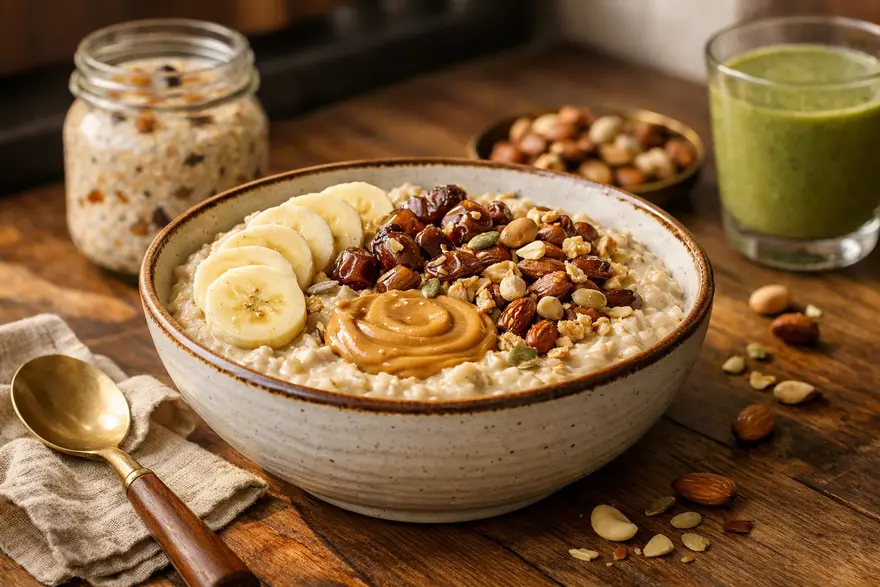
जर तुम्हाला आरोग्यदायी पद्धतीने वजन वाढवायचे असेल, तर ओट्स हा तुमच्या आहारात समाविष्ट करण्यासाठी एक चांगला अन्नपदार्थ ठरू शकतो. ते तयार करायला सोपे, परवडणारे, पोटभरीचे आणि पूर्ण फॅटचे दूध, दही, सुकामेवा, बिया, नट बटर आणि फळे यांसारख्या कॅलरीयुक्त घटकांसोबत सहज वापरता येतात. फक्त ओट्स खाल्ल्याने आपोआप वजन वाढत नाही. दिवसभरातील एकूण कॅलरी आणि पुरेशी प्रथिने वाढतील अशा पद्धतीने ते खाणे मदत करते. त्यामुळे आरोग्यदायी वजन वाढीसाठी ओट्स उपयुक्त ठरतात, विशेषतः तुम्हाला ताकद वाढवायची असेल आणि त्याच वेळी स्नायू वाढीस मदत करायची असेल तर. वजन वाढीसाठी ओट्स चांगले आहेत का? होय, कॅलरी आधिक्य असलेल्या आहाराचा भाग म्हणून ओट्स खाल्ल्यास ते वजन वाढीस मदत करू शकतात. त्यात कार्बोहायड्रेट्स भरपूर असतात, काही प्रमाणात प्रथिने असतात आणि ते जास्त कॅलरी असलेल्या अन्नपदार्थांसोबत चांगले जुळतात. म्हणूनच अनेक लोक विचारतात, वजन वाढीसाठी ओट्स चांगले आहेत का. उत्तर होय आहे, पण ते कसे तयार करता हे महत्त्वाचे आहे. पाण्यात केलेला साधा बाऊल हा पूर्ण फॅटचे दूध, पीनट बटर, खजूर, सुकामेवा आणि बिया घालून केलेल्या ओट्सपेक्षा खूप हलका असतो. वजन वाढीसाठी ओट्स का चांगले आहेत? काही सोप्या कारणांमुळे ओट्स वजन वाढीसाठी चांगले काम करतात. ते संमिश्र कार्बोहायड्रेट्समधून स्थिर ऊर्जा देतात. त्यात फायबरही असते, जे पचनास मदत करते, तसेच मध्यम प्रमाणात प्रथिने असतात. सर्वात महत्त्वाचे म्हणजे ओट्स लवचिक आहेत. कमी कष्टात त्यांचे पोरिज, ओव्हरनाईट ओट्स, शेक, स्मूदी, स्नॅक बाऊल किंवा खारट पदार्थ बनवता येतात. तुम्ही स्नायू वाढवण्याचा प्रयत्न करत असाल, तरही ते उपयुक्त आहेत, कारण दूध, दही, पनीर, ग्रीक दही आणि व्हे प्रोटीन यांसारख्या प्रथिनयुक्त पदार्थांसोबत ओट्स चांगले जुळतात. तुम्ही नियमित व्यायाम करत असाल, तर हे संयोजन रिकव्हरीला मदत करू शकते आणि दिवसभर पुरेशा कॅलरी खाण्यास मदत करू शकते. वजन वाढीसाठी सर्वोत्तम ओट्स तुम्ही वजन वाढीसाठी सर्वोत्तम ओट्स निवडत असाल, तर मुख्य पर्याय असे आहेत: रोल्ड ओट्स: वजन वाढीसाठी साधारणपणे सर्वोत्तम ओट्स. ते मऊ शिजतात, चवीला चांगले लागतात आणि पोरिज, ओव्हरनाईट ओट्स आणि शेकमध्ये चांगले वापरता येतात. स्टील-कट ओट्स: कमी प्रक्रिया केलेले आणि थोडे अधिक चिवट. ते पोटभरीचे असतात आणि भरगच्च बाऊलसाठी चांगले असतात. इन्स्टंट ओट्स: सोयीस्कर आणि पटकन तयार होणारे. कॅलरीयुक्त टॉपिंग्स घातल्यास हेही चांगले काम करू शकतात. उच्च-प्रथिनयुक्त ओट्स मिश्रणे: एका जेवणात जास्त प्रथिने हवी असतील तर उपयुक्त, पण वाढीव साखर आणि परिमाणासाठी नेहमी लेबल तपासा. बहुतेक लोकांसाठी रोल्ड ओट्स हा सर्वात सोपा आणि व्यावहारिक पर्याय आहे. वजन वाढीसाठी ओट्सचे पौष्टिक मूल्य ओट्स पोषकद्रव्यांनी समृद्ध असतात, म्हणजेच ते कॅलरींसोबत उपयुक्त पोषणही देतात. त्यात ऊर्जेसाठी कार्बोहायड्रेट्स, पचनासाठी फायबर, काही प्रमाणात प्रथिने आणि मॅग्नेशियम, लोह आणि मॅंगनीजसारखी खनिजे असतात. साध्या ओट्समधील अंदाजे पोषण रोल्ड ओट्सच्या कोरड्या 40 g सर्व्हिंगमधून साधारणपणे मिळते: 150 kcal 27 g कार्बोहायड्रेट्स 5 g प्रथिने 3 g चरबी 4 g फायबर हा एक चांगला आधार आहे, पण वजन वाढीसाठी खरी किंमत तुम्ही त्यात काय घालता यावर अवलंबून असते. वजन वाढीसाठी ओट्स अधिक चांगले कशामुळे होतात? वजन वाढीसाठी ओट्स अधिक उपयुक्त करण्यासाठी, त्यांना खालील पदार्थांसोबत खा: पूर्ण फॅटचे दूध पूर्ण फॅटचे दही किंवा ग्रीक दही पीनट बटर किंवा बदाम बटर खजूर, मनुके, अंजीर आणि इतर सुकी फळे बदाम, अक्रोड, काजू आणि बिया केळी, आंबा, चिकू किंवा अॅव्होकॅडो मध किंवा गूळ व्हे प्रोटीन किंवा स्किम्ड मिल्क पावडर नारळाचे काप किंवा सुक्या नारळाचा कीस वजन वाढीसाठी ओट्स कसे वापरावे: 12 उच्च-कॅलरी ओट्स पाककृती 1. पूर्ण फॅटचे दूध, केळी आणि पीनट बटरसह ओट्स साहित्य 1/2 cup रोल्ड ओट्स 300 ml पूर्ण फॅटचे दूध 1 केळी 1 to 2 tbsp पीनट बटर कसे बनवावे ओट्स दुधात मऊ होईपर्यंत शिजवा. वर केळीचे काप घाला आणि पीनट बटर मिसळा. वजन वाढीसाठी ते कसे मदत करते हा एका जेवणात कार्बोहायड्रेट्स, चरबी आणि प्रथिने असलेला साधा उच्च-कॅलरी बाऊल आहे. 2. दही, सुकामेवा आणि मधासह ओव्हरनाईट ओट्स साहित्य 1/2 cup रोल्ड ओट्स 1/2 cup पूर्ण फॅटचे दही 1/2 cup दूध 1 tbsp मध चिरलेले बदाम आणि अक्रोड कसे बनवावे सर्व काही एका जारमध्ये मिसळा आणि रात्रभर फ्रिजमध्ये ठेवा. वजन वाढीसाठी ते कसे मदत करते हे सकाळी खायला सोपे असते आणि फार जड न वाटता अतिरिक्त कॅलरी देते. 3. आंबा ओट्स स्मूदी साहित्य 1/2 cup ओट्स 1 पिकलेला आंबा 250 ml पूर्ण फॅटचे दूध 1 tbsp चिया बिया 1 tbsp नट बटर कसे बनवावे मऊसर होईपर्यंत ब्लेंड करा. वजन वाढीसाठी ते कसे मदत करते तुमची भूक कमी असेल आणि कॅलरी पिणे तुम्हाला अधिक सोयीचे वाटत असेल, तर हे उपयुक्त आहे. 4. चॉकलेट प्रोटीन ओट्स साहित्य 1/2 cup ओट्स 300 ml दूध 1 स्कूप चॉकलेट प्रोटीन पावडर 1 tsp कोको पावडर 1 tbsp पीनट बटर कसे बनवावे आधी ओट्स शिजवा. ते थोडे थंड होऊ द्या, नंतर त्यात प्रोटीन पावडर आणि पीनट बटर मिसळा. वजन वाढीसाठी ते कसे मदत करते व्यायामानंतर हे चांगले काम करते कारण यामुळे कॅलरी आणि प्रथिने दोन्ही मिळतात. 5. सुकामेवा ओट्स पोरिज साहित्य 1/2 cup ओट्स 300 ml पूर्ण फॅटचे दूध चिरलेले खजूर मनुके काजू वेलचीची चिमूटभर पूड कसे बनवावे ओट्स दुधात शिजवा आणि त्यात सुकामेवा मिसळा. वजन वाढीसाठी ते कसे मदत करते सुकी फळे कॅलरीचे प्रमाण पटकन वाढवतात आणि बाऊल अधिक पोटभरीचा करतात. 6. केळी खजूर ओट्स शेक साहित्य 1/2 cup ओट्स 1 केळी 3 to 4 खजूर 250 to 300 ml दूध 1 tbsp बदाम बटर कसे बनवावे सर्व साहित्य मऊसर होईपर्यंत ब्लेंड करा. वजन वाढीसाठी ते कसे मदत करते तुम्हाला झटपट स्नॅक हवा असेल, तेव्हा वजन वाढीसाठी ओट्स घेण्याचा हा सर्वात सोपा मार्गांपैकी एक आहे. 7. नट बटर आणि बियांसह ओट्स साहित्य 1/2 cup ओट्स 300 ml दूध 1 tbsp पीनट किंवा बदाम बटर 1 tbsp चिया किंवा जवसाच्या बिया 1 tsp मध कसे बनवावे ओट्स शिजवा आणि वाढण्यापूर्वी टॉपिंग्स घाला. वजन वाढीसाठी ते कसे मदत करते नट बटर आणि बियांमधील आरोग्यदायी चरबी जेवण अधिक कॅलरीयुक्त बनवते. 8. मनुक्यांसह सफरचंद दालचिनी ओट्स साहित्य 1/2 cup ओट्स 300 ml दूध 1 चिरलेले सफरचंद 2 tbsp मनुके दालचिनीची चिमूटभर पूड 1 tbsp चिरलेले अक्रोड कसे बनवावे ओट्स आणि दूध एकत्र शिजवा. सफरचंद, मनुके, दालचिनी आणि अक्रोड घाला. वजन वाढीसाठी ते कसे मदत करते यातून कार्बोहायड्रेट्स, फायबर आणि मनुके व अक्रोडमधून अतिरिक्त कॅलरी यांचे चांगले मिश्रण मिळते. 9. ग्रॅनोला टॉपिंगसह ओट्स आणि दह्याचा बाऊल साहित्य 1/2 cup भिजवलेले ओट्स 1 cup पूर्ण फॅटचे दही 2 tbsp ग्रॅनोला 1 tbsp मिश्र बिया 1 tsp मध कसे बनवावे ओट्स आणि दही मिसळा. वर ग्रॅनोला, बिया आणि मध घाला. वजन वाढीसाठी ते कसे मदत करते हे झटपट तयार होते, ऊर्जा जास्त देते आणि स्नॅक म्हणून सोबत नेणे सोपे असते. 10. पनीरसह खारे मसाला ओट्स साहित्य 1/2 cup ओट्स 1 tsp तूप कांदा, टोमॅटो आणि मटार 50 to 75 g पनीर मीठ आणि मसाले कसे बनवावे भाज्या तुपात शिजवा, ओट्स आणि पाणी घाला, नंतर पनीर मिसळा. वजन वाढीसाठी ते कसे मदत करते वाढीव चरबी आणि प्रथिनांसह हा संतुलित खारट पर्याय आहे. 11. तूप आणि भाज्यांसह ओट्स खिचडी साहित्य 1/2 cup ओट्स मूग डाळ 1 to 2 tsp तूप मिश्र भाज्या सौम्य मसाले कसे बनवावे सर्व काही एकत्र मऊ होईपर्यंत शिजवा. वजन वाढीसाठी ते कसे मदत करते गोड ओट्सऐवजी गरम खारट जेवण आवडत असेल, तर हे चांगले काम करते. 12. उच्च-कॅलरी ओट्स स्मूदी बाऊल साहित्य 1/2 cup ओट्स 1 गोठवलेले केळी 1/2 cup दही 1 tbsp पीनट बटर 1 tbsp भोपळ्याच्या बिया चिरलेले खजूर कसे बनवावे बेस घट्ट ब्लेंड करा आणि वर खजूर व बिया घाला. वजन वाढीसाठी ते कसे मदत करते हे कॅलरीयुक्त, समाधानकारक आणि तुमच्या आवडीनुसार बदलायला सोपे आहे. ओट्ससाठी सर्वोत्तम उच्च-कॅलरी अॅड-इन्स तुमचे ओट्स अधिक कॅलरीयुक्त बनवायचे असतील, तर हे अॅड-इन्स सर्वाधिक मदत करतात: पूर्ण फॅटचे दूध पीनट बटर किंवा बदाम बटर चिरलेले बदाम, अक्रोड किंवा काजू चिया, जवस किंवा भोपळ्याच्या बिया खजूर, मनुके, अंजीर आणि जर्दाळू केळी, आंबा, चिकू किंवा अॅव्होकॅडो ग्रीक दही किंवा चक्का दही व्हे प्रोटीन मध किंवा गूळ नारळाचे काप हे घटक खूप मोठा भाग न खाता कॅलरी वाढवतात. वजन वाढीसाठी तुमच्या रोजच्या आहारात ओट्स कसे घालावेत प्रत्येक जेवणात ओट्स खाण्याची गरज नाही. दिवसातून एक किंवा दोन सर्व्हिंग्स तुमच्या दिनचर्येत चांगल्या प्रकारे बसू शकतात. तुम्ही हे करून पाहू शकता: नाश्त्यात ओट्स पोरिज मधल्या सकाळच्या जेवणासाठी ओव्हरनाईट ओट्स व्यायामानंतर ओट्स स्मूदी संध्याकाळच्या जेवणासाठी खारे ओट्स भूक कमी असेल तर झोपण्यापूर्वी ओट्ससह कॅलरीयुक्त शेक एका दिवशी खूप जास्त खाण्यापेक्षा सातत्य अधिक महत्त्वाचे आहे. ओट्स स्नायू वाढीस मदत करू शकतात का? ओट्स स्नायू वाढीस मदत करू शकतात, पण ते एकटे पुरेसे नाहीत. ते व्यायामासाठी ऊर्जा देणारे कार्बोहायड्रेट्स देतात आणि स्नायूंच्या दुरुस्तीला मदत करणाऱ्या प्रथिनयुक्त पदार्थांसोबत चांगले जुळतात. तुमचे उद्दिष्ट स्नायू वाढवणे असेल, तर ओट्स दूध, दही, व्हे प्रोटीन, सुकामेवा, बिया आणि फळांसोबत वापरा. त्यासोबत नियमित शक्तीवर्धक व्यायाम आणि दिवसभर पुरेशी प्रथिने घ्या. फक्त ओट्सवर अवलंबून राहण्यापेक्षा हा खूप चांगला दृष्टिकोन आहे. वजन वाढीसाठी खूप जास्त ओट्स खाण्याचे संभाव्य धोके ओट्स आरोग्यदायी आहेत, पण खूप जास्त प्रमाणात आणि खूप पटकन खाल्ल्यास समस्या होऊ शकतात. विशेषतः तुमच्या नेहमीच्या आहारात फायबर कमी असेल, तर पोट फुगणे, गॅस होणे किंवा खूप भरल्यासारखे वाटणे होऊ शकते. ओट्सचे मोठे भाग इतर अन्नपदार्थांसाठी तुमची भूकही कमी करू शकतात, ज्यामुळे दिवसभर पुरेशा कॅलरी खाणे कठीण होऊ शकते. म्हणूनच सेवन हळूहळू वाढवणे आणि ओट्स इतर विविध अन्नपदार्थांसोबत एकत्र खाणे चांगले. तुम्हाला सीलिएक रोग किंवा ग्लूटेन संवेदनशीलता असल्यास, गरजेनुसार प्रमाणित ग्लूटेन-मुक्त ओट्स निवडा. वजन वाढीसाठी ओट्स सर्वोत्तम अन्न आहे का? आरोग्यदायी वजन वाढीसाठी ओट्स हे चांगल्या अन्नपदार्थांपैकी एक आहेत, पण तेच एकमेव उत्तर नाही. दूध, प्रथिने, सुकामेवा, बिया आणि फळे यांसोबत ज्यावर तुम्ही आहार तयार करता असा आधारभूत अन्नपदार्थ म्हणून ते उत्तम काम करतात. म्हणून, तुम्ही वजन वाढीसाठी सर्वोत्तम ओट्स कोणते असा विचार करत असाल, तर अधिक उपयुक्त प्रश्न असा आहे की तुम्ही ते कसे तयार करता. संतुलित कॅलरी आधिक्य, पुरेशी प्रथिने, चांगली झोप आणि नियमित व्यायाम हे कोणत्याही एका अन्नपदार्थापेक्षा खूप महत्त्वाचे आहेत. निष्कर्ष: वजन वाढीसाठी ओट्सबद्दल अंतिम विचार ओट्स हे आरोग्यदायी वजन वाढवण्याच्या योजनेचा अतिशय व्यावहारिक भाग ठरू शकतात. ते तयार करायला सोपे, आवडीनुसार बदलायला सोपे आणि उच्च-कॅलरी घटकांसोबत एकत्र करायला सोपे आहेत. त्यामुळे तुम्हाला गोड बाऊल, शेक, स्मूदी किंवा खारे पदार्थ आवडत असले तरी ते उपयुक्त ठरतात. मुख्य गोष्ट म्हणजे तुमचे ओट्स ऊर्जायुक्त आणि संतुलित बनवणे. प्रत्येक सर्व्हिंग उपयोगी ठरावी यासाठी पूर्ण फॅटचे दूध, नट बटर, फळे, सुकामेवा, बिया, दही किंवा प्रोटीन पावडर वापरा. पोषणाबद्दल अधिक व्यापक माहिती हवी असल्यास, तुम्ही ओट्सचे फायदे या आमच्या लेखाचाही अभ्यास करू शकता. तुम्ही वजन वाढवण्याचा प्रयत्न करत असाल पण त्यासोबत भूक कमी असणे, पचनाचा त्रास, थकवा किंवा अस्पष्ट वजन घटणे असेल, तर डॉक्टरांशी बोलणे योग्य ठरेल. मेट्रोपोलिस हेल्थकेअर तुमचे आरोग्य अधिक चांगल्या प्रकारे समजून घेण्यासाठी आणि माहितीपूर्ण निर्णय घेण्यासाठी विविध निदान चाचण्या, तज्ज्ञांचे सहाय्य आणि सोयीस्कर घरून नमुना संकलन सेवा देते. वारंवार विचारले जाणारे प्रश्न वजन वाढीसाठी ओट्स चांगले आहेत का? होय, कॅलरी आधिक्य असलेल्या आहारात आणि दूध, नट बटर, सुकामेवा, बिया व सुकी फळे यांसारख्या कॅलरीयुक्त पदार्थांसोबत खाल्ल्यास ओट्स वजन वाढीस मदत करू शकतात. चांगल्या वजन वाढीसाठी मी ओट्समध्ये ड्रॅगन फ्रूट घालू शकतो का? होय, चव, फायबर आणि विविधतेसाठी तुम्ही ओट्समध्ये ड्रॅगन फ्रूट घालू शकता. मात्र, ड्रॅगन फ्रूट फारसे कॅलरीयुक्त नसते, त्यामुळे चांगल्या वजन वाढीसाठी ते दूध, दही, नट बटर, बिया किंवा सुकामेव्यांसोबत खा. मी माझे ओट्स अधिक कॅलरीयुक्त कसे बनवू शकतो? तुम्ही खालील गोष्टी घालून ओट्स अधिक कॅलरीयुक्त बनवू शकता: पूर्ण फॅटचे दूध पीनट बटर किंवा बदाम बटर सुकामेवा आणि बिया केळी, आंबा किंवा खजूर पूर्ण फॅटचे दही व्हे प्रोटीन मध किंवा गूळ नारळाचे काप वजन वाढीसाठी मी ओट्स किती वेळा खावेत? दिवसातून एक किंवा दोन सर्व्हिंग्स अनेक लोकांसाठी चांगल्या प्रकारे काम करू शकतात. योग्य प्रमाण तुमची भूक, तुमचा एकूण आहार आणि तुमचे वजन वाढीचे उद्दिष्ट यावर अवलंबून असते. खूप जास्त ओट्स खाल्ल्यास पोट फुगू शकते का? होय. ओट्समध्ये फायबर जास्त असते, त्यामुळे खूप जास्त आणि खूप पटकन खाल्ल्यास पोट फुगणे, गॅस होणे किंवा खूप भरल्यासारखे वाटणे होऊ शकते. मध्यम प्रमाणापासून सुरुवात करा आणि हळूहळू वाढवा. वजन वाढीसाठी काही खाऱ्या ओट्स पाककृती कोणत्या? काही चांगले खारे पर्याय असे आहेत: पनीरसह मसाला ओट्स तुपासह ओट्स खिचडी शेंगदाण्यांसह ओट्स उपमा भाज्यांसह चीजयुक्त ओट्स ओट्स आणि डाळ पोरिज वजन वाढीसाठी ओट्स सर्वोत्तम अन्न आहे का? आरोग्यदायी वजन वाढीसाठी ओट्स हे चांगल्या अन्नपदार्थांपैकी एक आहेत, पण ते एकमेव नाहीत. एकूण उच्च-कॅलरी, संतुलित आहारात त्यांचा समावेश केल्यास ते सर्वोत्तम काम करतात. ओट्स स्नायू वाढीस मदत करू शकतात का? होय, ते व्यायामासाठी ऊर्जा देऊन स्नायू वाढीस मदत करू शकतात. पुरेशी प्रथिने आणि योग्य शक्तीवर्धक व्यायामाच्या दिनक्रमासोबत घेतल्यास ते सर्वोत्तम काम करतात. वजन वाढीसाठी सर्वोत्तम ओट्स कोणते आहेत? रोल्ड ओट्स साधारणपणे वजन वाढीसाठी सर्वोत्तम ओट्स असतात, कारण ते शिजवायला सोपे, पचायला सोपे आणि गोड व खाऱ्या दोन्ही पाककृतींमध्ये चांगले वापरता येतात.
मनुक्याच्या पाण्याचे 8 फायदे आणि ते योग्य पद्धतीने कसे बनवावे
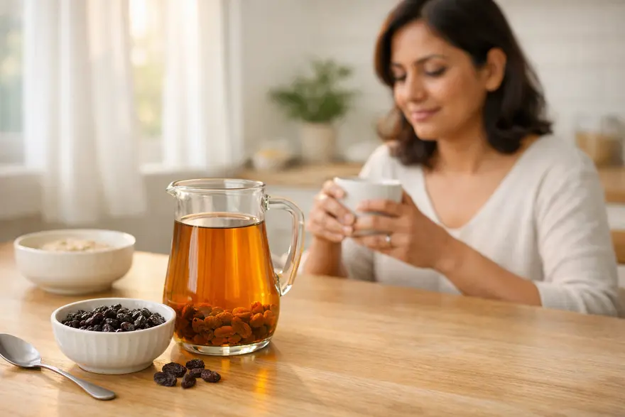
मनुक्याचे पाणी हे रात्रभर पाण्यात मनुके भिजवून तयार केले जाणारे एक साधे घरगुती पेय आहे. ते तयार करणे सोपे आणि चवीला हलके गोड असल्यामुळे अनेक लोक ते त्यांच्या सकाळच्या दिनक्रमात समाविष्ट करतात. तुम्ही ऑनलाइन मनुक्याच्या पाण्याच्या फायद्यांबद्दल अनेक जोरदार दावे पाहिले असतील. प्रत्यक्षात, ते संतुलित आहाराचा उपयुक्त भाग असू शकते, पण ते कोणताही चमत्कारी उपाय नाही. पाण्यात भिजवलेल्या मनुक्यांचे फायदे हे झटपट उपाय म्हणून नव्हे, तर सौम्य पोषणात्मक आधार म्हणून समजून घेणे योग्य आहे. मनुक्याचे पाणी म्हणजे काय? मनुक्याचे पाणी, ज्याला किशमिश पाणी असेही म्हणतात, हे मनुके अनेक तास, साधारणपणे रात्रभर, पाण्यात भिजवल्यानंतर उरलेले द्रव असते. काही लोक मनुके गाळून फक्त पाणी पितात, तर काही जण भिजवलेली मनुकेही खातात. मनुके ही वाळवलेली द्राक्षे असल्यामुळे या पेयात फळात आढळणारी काही पोषकद्रव्ये आणि वनस्पती संयुगे असू शकतात. त्याच वेळी, फायबरचा मोठा भाग मनुक्यांमध्येच राहतो. म्हणूनच मनुक्याचे पाणी हे संपूर्ण फळाचा पूर्ण पर्याय नसून एक साधे आरोग्यदायी पेय म्हणून पाहणे योग्य आहे. तुम्हाला काळ्या मनुक्याच्या पाण्याचे फायदे यासारखे शोधही दिसू शकतात. या पेयासाठी काळी मनुके हा लोकप्रिय पर्याय आहे, पण तुम्ही काळी, तपकिरी किंवा सोनेरी मनुके वापरली तरी मूलभूत कल्पना तीच राहते. मनुक्याच्या पाण्याचे पोषण एका दृष्टीक्षेपात मनुक्याच्या पाण्याचे आकर्षण मनुक्यांमध्ये नैसर्गिकरित्या असलेल्या पोषकद्रव्यांमुळे आहे. यात समावेश होतो: नैसर्गिक साखर लोह पोटॅशियम अँटिऑक्सिडंट्स फायबरचे थोडे प्रमाण, तरीही जास्त फायबर भिजवलेल्या मनुक्यांमध्येच राहते फक्त पाण्यातील अचूक पोषण तुम्ही किती मनुके वापरता, किती वेळ भिजवता आणि ते फळही खाता का यावर अवलंबून बदलू शकते. मनुक्याच्या पाण्याचे 8 फायदे मनुक्याच्या पाण्यावरच संशोधन मर्यादित असले, तरी मनुक्यांमध्ये तुमच्या एकूण आरोग्यास आधार देणारी पोषकद्रव्ये असतात. ते तुमच्या दिनक्रमात समजूतदारपणे समाविष्ट केल्यास मनुक्याच्या पाण्याचे आठ संभाव्य फायदे येथे दिले आहेत. 1. पचनास मदत करू शकते मनुक्यांमध्ये फायबर असते आणि भिजवल्यामुळे ती मऊ व खाण्यास सोपी होतात. तुम्ही पाणी पिण्यासोबत भिजवलेली मनुकेही खाल्ल्यास, फायबरयुक्त आहाराचा भाग म्हणून ते पचन अधिक सुरळीत ठेवण्यास मदत करू शकते. याचा अर्थ असा नाही की फक्त मनुक्याचे पाणी दीर्घकाळ चालणाऱ्या बद्धकोष्ठतेवर उपचार करू शकते. पण पुरेसे द्रव, फळे, भाज्या आणि संपूर्ण धान्यांसोबत घेतल्यास ते आतड्यांसाठी अनुकूल दिनक्रमात बसू शकते. 2. लोह सेवनास मदत करू शकते मनुक्यांमध्ये लोह असते, जे तुमच्या शरीराला हिमोग्लोबिन तयार करण्यासाठी आणि ऑक्सिजन वाहून नेण्यासाठी आवश्यक असते. त्यामुळे लोह असलेले साधे अन्नपदार्थ शोधणाऱ्या लोकांसाठी मनुक्याचे पाणी हा लोकप्रिय पर्याय आहे. तरीही, वास्तववादी राहणे महत्त्वाचे आहे. फक्त मनुक्याचे पाणी हे अशक्तपणावर उपचार नाही. तुम्हाला थकवा, अशक्तपणा, धाप लागणे जाणवत असेल किंवा लोह कमी असल्याची इतर लक्षणे दिसत असतील, तर योग्य तपासणी करणे ही पुढची योग्य पायरी आहे. 3. नैसर्गिक ऊर्जा देते मनुक्यांमध्ये नैसर्गिकरित्या ग्लुकोज आणि फ्रुक्टोज असतात, त्यामुळे मनुक्याचे पाणी हलकी ऊर्जा देऊ शकते. म्हणूनच अनेक लोक ते सकाळी पिणे पसंत करतात. तुम्हाला साधे आणि हलके गोड काहीतरी हवे असल्यास, अतिगोड पेयांपेक्षा हा चांगला पर्याय असू शकतो. पण परिमाणावर नियंत्रण तरीही महत्त्वाचे आहे, कारण मनुक्यांमध्ये नैसर्गिक साखर जास्त असते. 4. अँटिऑक्सिडंट आधार देते मनुक्यांमध्ये अँटिऑक्सिडंट संयुगे असतात, जी तुमच्या पेशींना ऑक्सिडेटिव्ह ताणापासून संरक्षण देण्यास मदत करतात. मनुक्यांच्या पोषणात्मक ताकदींपैकी हा एक सर्वाधिक चर्चिला जाणारा मुद्दा आहे. याचा अर्थ असा नाही की मनुक्याचे पाणी स्वतःहून रोग टाळू शकते. पण फळे, भाज्या, कडधान्ये, सुकामेवा आणि संपूर्ण धान्ये असलेल्या विविध आहाराचा तो एक छोटा भाग असू शकतो. 5. हृदयाच्या आरोग्यास मदत करू शकते मनुक्यांमध्ये पोटॅशियम असते, जे संतुलित आहारात उपयुक्त खनिज आहे. पोटॅशियम असलेले अन्नपदार्थ शरीराच्या सामान्य कार्यांना आधार देऊ शकतात आणि हृदयाबाबत जागरूक आहारपद्धतीत चांगले बसू शकतात. हा फायदा तुमच्या एकूण जीवनशैलीवरही अवलंबून असतो. मनुक्याचे पाणी अतिरिक्त मीठ, अपुरी झोप, निष्क्रियता किंवा असंतुलित आहाराचे परिणाम रद्द करणार नाही. त्याला उपाय नव्हे, तर आधार म्हणून पाहा. 6. हाडांच्या आरोग्यास मदत करू शकते मनुक्यांमध्ये सामान्य आरोग्यात, त्यात हाडांच्या आरोग्यातही, भूमिका बजावणारी खनिजे असतात. म्हणूनच आरोग्यदायी वृद्धत्व आणि रोजच्या पोषणाबाबतच्या चर्चांमध्ये मनुक्यांचा समावेश अनेकदा केला जातो. तरीही, हाडांची ताकद ही कॅल्शियम, व्हिटॅमिन D, प्रथिने, हालचालीची पातळी आणि वय अशा अनेक घटकांवर अवलंबून असते. मनुक्याचे पाणी फक्त छोटा सहाय्यक भाग बजावू शकते. 7. त्वचेच्या आरोग्यास मदत करू शकते हायड्रेशन आणि अँटिऑक्सिडंटचे सेवन या दोन्ही गोष्टी एकूण त्वचेच्या आरोग्याशी संबंधित आहेत. विशेषतः साखरयुक्त पर्यायांऐवजी हलके पेय निवडण्यास ते मदत करत असेल, तर मनुक्याचे पाणी साध्या पद्धतीने याला आधार देऊ शकते. येथे अति दावे न करणे चांगले. मनुक्याचे पाणी हे मुरुमांवरील थेट उपचार किंवा वृद्धत्वरोधक उपाय नाही. चांगले त्वचा आरोग्य अजूनही झोप, हायड्रेशन, पोषण आणि गरजेनुसार वैद्यकीय काळजी यावर अवलंबून असते. 8. आरोग्यदायी दिनक्रमाचा साधा भाग होऊ शकते मनुक्याच्या पाण्याचा सर्वात मोठा फायदा म्हणजे त्याची व्यावहारिकता. ते सोपे, कमी खर्चिक आणि दिनक्रमात समाविष्ट करायला सहज आहे. ते पिण्यामुळे तुमचा दिवस सजग आणि आरोग्यदायी निवडीने सुरू होत असेल, तर तो आधीच एक उपयुक्त फायदा आहे. काळ्या मनुक्याच्या पाण्याचे फायदे शोधणारे अनेक लोक प्रत्यक्षात सहज पाळता येतील अशा दैनंदिन सवयी शोधत असतात. मध्यम प्रमाणात वापरल्यास मनुक्याचे पाणी अशा सवयींपैकी एक असू शकते. मनुक्याचे पाणी योग्य पद्धतीने कसे बनवावे मनुक्याचे पाणी कसे बनवावे हे जाणून घ्यायचे असल्यास, पद्धत सोपी आणि स्वच्छ ठेवा. चांगल्या प्रतीची मनुके निवडा. तुम्ही काळी, तपकिरी किंवा सोनेरी मनुके वापरू शकता. ती स्वच्छ वाहत्या पाण्याखाली नीट धुवा. सुमारे 1 cup मनुके 2 cups पाण्यात घाला. तुम्ही आचेवरून उतरवलेल्या उकळलेल्या पाण्यात मनुके घालू शकता किंवा स्वच्छ पिण्याचे पाणी वापरून रात्रभर भिजवू शकता. ती 8 to 12 hours भिजू द्या. सकाळी, तुम्हाला स्वच्छ मनुक्याचे पाणी हवे असल्यास द्रव गाळून घ्या. ते साधे किंवा किंचित कोमट प्या. तुमच्या पचनाला चालत असल्यास तुम्ही भिजवलेली मनुकेही खाऊ शकता. मनुक्याच्या पाण्याचा जास्तीत जास्त फायदा घेण्यासाठी टिप्स साखर, मध किंवा गोड सिरप घालू नका तुमचे परिमाण मध्यम ठेवा भिजवण्यापूर्वी मनुके नीट धुवा अधिक फायबर हवे असल्यास भिजवलेली मनुकेही खा जेवणाचा पर्याय म्हणून वापरण्याऐवजी संतुलित नाश्त्यासोबत घ्या डिटॉक्सचा झटपट मार्ग म्हणून नव्हे, तर आरोग्यदायी दिनक्रमाचा भाग म्हणून वापरा तुम्ही विशेषतः काळ्या मनुक्याच्या पाण्याचे फायदे जाणून घेत असाल, तर फार मोठ्या परिणामांची अपेक्षा करण्याऐवजी चांगल्या प्रतीची मनुके आणि समजूतदार परिमाण यावर लक्ष द्या मनुक्याचे पाणी पिण्याची सर्वोत्तम वेळ कोणती? अनेक लोक सकाळी रिकाम्या पोटी मनुक्याचे पाणी पिणे पसंत करतात. याचे मुख्य कारण म्हणजे ते दैनंदिन दिनक्रमात सहज बसते आणि नाश्त्यापूर्वी हलके वाटते. मात्र, सकाळच एकमेव योग्य वेळ आहे याचा ठोस पुरावा नाही. तुम्हाला अधिक सोयीचे वाटत असल्यास तुम्ही दिवसाच्या दुसऱ्या वेळीही ते पिऊ शकता. योग्य वेळेपेक्षा मध्यम प्रमाण आणि सातत्य अधिक महत्त्वाचे आहे. टाळावयाच्या सामान्य चुका खूप जास्त मनुके वापरणे आणि जास्त म्हणजे अधिक चांगले असे गृहीत धरणे ही एक सामान्य चूक आहे. त्यामुळे तुम्हाला जाणवण्यापेक्षा साखरेचे सेवन अधिक वाढू शकते. दुसरी चूक म्हणजे झटपट परिणामांची अपेक्षा करणे. मनुक्याचे पाणी तुमचे पचन, त्वचा, वजन किंवा ऊर्जा एका रात्रीत बदलण्याची शक्यता नाही. काही लोक हे पेय बनवतात आणि प्रत्येक वेळी भिजवलेली मनुके फेकून देतात. फायबरचा मोठा भाग फळातच राहतो, त्यामुळे तुम्ही ती कधीच खाल्ली नाहीत तर पोषणमूल्याचा काही भाग चुकू शकतो. मनुक्याचे पाणी वैद्यकीय सल्ल्याचा पर्याय म्हणून न वापरणेही महत्त्वाचे आहे. तुम्हाला थकवा, अस्पष्ट वजनबदल, पचनाशी संबंधित लक्षणे किंवा रक्तातील साखरेबद्दल चिंता असल्यास, योग्य तपासण्या करून घेणे चांगले. मनुक्याच्या पाण्याबाबत कोणांनी काळजी घ्यावी? मध्यम प्रमाणात घेतल्यास मनुक्याचे पाणी अनेक लोकांसाठी सुरक्षित असते, पण काही गटांनी सावध राहावे. तुम्हाला मधुमेह, प्रीडायबिटीज असेल किंवा रक्तातील साखर पाहणे आवश्यक असेल, तर लक्षात ठेवा की मनुक्यांमध्ये नैसर्गिक साखर केंद्रित स्वरूपात असते. ती नेहमीच टाळावी लागते असे नाही, पण परिमाण महत्त्वाचे असते. तुमचे पोट संवेदनशील असेल, तर खूप जास्त मनुक्याचे पाणी किंवा खूप जास्त भिजवलेली मनुके घेतल्यास पोट फुगणे, गॅस किंवा जुलाब होऊ शकतात. तुम्हाला द्राक्षे किंवा सुकी फळे यांची ज्ञात अॅलर्जी असल्यास, डॉक्टरांनी अन्यथा सल्ला दिल्याशिवाय ते टाळा. मनुक्याचे पाणी विरुद्ध भिजवलेली मनुके खाणे मनुक्याचे पाणी सोयीस्कर आहे, पण भिजवलेली मनुके साधारणपणे अधिक पूर्ण पोषण देतात, कारण फायबर फळातच राहते. फक्त पाणी प्यायल्यास तुम्हाला काही चव आणि काही विरघळलेली पोषकद्रव्ये मिळू शकतात, पण मनुके खाल्ल्याने ही सवय अधिक पोटभरीची आणि पचनासाठी संभाव्यतः अधिक उपयुक्त होऊ शकते. म्हणून तुम्हाला त्याची चव आवडत असेल आणि ती चांगली सहन होत असतील, तर फक्त द्रव निवडण्यापेक्षा दोन्ही घेणे अधिक संतुलित पर्याय ठरू शकतो. निष्कर्ष मनुक्याचे पाणी तुमच्या दैनंदिन दिनक्रमात एक साधी आणि समजूतदार भर ठरू शकते. मध्यम प्रमाणात वापरल्यास ते पचन, ऊर्जा, अँटिऑक्सिडंट सेवन आणि एकूण आरोग्यदायी जीवनशैलीला आधार देऊ शकते. मुख्य गोष्ट म्हणजे त्याला सर्व आजारांवरील उपाय नव्हे, तर एक छोटी आरोग्यदायी सवय म्हणून पाहणे. पौष्टिक अन्नपदार्थ आणि आरोग्यदायी दिनक्रमांसोबत प्रतिबंधात्मक आरोग्य तपासण्या तुमच्या शरीरात काय घडत आहे याबद्दल माहिती ठेवण्यास मदत करतात. तुम्हाला नियमित रक्त तपासण्या, पूर्ण शरीर तपासण्या किंवा सतत आरोग्य निरीक्षण हवे असले, तरी मेट्रोपोलिस हेल्थकेअर अचूक निकाल, प्रगत निदान कौशल्य, 4,000+ चाचण्या आणि विस्तृत नेटवर्कद्वारे सोयीस्कर घरून नमुना संकलनासह तुम्हाला साथ देते. तुम्ही फळांवर आधारित अधिक आरोग्यदायी सवयी जाणून घेत असाल, तर व्यापक प्रतिबंधात्मक आरोग्य प्रवासाचा भाग म्हणून तुम्ही द्राक्षांचे फायदे याबद्दलही वाचू शकता. मनुक्याच्या पाण्याबद्दल वारंवार विचारले जाणारे प्रश्न मी रोज मनुक्याचे पाणी पिऊ शकतो का? होय, ते तुमच्या शरीराला आणि एकूण आहाराला अनुकूल असेल, तर तुम्ही मध्यम प्रमाणात रोज मनुक्याचे पाणी पिऊ शकता. फक्त परिमाण समजूतदार ठेवा, विशेषतः तुम्ही साखरेचे सेवन पाहत असाल तर. सर्वोत्तम परिणामांसाठी मनुके पाण्यात किती वेळ भिजवावीत? मनुके किमान 8 hours भिजवा. रात्रभर भिजवणे हा सर्वात व्यावहारिक पर्याय आहे आणि सामान्यतः वापरला जातो. मनुक्याचे पाणी वजन कमी करण्यात मदत करू शकते का? मनुक्याचे पाणी स्वतःहून वजन कमी करणारे पेय नाही. ते तुम्हाला साखरयुक्त पेये किंवा विचार न करता खाणे टाळण्यास मदत करत असेल, तर ते आरोग्यदायी दिनक्रमात बसू शकते, पण वजन नियंत्रण अजूनही तुमचा पूर्ण आहार, हालचाल, झोप आणि एकूण कॅलरी संतुलन यावर अवलंबून असते. मी माझ्या त्वचेसाठी मनुक्याचे पाणी वापरू शकतो का? मनुक्याचे पाणी त्वचेला अप्रत्यक्षरीत्या आधार देऊ शकते, कारण ते हायड्रेशनमध्ये योगदान देते आणि मनुक्यांमधून अँटिऑक्सिडंट संयुगे देते. पण ते त्वचेची काळजी, संतुलित पोषण किंवा त्वचेच्या समस्यांसाठी वैद्यकीय उपचारांचा पर्याय नाही. मी मनुक्याचे पाणी किती वेळा घ्यावे? बहुतेक लोकांसाठी आठवड्यातून काही वेळा किंवा दिवसातून एकदा मध्यम प्रमाणात घेणे योग्य ठरू शकते. मोठ्या प्रमाणाची गरज नाही. तुम्हाला रक्तातील साखरेबद्दल चिंता किंवा पचनाची संवेदनशीलता असल्यास, अधिक काळजी घेणे चांगले.
कोळंबीचे फायदे: पोषण, प्रथिनांचे प्रमाण आणि हृदयाचे आरोग्य
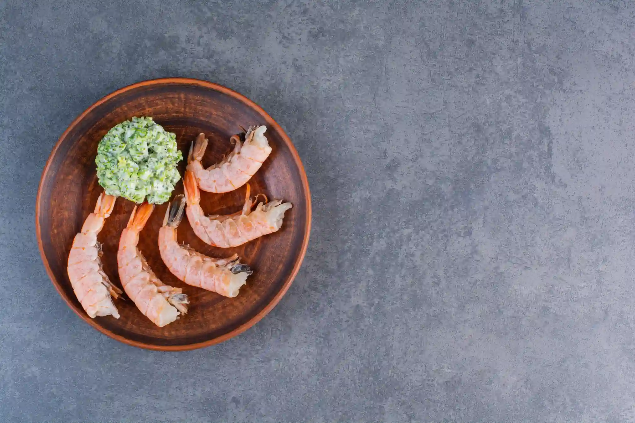
कोळंबी हा जगभरात सर्वाधिक खाल्ल्या जाणाऱ्या समुद्री अन्नपदार्थांपैकी एक आहे, आणि त्याचे कारण समजणे सोपे आहे. ती पटकन शिजते, स्वयंपाकात अनेक प्रकारे वापरता येते आणि निरोगी शरीराला आधार देणाऱ्या पोषकद्रव्यांनी समृद्ध असते. तुम्ही ती स्टर-फ्राय, सलाड किंवा साध्या ग्रिल्ड पद्धतीने खात असाल, तरी कोळंबी कमी कॅलरीच्या छोट्या पदार्थात भरपूर पोषणमूल्य देते. तुम्हाला कोळंबीचे फायदे, त्यात काय असते आणि ती तुमच्या आहारासाठी योग्य आहे का याबद्दल जाणून घ्यायचे असल्यास, या मार्गदर्शिकेत तुम्हाला आवश्यक असलेली सर्व माहिती दिली आहे. प्रथिने आणि हृदयाचे आरोग्य यापासून वजन नियंत्रण आणि अन्नसुरक्षेपर्यंत, कोळंबी काय देऊ शकते याचा येथे सविस्तर आढावा दिला आहे. कोळंबी म्हणजे काय? कोळंबी हे जगभरातील समुद्र, नद्या आणि तलावांमध्ये आढळणारे छोटे जलचर कवचधारी प्राणी आहेत. खेकडे आणि लॉब्स्टर यांच्यासह ती क्रस्टेशियन कुटुंबात येते आणि जागतिक स्तरावर सर्वाधिक खाल्ल्या जाणाऱ्या समुद्री अन्नपदार्थांपैकी एक आहे. तिची सौम्य, किंचित गोड चव आणि मसाले व चवदार पदार्थ चांगल्या प्रकारे शोषून घेण्याची क्षमता यामुळे अनेक खाद्यसंस्कृतींमध्ये कोळंबी लोकप्रिय आहे. ती ताजी, गोठवलेली, कच्ची आणि शिजवलेली अशा अनेक स्वरूपांत उपलब्ध असते आणि लहान आकारापासून जम्बो आकारापर्यंत मिळते. प्रॉन विरुद्ध श्रिंप दैनंदिन बोलण्यात "प्रॉन" आणि "श्रिंप" हे शब्द अनेकदा एकमेकांच्या जागी वापरले जातात, आणि भारत व युनायटेड किंगडमसह जगातील अनेक भागांत "प्रॉन" हा शब्द अधिक सामान्यपणे वापरला जातो. मात्र, जैविकदृष्ट्या ते पूर्णपणे सारखे नाहीत. श्रिंप प्लिओसायमाटा उपगणात येतात, तर प्रॉन डेंड्रोब्रँकिएटा उपगणात येतात. रचनात्मकदृष्ट्या, प्रॉन साधारणपणे मोठे असतात, त्यांचे पाय लांब असतात आणि शरीराची रचना किंचित वेगळी असते. पोषण आणि स्वयंपाकाच्या दृष्टीने फरक फार कमी आहे. व्यावहारिकदृष्ट्या, आरोग्य फायदे आणि खाद्य वापराच्या बाबतीत दोन्ही समान मानता येतात. कोळंबीचे पौष्टिक मूल्य कोळंबीतील पोषणघटक तिचा आकार, प्रजाती आणि शिजवण्याची पद्धत यानुसार थोडे बदलू शकतात. वाफवलेली किंवा उकडलेली कोळंबी तळलेल्या प्रकारांच्या तुलनेत तिची नैसर्गिक पोषकद्रव्ये अधिक राखून ठेवते, कारण तळलेल्या पदार्थांत अतिरिक्त चरबी आणि कॅलरी वाढतात. खालील आकडे प्रमाणित शिजवलेल्या सर्व्हिंगसाठी अंदाजे मूल्ये दर्शवतात. प्रत्येक सर्व्हिंगमधील कोळंबीचे पोषण खालील तक्त्यात शिजवलेल्या कोळंबीच्या 3-ounce किंवा 85-gram सर्व्हिंगचे अंदाजे पोषणमूल्य दाखवले आहे: पोषकद्रव्य प्रत्येक सर्व्हिंगमधील प्रमाण (85g) कॅलरी ~84 kcal प्रथिने ~20 g चरबी ~0.9 g कार्बोहायड्रेट्स 0 g कोलेस्टेरॉल ~161 mg सोडियम ~94 mg सेलेनियम ~38 mcg व्हिटॅमिन B12 ~1.3 mcg आयोडीन ~13 mcg झिंक ~1.4 mg ओमेगा-3 फॅटी अॅसिड्स ~0.3 g पोषणाच्या दृष्टीने कोळंबी वेगळी का ठरते? पोषणाच्या दृष्टीने कोळंबी विशेषतः उपयुक्त ठरण्याचे कारण म्हणजे जास्त प्रथिने आणि अतिशय कमी कॅलरी यांचे संयोजन. 90 पेक्षा कमी कॅलरी देणाऱ्या एका सर्व्हिंगमधून सुमारे 20 grams प्रथिने मिळतात. त्यामुळे, विशेषतः कॅलरीचे सेवन फार वाढवता चांगले खायचे असलेल्या लोकांसाठी, कोळंबी हा उपलब्ध पोषकद्रव्यांनी समृद्ध लीन प्रोटीन पर्यायांपैकी एक ठरतो. कोळंबीत संतृप्त चरबीही कमी असते, त्यामुळे संतुलित आहारात लीन समुद्री अन्न म्हणून तिचे मूल्य आणखी वाढते. कोळंबीचे प्रमुख आरोग्य फायदे उच्च-प्रथिनयुक्त अन्न म्हणून कोळंबी प्रथिने ही तुमच्या शरीराला आवश्यक असलेल्या सर्वात महत्त्वाच्या मोठ्या पोषकद्रव्यांपैकी एक आहेत. ती स्नायूंची दुरुस्ती आणि वाढ यांना आधार देतात, जेवणानंतर तुम्हाला जास्त वेळ पोटभरल्यासारखे वाटण्यास मदत करतात आणि हार्मोन निर्मितीपासून रोगप्रतिकारक कार्यापर्यंत जवळजवळ प्रत्येक जैविक प्रक्रियेत भूमिका बजावतात. कोळंबी ही लीन, उच्च-गुणवत्तेच्या प्रथिनांचा उत्कृष्ट स्रोत आहे. एका 3-ounce सर्व्हिंगमधून अतिशय कमी चरबीसह सुमारे 20 grams प्रथिने मिळतात. त्यामुळे वजन नियंत्रित करणे, सक्रिय राहणे किंवा फक्त अधिक संतुलित पद्धतीने खाणे असे कोणतेही उद्दिष्ट असले, तरी प्रथिनांचे सेवन प्राधान्य असलेल्या कोणत्याही आहार योजनेत कोळंबी व्यावहारिक भर ठरते. कोळंबी आणि हृदयाचे आरोग्य कोळंबी आणि हृदयाच्या आरोग्याचा संबंध सुरुवातीला वाटतो त्यापेक्षा अधिक सूक्ष्म आहे. कोळंबीत कोलेस्टेरॉल असले, तरी निरोगी व्यक्तींमध्ये कोळंबी खाणे आणि हृदयरोगाचा वाढलेला धोका यांच्यात संशोधनाने सातत्यपूर्ण संबंध आढळलेला नाही. प्रत्यक्षात, कोळंबीत हृदयाच्या आरोग्याला सक्रियपणे आधार देणारी अनेक पोषकद्रव्ये असतात. कोळंबी ओमेगा-3 फॅटी अॅसिड्स देते, जी निरोगी रक्तदाबास मदत करतात आणि दाह कमी करण्याशी संबंधित आहेत. त्यात संतृप्त चरबीही कमी असते, जी रक्तातील कोलेस्टेरॉलची पातळी वाढवण्याशी अधिक थेट संबंधित असलेली चरबी आहे. कोळंबीत आढळणाऱ्या अॅस्टॅझॅन्थिन या अँटिऑक्सिडंटचा धमनींच्या भिंती मजबूत करण्याची आणि एचडीएल (HDL), म्हणजेच तथाकथित "चांगले" कोलेस्टेरॉल, याची आरोग्यदायी पातळी राखण्याची संभाव्य भूमिका यावर अभ्यास झाला आहे. एक महत्त्वाची गोष्ट म्हणजे शिजवण्याची पद्धत. ग्रिल्ड, वाफवलेली, उकडलेली किंवा हलकी परतलेली कोळंबी तिचे हृदयास आधार देणारे गुणधर्म राखून ठेवते. डीप-फ्राय केलेली किंवा जड क्रीम सॉसमध्ये तयार केलेली कोळंबी खूप जास्त संतृप्त चरबी आणि कॅलरी मिळवते, ज्यामुळे हे फायदे कमी होऊ शकतात. वजन कमी करण्यासाठी कोळंबी तुम्ही वजन नियंत्रित करण्याचा प्रयत्न करत असाल, तर कोळंबी तुमच्या जेवणात समाविष्ट करण्यासाठी अतिशय व्यावहारिक अन्नपदार्थ ठरू शकते. तिच्यातील जास्त प्रथिने तृप्तीला आधार देतात, म्हणजे ती खाल्ल्यानंतर तुम्हाला जास्त वेळ पोटभरल्यासारखे वाटू शकते, ज्यामुळे दिवसभरातील एकूण कॅलरी सेवन कमी करण्यास मदत होऊ शकते. 3-ounce सर्व्हिंगमध्ये 90 पेक्षा कमी कॅलरी आणि जवळजवळ कार्बोहायड्रेट्स किंवा चरबी नसल्यामुळे, कोळंबी कॅलरीबाबत जागरूक आहार योजनांमध्ये सहज बसते. भाज्या आणि संपूर्ण धान्यांसोबत घेतल्यास, पोषणात तडजोड न करता ती समाधानकारक, संतुलित जेवण बनवते. कोळंबीतील अँटिऑक्सिडंट्स कोळंबीत अॅस्टॅझॅन्थिन नावाचा अँटिऑक्सिडंट असतो, जो नैसर्गिकरित्या आढळणारा कॅरोटेनॉइड रंगद्रव्य आहे आणि शिजवल्यावर कोळंबीला तिचा वैशिष्ट्यपूर्ण गुलाबीसर-लाल रंग देतो. अॅस्टॅझॅन्थिन हे कोळंबी खाणाऱ्या सूक्ष्मशैवालांद्वारे तयार होते आणि त्यांच्या ऊतींमध्ये साठते. अँटिऑक्सिडंट म्हणून, अॅस्टॅझॅन्थिन तुमच्या पेशींना फ्री रॅडिकल्समुळे होणाऱ्या नुकसानीपासून संरक्षण करण्यास मदत करते. फ्री रॅडिकल्स या अस्थिर रेणूंना दाह आणि दीर्घकालीन आरोग्य समस्यांशी जोडले जाते. संशोधनानुसार ते मेंदू आणि हृदयाच्या आरोग्यास आधार देण्यात भूमिका बजावू शकते, मात्र बहुतेक संशोधन सामान्य कोळंबी सर्व्हिंगमध्ये आढळणाऱ्या प्रमाणाऐवजी संकेंद्रित पूरकांवर केले गेले आहे. कोळंबीचे अँटिऑक्सिडंट योगदान अर्थपूर्ण आहे, पण ते व्यापक, विविध आहाराचा भाग म्हणून समजणे चांगले. कोळंबीतील जीवनसत्त्वे आणि खनिजे प्रथिने आणि अँटिऑक्सिडंट्सव्यतिरिक्त, कोळंबी उपयुक्त जीवनसत्त्वे आणि खनिजे देते: सेलेनियम: कोळंबी सेलेनियमचा विशेषतः चांगला स्रोत आहे. हे खनिज अँटिऑक्सिडंट एन्झाइम्स तयार करण्यास मदत करते आणि रोगप्रतिकारक कार्य व थायरॉइड आरोग्यास आधार देते. आयोडीन: कोळंबी आयोडीनच्या चांगल्या आहार स्रोतांपैकी एक आहे. हे खनिज निरोगी थायरॉइड कार्य आणि मेंदूच्या विकासासाठी आवश्यक आहे. व्हिटॅमिन B12: हे जीवनसत्त्व मज्जातंतूंचे आरोग्य, लाल रक्तपेशींची निर्मिती आणि संज्ञानात्मक कार्यासाठी महत्त्वाचे आहे. कोळंबी दैनंदिन B12 गरजांमध्ये अर्थपूर्ण योगदान देते. लोह: लोह लाल रक्तपेशींच्या निर्मितीस मदत करते आणि संपूर्ण शरीरात ऑक्सिजन वाहून नेण्यास मदत करते. झिंक: झिंक रोगप्रतिकारक संरक्षण, जखम भरून येणे आणि पेशींची वाढ यांत महत्त्वाची भूमिका बजावते. एकत्रितपणे, संतुलित आहाराचा भाग म्हणून खाल्ल्यास ही सूक्ष्मपोषकद्रव्ये कोळंबीला एकूण आरोग्यासाठी खरोखरच सहाय्यक अन्न बनवतात. कोळंबी खाण्यास सुरक्षित आहे का? बहुतेक लोकांसाठी कोळंबी सुरक्षित आणि पौष्टिक आहे. मात्र, ती तुमच्या आहाराचा नियमित भाग करण्यापूर्वी काही बाबी जाणून घेणे योग्य आहे. कोळंबी आणि कोलेस्टेरॉल कोळंबीत आहारातील कोलेस्टेरॉल तुलनेने जास्त असते, सुमारे 3-ounce सर्व्हिंगमधून 161 milligrams. त्यामुळे पूर्वीपासून तिच्या हृदयाच्या आरोग्यावर होणाऱ्या परिणामाबद्दल चिंता व्यक्त केली जात होती. मात्र, सध्याचे संशोधन अधिक संतुलित चित्र दाखवते. बहुतेक लोकांसाठी, आहारातील कोलेस्टेरॉलचा रक्तातील कोलेस्टेरॉल पातळीवर मध्यम परिणाम होतो. कारण अन्नातून किती कोलेस्टेरॉल मिळते यानुसार यकृत स्वतः किती कोलेस्टेरॉल तयार करायचे हे समायोजित करते. त्यामुळे, योग्य प्रमाणात कोळंबी खाणे बहुतेक निरोगी व्यक्तींसाठी समस्याजनक मानले जात नाही. तरीही, विशिष्ट लिपिड विकार असलेल्या किंवा डॉक्टरांनी कोलेस्टेरॉलचे सेवन पाहण्याचा सल्ला दिलेल्या लोकांनी कोळंबीचे सेवन लक्षणीयरीत्या वाढवण्यापूर्वी त्यांच्या आरोग्यसेवा पुरवठादाराशी बोलावे. एकूण आहाराची गुणवत्ता आणि शिजवण्याची पद्धत हे सर्वात महत्त्वाचे घटक राहतात. कोळंबीची अॅलर्जी आणि कोणांनी काळजी घ्यावी? कोळंबीसह शेलफिश हे सर्वात सामान्य अन्न अॅलर्जन्सपैकी आहेत. कोळंबीची अॅलर्जी शेलफिशमध्ये आढळणाऱ्या काही प्रथिनांवरील प्रतिक्रियेमुळे होते, त्यापैकी सर्वात सामान्य म्हणजे ट्रोपोमायोसिन नावाचे प्रथिन. कोळंबी अॅलर्जीची लक्षणे सौम्य ते गंभीर असू शकतात आणि त्यात समावेश असू शकतो: तोंड किंवा घशात खाज येणे किंवा मुंग्या येणे हायव्ह्ज किंवा पुरळ यांसारख्या त्वचेच्या प्रतिक्रिया मळमळ, उलट्या किंवा जुलाब यांसह पचनाचा त्रास चेहरा, ओठ किंवा जीभ सुजणे श्वास घेण्यास त्रास गंभीर प्रकरणांमध्ये, अॅनाफायलॅक्सिस, जी जीवघेणी प्रतिक्रिया असून तातडीच्या वैद्यकीय लक्षाची गरज असते तुम्हाला शेलफिश अॅलर्जीचा संशय असल्यास, डॉक्टरांशी बोलणे महत्त्वाचे आहे. कोळंबी अॅलर्जी चाचणी, जी साधारणपणे स्किन प्रिक टेस्ट किंवा रक्त तपासणी म्हणून केली जाते, तुम्हाला अॅलर्जी आहे का हे निश्चित करण्यात आणि त्यानुसार आहार निवडीचे मार्गदर्शन करण्यात मदत करू शकते. एकदा खात्री झाल्यावर, प्रतिक्रिया टाळण्याचा एकमेव मार्ग म्हणजे कोळंबी आणि इतर शेलफिश पूर्णपणे टाळणे. काही प्रकरणांमध्ये, अतिसंवेदनशील व्यक्तींमध्ये कोळंबी शिजवताना निघणारी वाफही प्रतिक्रिया निर्माण करू शकते. गर्भधारणेदरम्यान कोळंबी कोळंबी कमी-पारा असलेल्या समुद्री अन्नात वर्गीकृत केली जाते, त्यामुळे स्वॉर्डफिश किंवा किंग मॅकरेलसारख्या मोठ्या माशांच्या तुलनेत गर्भधारणेदरम्यान ती सुरक्षित समुद्री अन्न पर्यायांपैकी एक ठरते. आयोडीन आणि व्हिटॅमिन B12 यांसह कोळंबीतील पोषकद्रव्ये गर्भाच्या विकासालाही आधार देतात. तरीही, अन्नातून होणाऱ्या आजाराचा धोका कमी करण्यासाठी गर्भधारणेदरम्यान कोळंबी पूर्णपणे शिजवलेली असावी. कच्ची किंवा अर्धवट शिजलेली कोळंबी पूर्णपणे टाळावी. कोळंबीसह कमी-पारा समुद्री अन्नाचे आठवड्याला दोन ते तीन लहान सर्व्हिंग्स घेणे गर्भधारणेदरम्यान साधारणपणे योग्य मानले जाते, पण तुमच्या वैयक्तिक आरोग्यस्थितीनुसार तुमच्या डॉक्टर किंवा मिडवाइफचा सल्ला पाळणे नेहमीच चांगले. फार्म्ड विरुद्ध वाइल्ड कोळंबी बाजारात उपलब्ध बहुतेक कोळंबी फार्म्ड असते, आणि स्रोत व हाताळणी पद्धतींमध्ये मोठा फरक असू शकतो. फार्म्ड कोळंबीबद्दलच्या काही चिंता प्रतिजैविकांच्या संभाव्य वापराशी किंवा दूषित घटकांच्या उपस्थितीशी संबंधित असतात. प्रतिष्ठित पुरवठादारांकडून मिळालेली कोळंबी साधारणपणे सुरक्षित असली, तरी तुमची कोळंबी कुठून येते याकडे लक्ष देणे योग्य आहे. प्रतिजैविकांच्या संपर्काच्या दृष्टीने वाइल्ड-कॉट कोळंबी साधारणपणे अधिक स्वच्छ पर्याय मानली जाते. फार्म्ड कोळंबी खरेदी करताना विश्वासार्ह, व्यवस्थित नियमन असलेल्या पुरवठादारांकडील उत्पादने निवडा आणि उपलब्ध असल्यास संबंधित गुणवत्ता प्रमाणपत्रे पाहा. योग्य साठवण आणि पूर्णपणे शिजवणे यामुळे अन्नसुरक्षेचे कोणतेही धोके आणखी कमी होतात. चांगल्या प्रतीची कोळंबी कशी निवडावी? ताजी, चांगल्या प्रतीची कोळंबी निवडल्याने चव आणि सुरक्षितता दोन्हीमध्ये अर्थपूर्ण फरक पडतो. कोळंबी खरेदी करताना काय पाहावे? वास: ताज्या कच्च्या कोळंबीला सौम्य, समुद्रासारखा किंवा किंचित खारट वास असावा. फार तीव्र मासळीचा किंवा अमोनियासारखा वास हा कोळंबी खराब झाल्याचे लक्षण आहे. पोत: कच्ची कोळंबी घट्ट वाटली पाहिजे. मऊ किंवा लगद्यासारखी कोळंबी गुणवत्ता कमी झाल्याचे लक्षण आहे. दिसणे: कवच पारदर्शक आणि करडट-हिरवे, गुलाबीसर किंवा हलके तपकिरी रंगाचे असावे. कवचावरील काळे डाग किंवा गडद झालेल्या कडा खराब होण्याचे संकेत देऊ शकतात. थंड ठेवणे: योग्य रीतीने थंड ठेवलेली किंवा ताज्या बर्फाच्या जाड थरावर ठेवलेली कोळंबीच खरेदी करा. स्रोत: कोळंबीचा उगम आणि हाताळणी पद्धती सांगू शकणाऱ्या प्रतिष्ठित पुरवठादाराकडून खरेदी करा. गोठवलेली विरुद्ध ताजी: आधी गोठवलेली आणि योग्य प्रकारे वितळवलेली कोळंबी ताजी कोळंबीइतकीच पौष्टिक असू शकते. गोठवलेली खरेदी करताना फ्रीझर बर्न किंवा बर्फाचे स्फटिक दिसतात का ते तपासा, कारण त्याचा गुणवत्तेवर परिणाम होऊ शकतो. जास्तीत जास्त पोषकद्रव्यांसाठी कोळंबी कशी शिजवावी? तुम्ही कोळंबी कशी शिजवता याचा अंतिम पदार्थ किती पौष्टिक असेल यावर थेट परिणाम होतो. खालील टप्पे पाळल्यास तुम्हाला या समुद्री अन्नाचा जास्तीत जास्त फायदा मिळू शकतो: सुरक्षितपणे वितळवा: गोठवलेली कोळंबी वापरत असल्यास, ती रात्रभर फ्रिजमध्ये वितळवा किंवा सीलबंद पिशवीत ठेवून थंड पाण्याच्या बाऊलमध्ये ठेवा. खोलीच्या तापमानावर दीर्घकाळ वितळवणे टाळा. योग्य प्रकारे स्वच्छ करून तयार करा: शिजवण्यापूर्वी कवच काढा आणि पाठीकडून कापून गडद शीर बाहेर काढून कोळंबी डिव्हेन करा. हलक्या शिजवण्याच्या पद्धती निवडा: कोळंबी वाफवा, ग्रिल करा, हलकी परता किंवा उकडा. या पद्धती अधिक पोषकद्रव्ये टिकवून ठेवतात आणि कॅलरी व चरबीचे प्रमाण कमी ठेवतात. डीप-फ्राय करणे आणि जड सॉस टाळा: डीप-फ्राय करणे किंवा भरपूर क्रीम असलेल्या सॉसमध्ये शिजवणे यामुळे संतृप्त चरबी आणि कॅलरी खूप वाढतात, ज्यामुळे पदार्थाचे एकूण आरोग्य मूल्य कमी होते. भाज्या आणि संपूर्ण धान्यांसोबत खा: रंगीबेरंगी भाज्या आणि ब्राऊन राइस किंवा क्विनोआसारख्या संपूर्ण धान्यांसोबत कोळंबी दिल्यास अधिक संतुलित, पोषकद्रव्यांनी समृद्ध जेवण तयार होते. पूर्णपणे शिजवा: कोळंबीचे अंतर्गत तापमान सुमारे 63 degrees Celsius पर्यंत पोहोचले पाहिजे. योग्यरीत्या शिजवलेली कोळंबी गुलाबी आणि अपारदर्शक होते आणि स्पर्शाला घट्ट वाटते. तुमच्या आहारात कोळंबी घालण्याचे सोपे मार्ग कोळंबी अतिशय बहुपयोगी आहे आणि रोजच्या जेवणात समाविष्ट करणे सोपे आहे. काही सोप्या कल्पना येथे दिल्या आहेत: स्टर-फ्राइज: हंगामी भाज्या, लसूण, आले आणि हलका सॉस यांसोबत कोळंबी मिसळा आणि ब्राऊन राइस किंवा नूडल्सवर वाढा. सूप: अतिरिक्त प्रथिनांसाठी हलक्या ब्रॉथमध्ये, डाळीच्या सूपमध्ये किंवा टोमॅटो-आधारित पदार्थांमध्ये कोळंबी घाला. सलाड: साध्या लिंबू ड्रेसिंगसह मिश्र हिरव्या भाज्यांवर ग्रिल्ड किंवा उकडलेली कोळंबी चांगली लागते. राइस बाऊल्स: हलकी परतलेली कोळंबी संपूर्ण धान्याच्या भात, काकडी, अॅव्होकॅडो आणि कमी-सोडियम सीझनिंगच्या हलक्या थेंबांसोबत एकत्र करा. टॅकोस: लहान संपूर्ण धान्याच्या रॅप्समध्ये मसालेदार कोळंबी, किसलेला कोबी आणि दही-आधारित सॉस भरा. ग्रिल्ड स्क्यूअर्स: कोळंबी कॅप्सिकम, झुकिनी आणि चेरी टोमॅटो यांसारख्या भाज्यांसह स्क्यूअर्सवर लावा आणि हलक्या, चविष्ट जेवणासाठी ग्रिल करा. कोळंबी खाण्याबाबत कधी सावध राहावे? कोळंबी बहुतेक लोकांसाठी सुरक्षित आणि फायदेशीर असली, तरी काही परिस्थितींमध्ये अतिरिक्त काळजी किंवा वैद्यकीय मार्गदर्शन योग्य ठरते: निश्चित शेलफिश अॅलर्जी: तुम्हाला कोळंबी किंवा शेलफिश अॅलर्जीचे निदान झाले असल्यास, तयारीत कोळंबी वापरली असण्याची शक्यता असलेल्या पदार्थांसह कोळंबीचे सर्व प्रकार पूर्णपणे टाळा. समुद्री अन्न खाल्ल्यानंतर प्रतिक्रिया: कोळंबी खाल्ल्यानंतर खाज, सूज, पचनाचा त्रास किंवा श्वास घेण्यास त्रास जाणवला, तर तातडीने तुमच्या डॉक्टरांचा सल्ला घ्या. कोळंबी अॅलर्जी चाचणी अॅलर्जी आहे का हे स्पष्ट करण्यात मदत करू शकते. कोलेस्टेरॉल किंवा हृदयविकार: तुमच्या डॉक्टरांनी आहारातील कोलेस्टेरॉल किंवा चरबीचे सेवन नियंत्रित करण्याचा सल्ला दिला असल्यास, कोळंबी खाण्याची योग्य वारंवारता आणि भागाचे प्रमाण याबद्दल चर्चा करा. गर्भधारणेसंबंधी मार्गदर्शन: गर्भधारणेदरम्यान समुद्री अन्न सेवनाबाबत, विशेषतः वारंवारता आणि तयारी याबद्दल, तुमच्या डॉक्टरांचा सल्ला नेहमी पाळा. घराबाहेर खाणे: बाहेर जेवताना कोळंबीचा स्रोत किंवा शिजवण्याची पद्धत जाणून घेणे कठीण असू शकते. तुम्हाला अॅलर्जी किंवा आरोग्याबाबत चिंता असल्यास, ऑर्डर देण्यापूर्वी रेस्टॉरंट कर्मचाऱ्यांशी तपासा. कोळंबीच्या फायद्यांबद्दल अंतिम विचार कोळंबी हा खरोखरच पौष्टिक समुद्री अन्न पर्याय आहे, जो निरोगी, संतुलित आहाराला आधार देऊ शकतो. कमी कॅलरी आणि कमी चरबी असलेल्या पॅकेजमध्ये ती लीन प्रथिने, आवश्यक जीवनसत्त्वे व खनिजे, हृदयाला आधार देणारी ओमेगा-3 फॅटी अॅसिड्स आणि अँटिऑक्सिडंट्स देते. तुम्ही वजन नियंत्रण, हृदयाचे आरोग्य किंवा फक्त विविध आणि पौष्टिक आहारावर लक्ष केंद्रित करत असाल, तरी कोळंबी तुमच्या जेवणात व्यावहारिक आणि आनंददायी भर ठरू शकते. कोणत्याही अन्नपदार्थाप्रमाणेच, फायदे तुम्ही ती कशी तयार करता आणि किती प्रमाणात खाता यावर अवलंबून असतात. कोळंबी ग्रिल करणे, वाफवणे किंवा हलकी परतणे आणि पौष्टिक घटकांसोबत खाणे यामुळे सातत्याने सर्वोत्तम पोषण परिणाम मिळतात. स्रोत, संभाव्य अॅलर्जन्स आणि वैयक्तिक आरोग्य गरजा याकडे लक्ष दिल्यास तुम्ही कोळंबी सुरक्षितपणे आणि आत्मविश्वासाने खाऊ शकता. चांगले खाणे हे निरोगी राहण्याचा एक भाग आहे. नियमित आरोग्य निरीक्षण हा दुसरा भाग आहे. मेट्रोपोलिस हेल्थकेअरमध्ये, हृदयाच्या आरोग्याचे मार्कर्स, थायरॉइड कार्य, जीवनसत्त्वांची पातळी आणि बरेच काही तपासणाऱ्या पॅनेल्ससह 4,000 पेक्षा अधिक चाचण्यांची उपलब्धता वापरून तुम्ही तुमच्या आरोग्याबाबत सक्रिय दृष्टिकोन ठेवू शकता. तुम्हाला कोलेस्टेरॉलचे निरीक्षण करायचे असेल, पोषकद्रव्यांच्या कमतरता तपासायच्या असतील किंवा पूर्ण शरीर तपासणी बुक करायची असेल, तरी मेट्रोपोलिस वेबसाइट, अॅप, फोन किंवा व्हॉट्सअॅपद्वारे सोपी बुकिंग आणि 10,000 पेक्षा अधिक टचपॉइंट्सच्या नेटवर्कमध्ये उपलब्ध सोयीस्कर घरून नमुना संकलनाद्वारे हे सोपे करते. अचूक निकाल, जलद मिळणारे, जेणेकरून योग्य आरोग्य निर्णय घेण्यासाठी आवश्यक माहिती तुमच्याकडे नेहमी असेल. वारंवार विचारले जाणारे प्रश्न श्रिंपचा अर्थ काय आहे? श्रिंप हे जगभरातील खाऱ्या आणि गोड्या पाण्याच्या वातावरणात आढळणारे छोटे जलचर क्रस्टेशियन आहेत. ते खेकडे आणि लॉब्स्टर यांच्याच कुटुंबात येतात आणि जागतिक स्तरावर सर्वाधिक खाल्ल्या जाणाऱ्या समुद्री अन्नपदार्थांपैकी एक आहेत. श्रिंप त्यांच्या सौम्य चवी, घट्ट पोत आणि उच्च पोषणमूल्यासाठी ओळखले जातात. कोळंबी तुमच्या हृदयासाठी चांगली आहे का? होय, आरोग्यदायी पद्धतीने तयार केल्यास कोळंबी हृदयाच्या आरोग्यास आधार देऊ शकते. त्यात ओमेगा-3 फॅटी अॅसिड्स असतात, जी निरोगी रक्तदाब आणि कमी दाहाशी संबंधित आहेत, आणि त्यात संतृप्त चरबी कमी असते. कोळंबीत आहारातील कोलेस्टेरॉल जास्त असले, तरी सध्याचे संशोधन सूचित करते की मध्यम प्रमाणात सेवन केल्यास बहुतेक लोकांमध्ये रक्तातील कोलेस्टेरॉलवर नकारात्मक परिणाम होत नाही. हे फायदे टिकवण्यासाठी डीप-फ्राय करणे किंवा जड सॉस टाळा. कोळंबी वजन कमी करण्यात मदत करू शकते का? वजनाबाबत जागरूक आहारात कोळंबी उपयुक्त भर ठरू शकते. ती कॅलरी आणि चरबीमध्ये अतिशय कमी असते, तर प्रथिनांचे भरपूर प्रमाण देते, ज्यामुळे तुम्हाला जास्त वेळ पोटभरल्यासारखे वाटते. भाज्या आणि संपूर्ण धान्यांसोबत जेवणात कोळंबीचा समावेश केल्यास कॅलरी सेवन नियंत्रित करण्यासाठी संतुलित, समाधानकारक दृष्टिकोनाला आधार मिळू शकतो. कोळंबीत प्रथिने जास्त असतात का? होय. शिजवलेल्या कोळंबीच्या 3-ounce सर्व्हिंगमधून अंदाजे 20 grams प्रथिने मिळतात, त्यामुळे ती समुद्री अन्नातून उपलब्ध चांगल्या लीन प्रोटीन स्रोतांपैकी एक ठरते. कोळंबीतील प्रथिने स्नायूंची देखभाल, तृप्ती आणि शरीराच्या अनेक आवश्यक कार्यांना आधार देतात. गर्भधारणेदरम्यान कोळंबी खाणे सुरक्षित आहे का? कोळंबी सामान्यतः गर्भधारणेदरम्यान अधिक सुरक्षित समुद्री अन्न पर्याय मानली जाते कारण ती कमी-पारा अन्न म्हणून वर्गीकृत आहे. ती आयोडीन आणि व्हिटॅमिन B12 देखील देते, जी गर्भाच्या विकासास आधार देतात. मात्र, अन्नातून होणाऱ्या आजाराचा कोणताही धोका टाळण्यासाठी गर्भधारणेदरम्यान कोळंबी नेहमी पूर्णपणे शिजवलेली असावी. तुमच्या डॉक्टर किंवा मिडवाइफचा आहार सल्ला नेहमी पाळा. प्रॉन विरुद्ध श्रिंप यात काय फरक आहे? दैनंदिन भाषेत प्रॉन आणि श्रिंप हे शब्द अनेकदा एकमेकांच्या जागी वापरले जात असले, तरी जैविकदृष्ट्या ते वेगवेगळ्या क्रस्टेशियनना संदर्भित करतात. प्रॉन साधारणपणे मोठे असतात, त्यांचे पाय लांब असतात आणि शरीररचना किंचित वेगळी असते. श्रिंप साधारणपणे लहान असतात आणि त्यांचे शरीर अधिक वाकलेले असते. प्रत्यक्ष वापरात, विशेषतः भारतीय आणि ब्रिटिश इंग्रजीत, "प्रॉन" हा शब्द अनेकदा दोन्हीसाठी वापरला जातो. पोषण आणि स्वयंपाकाच्या दृष्टीने दोन्ही अतिशय समान आहेत. कोळंबी अॅलर्जी चाचणी कधी आवश्यक असते? कोळंबी किंवा इतर शेलफिश खाल्ल्यानंतर खाज, हायव्ह्ज, सूज, पचनाचा त्रास किंवा श्वास घेण्यास त्रास यांसारखी लक्षणे जाणवल्यास, कोळंबी अॅलर्जी चाचणीबद्दल तुमच्या डॉक्टरांशी बोलण्याचा विचार करा. कोळंबी अॅलर्जी चाचणी, जी सामान्यतः स्किन प्रिक टेस्ट किंवा रक्त तपासणी म्हणून केली जाते, अॅलर्जी आहे का हे निश्चित करू शकते. शेलफिश अॅलर्जी निश्चित झाल्यास, संभाव्य गंभीर प्रतिक्रिया टाळण्यासाठी कोळंबी आणि संबंधित अन्नपदार्थ पूर्णपणे टाळणे आवश्यक आहे. कोळंबीत कोलेस्टेरॉल जास्त असते का? होय, कोळंबीत 3-ounce सर्व्हिंगमागे सुमारे 161 milligrams इतके आहारातील कोलेस्टेरॉल तुलनेने जास्त असते. मात्र, संशोधनानुसार बहुतेक निरोगी लोकांसाठी आहारातील कोलेस्टेरॉलचा रक्तातील कोलेस्टेरॉल पातळीवर थेट परिणाम मर्यादित असतो, कारण यकृत त्यानुसार स्वतःचे कोलेस्टेरॉल उत्पादन समायोजित करते. कोळंबीत संतृप्त चरबीही कमी असते, जी रक्तातील कोलेस्टेरॉल वाढवण्याशी अधिक जवळून संबंधित आहे. तुम्हाला विशिष्ट लिपिड स्थिती असल्यास किंवा आहारातील कोलेस्टेरॉल मर्यादित करण्याचा सल्ला मिळाला असल्यास, तुमच्या वैयक्तिक परिस्थितीसाठी योग्य दृष्टिकोनाबद्दल डॉक्टरांशी बोला.
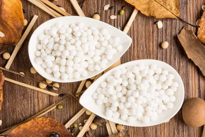
अल्फाल्फाचे फायदे: पोषण, उपयोग, सुरक्षितता आणि तुम्हाला काय माहीत असावे
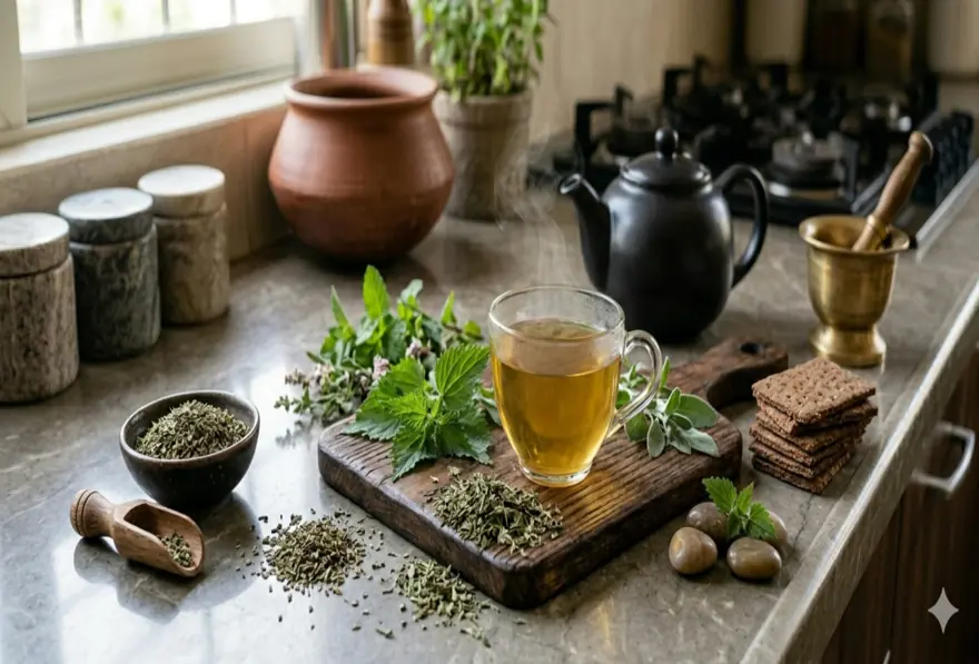
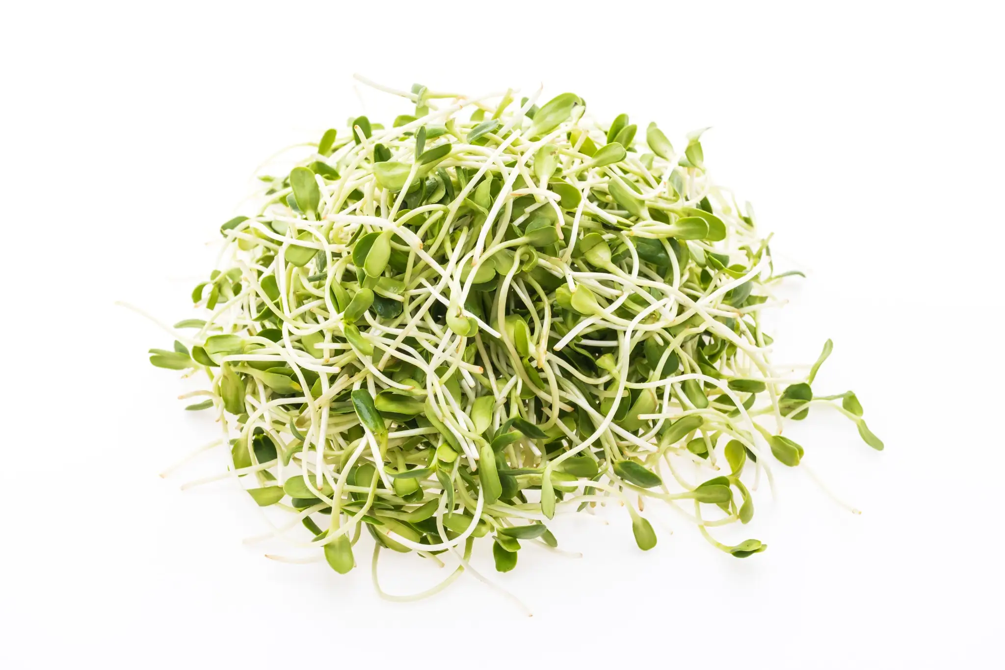
अल्फाल्फाची लागवड हजारो वर्षांपासून केली जाते, मुख्यतः पशुधनासाठी खाद्य पीक म्हणून. आजकाल लोक ते कोंब, चहा, पावडर आणि पूरकांच्या स्वरूपात अधिक प्रमाणात सेवन करू लागले आहेत. त्याचे आकर्षण त्याच्या समृद्ध पोषणघटकांमध्ये आणि सुरुवातीच्या संशोधनाने समर्थित केलेल्या संभाव्य आरोग्य फायद्यांमध्ये आहे. तरीही, अल्फाल्फा हे सर्व आजारांवर उपाय करणारे सुपरफूड नाही. हृदयाचे आरोग्य, रक्तातील साखरेचे संतुलन आणि अँटिऑक्सिडंट आधार यासाठी ते आशादायक दिसत असले, तरी ते प्रत्येकासाठी योग्य नाही. अल्फाल्फा म्हणजे काय, ते काय करू शकते आणि काय करू शकत नाही, तसेच ते सुरक्षितपणे कसे वापरावे हे समजून घेतल्यास तुम्हाला माहितीपूर्ण निवड करण्यात मदत होईल. अल्फाल्फा म्हणजे काय? अल्फाल्फा, ज्याला वैज्ञानिकदृष्ट्या मेडिकागो सटिवा म्हणतात, ही लेग्युम कुटुंबातील फुलझाड वनस्पती आहे. तिला ल्यूसर्न, बफेलो हर्ब किंवा पर्पल मेडिक असेही म्हणतात. दक्षिण आणि मध्य आशियातील काही भागांत मूळ असलेल्या या वनस्पतीची शतकानुशतके जगभर लागवड केली जाते, मुख्यतः घोडे आणि गुरांसाठी उच्च-पोषणयुक्त खाद्य म्हणून. गेल्या काही दशकांत, अल्फाल्फा त्याच्या पोषणमूल्यामुळे लोकांमध्ये लोकप्रिय झाले आहे. ते ताज्या कोंबांच्या स्वरूपात खाता येते, चहासारखे उकळून पिता येते, स्मूदी आणि पेयांमध्ये पावडर म्हणून घालता येते किंवा हर्बल पूरक म्हणून कॅप्सूल किंवा गोळीच्या स्वरूपात वापरता येते. अल्फाल्फाचे कोंब विरुद्ध अल्फाल्फा पूरक अल्फाल्फाचे कोंब हे लोक ही वनस्पती खाण्याचा सर्वात सामान्य मार्ग आहे. ते तरुण, अंकुरलेली बिया असतात, ज्या लवकर काढल्या जातात आणि कच्च्या खाल्ल्या जातात. त्यात कॅलरी कमी असतात, चव सौम्य असते आणि जेवणात सहज घालता येतात. दुसरीकडे, अल्फाल्फा पूरक वाळवलेल्या पानांपासून, बियांपासून किंवा अर्कांपासून बनवले जातात. ते गोळ्या, कॅप्सूल, पावडर किंवा द्रव टिंक्चरच्या स्वरूपात उपलब्ध असतात. पूरक अधिक संकेंद्रित असतात आणि अधिक सावधगिरीने वापरावेत, कारण मात्राविषयक मार्गदर्शन मर्यादित आहे आणि हर्बल पूरकांचे नियमन बदलते. अल्फाल्फाचे पोषण एका दृष्टीक्षेपात अल्फाल्फा ही पोषकद्रव्यांनी समृद्ध वनस्पती आहे, विशेषतः कोंबांच्या स्वरूपात. अल्फाल्फा कोंबांच्या एक कपमध्ये फक्त सुमारे 8 कॅलरी असतात, त्यामुळे ते तुमच्या आहारात अतिशय हलकी पण पौष्टिक भर ठरते. अल्फाल्फामध्ये आढळणाऱ्या महत्त्वाच्या पोषकद्रव्यांमध्ये समावेश आहे: व्हिटॅमिन K: रक्त गोठण्यास आणि हाडांच्या आरोग्यास आधार देते व्हिटॅमिन C: रोगप्रतिकारक कार्य आणि त्वचेच्या आरोग्यास मदत करते फोलेट: पेशींच्या कार्यासाठी आवश्यक आणि गर्भधारणेदरम्यान विशेषतः महत्त्वाचे तांबे: ऊर्जा निर्मिती आणि लोह चयापचयास आधार देते लोह: लाल रक्तपेशींच्या निर्मितीसाठी महत्त्वाचे मॅग्नेशियम: स्नायू, मज्जातंतू आणि हृदयाच्या कार्यात भूमिका बजावते वनस्पती संयुगे: सॅपोनीन्स, फ्लॅव्होनॉइड्स, फायटोस्टेरॉल्स, फायटोएस्ट्रोजेन्स, कुमॅरिन्स आणि अल्कलॉइड्स यांचा समावेश ही जैवसक्रिय वनस्पती संयुगे अल्फाल्फाने आरोग्य आणि वेलनेस संशोधनात इतके लक्ष वेधून घेण्याचे महत्त्वाचे कारण आहेत. अल्फाल्फाचे आरोग्य फायदे हृदयाच्या आरोग्यास मदत करू शकते अल्फाल्फाच्या सर्वाधिक अभ्यासलेल्या फायद्यांपैकी एक म्हणजे हृदय व रक्तवाहिन्यांच्या आरोग्यास मदत करण्यातील त्याची संभाव्य भूमिका. प्राण्यांवरील अभ्यासांनी दाखवले आहे की अल्फाल्फा एकूण कोलेस्टेरॉल, एलडीएल (LDL) म्हणजेच वाईट कोलेस्टेरॉल आणि ट्रायग्लिसराइड पातळी कमी करण्यास मदत करू शकते, तसेच एचडीएल (HDL) म्हणजेच चांगल्या कोलेस्टेरॉलला आधार देऊ शकते. हा परिणाम मोठ्या प्रमाणात सॅपोनीन्समुळे मानला जातो. ही वनस्पती संयुगे आतड्यात कोलेस्टेरॉलचे शोषण कमी करतात आणि नवीन कोलेस्टेरॉल निर्मितीत सहभागी असलेल्या संयुगांचे विसर्जन वाढवतात असे दिसते. मानवी संशोधन अजूनही मर्यादित असले, तरी सुरुवातीचे निष्कर्ष प्रोत्साहन देणारे आहेत. कोलेस्टेरॉल नियंत्रण तुमच्यासाठी चिंतेचा विषय असल्यास, कोणत्याही पूरकांसोबत आहार आणि जीवनशैलीची भूमिका याबद्दल तुमच्या डॉक्टरांशी बोला. रक्तातील साखरेचे संतुलन राखण्यास मदत करू शकते अल्फाल्फाचा रक्तातील साखर कमी करणारा घटक म्हणून पारंपरिक वापराचा इतिहास आहे. काही प्राण्यांवरील अभ्यास सुचवतात की ते रक्तातील चरबी आणि रक्तातील साखरेची पातळी कमी करून कार्डिओमेटाबॉलिक आरोग्य सुधारण्यास मदत करू शकते. त्यातील फायबरही ग्लुकोजचे रक्तप्रवाहात शोषण मंद करण्यास मदत करू शकते. मात्र, हे निष्कर्ष मुख्यतः प्राण्यांवरील आणि सुरुवातीच्या टप्प्यातील अभ्यासांमधून आले आहेत. ठोस निष्कर्ष काढण्यापूर्वी मानवांमध्ये अधिक संशोधनाची गरज आहे. तुम्ही मधुमेह किंवा रक्तातील साखरेशी संबंधित चिंता सांभाळत असाल, तर डॉक्टरांशी आधी बोलल्याशिवाय वैद्यकीय उपचारांच्या जागी अल्फाल्फाचा वापर करू नका. अँटिऑक्सिडंट आधार देते अल्फाल्फामध्ये फ्लॅव्होनॉइड्स आणि इतर फेनोलिक संयुगे असतात, जी शरीराला ऑक्सिडेटिव्ह ताणाचा सामना करण्यास मदत करतात. फ्री रॅडिकल्स हे अस्थिर रेणू रोजच्या शरीर प्रक्रियांमधून तसेच प्रदूषण, तंबाखूचा धूर आणि विषारी पदार्थांच्या संपर्कातून तयार होतात. ते कालांतराने पेशींना नुकसान करू शकतात आणि हृदयरोग, मधुमेह आणि काही कर्करोगांसारख्या स्थितींशी संबंधित आहेत. सुरुवातीचे संशोधन सुचवते की अल्फाल्फा फ्री रॅडिकल्सचे उत्पादन मर्यादित करून आणि शरीराच्या नैसर्गिक संरक्षणांना आधार देऊन त्यांच्यामुळे होणारे पेशींचे नुकसान कमी करण्यास मदत करू शकते. या पुराव्यांपैकी बहुतेक प्राण्यांवरील आणि पेशी अभ्यासांमधून आले आहेत, आणि मानवी संशोधन मर्यादित राहते. हाडांच्या आरोग्यास मदत करू शकते अल्फाल्फा हे व्हिटॅमिन K चे चांगले स्रोत आहे. हे पोषकद्रव्य हाडांच्या चयापचयात आणि कॅल्शियम नियमनात महत्त्वाची भूमिका बजावते. पुरेसे व्हिटॅमिन K सेवन चांगल्या हाडांच्या घनतेशी संबंधित आहे आणि विशेषतः वयानुसार दीर्घकालीन हाडांच्या आरोग्यास आधार देऊ शकते. अल्फाल्फा मॅग्नेशियम आणि इतर सूक्ष्म खनिजेही देते, जी एकूण सांगाडा आरोग्यास मदत करतात. मेनोपॉजची लक्षणे कमी करण्यास मदत करू शकते अल्फाल्फामध्ये फायटोएस्ट्रोजेन्स असतात, जी नैसर्गिकरित्या आढळणारी वनस्पती संयुगे असून त्यांची रचना एस्ट्रोजेन हार्मोनसारखी असते. मेनोपॉजदरम्यान एस्ट्रोजेनची पातळी कमी होते आणि त्यामुळे हॉट फ्लॅशेस, रात्री घाम येणे आणि मूड बदलणे अशी लक्षणे दिसू शकतात. काही सुरुवातीचे संशोधन सुचवते की फायटोएस्ट्रोजेनयुक्त वनस्पती ही लक्षणे कमी करण्यास मदत करू शकतात. एका छोट्या अभ्यासात सेज आणि अल्फाल्फाच्या संयोजनामुळे काही सहभागींमध्ये हॉट फ्लॅशेस आणि रात्री घाम येणे कमी झाले असे आढळले. मात्र, संशोधनाचा आधार लहान आहे आणि परिणाम सातत्यपूर्ण नाहीत. मेनोपॉजच्या लक्षणांसाठी अल्फाल्फाचा विचार करत असाल, तर आधी डॉक्टरांशी बोलणे चांगले, विशेषतः तुमच्या इतिहासात हार्मोन-संवेदनशील स्थिती असल्यास. अल्फाल्फा डिटॉक्सला मदत करू शकते का? अल्फाल्फाचा उल्लेख अनेकदा वेलनेस चर्चांमध्ये डिटॉक्सिफिकेशनच्या संदर्भात केला जातो. त्यात सर्वसाधारण आरोग्यास आधार देणारे अँटिऑक्सिडंट्स आणि पोषकद्रव्ये असली, तरी पुरावे प्रत्यक्षात काय दाखवतात याबद्दल स्पष्ट असणे महत्त्वाचे आहे. तुमच्या शरीरात आधीच यकृत आणि मूत्रपिंडांवर केंद्रित एक प्रगत डिटॉक्सिफिकेशन प्रणाली असते. निरोगी व्यक्तींमध्ये ही अवयवे रक्तातील अपशिष्ट पदार्थ सतत आणि प्रभावीपणे फिल्टर करून बाहेर टाकतात. अल्फाल्फासह कोणताही अन्नपदार्थ ही प्रक्रिया बदलू किंवा लक्षणीयरीत्या वाढवू शकत नाही. तरीही, अल्फाल्फासारखे अँटिऑक्सिडंट असलेले अन्नपदार्थ समाविष्ट असलेला पोषकद्रव्यांनी समृद्ध आहार एकूण पेशी आरोग्यास आधार देऊ शकतो आणि ऑक्सिडेटिव्ह ताण कमी करू शकतो. या अर्थाने, अल्फाल्फा आरोग्यदायी, संतुलित आहारात सहाय्यक भर ठरू शकते. त्याला डिटॉक्स उपाय किंवा वैद्यकीय काळजीचा पर्याय मानू नये. अल्फाल्फा आणि पचनाचे आरोग्य अल्फाल्फाचे कोंब हे हलके, सहज पचणारे अन्न आहे आणि तुमच्या पचनसंस्थेला सौम्य आधार देऊ शकते. जाणून घेण्यासारखे मुद्दे असे आहेत: अल्फाल्फाचे कोंब कॅलरीमध्ये अतिशय कमी आणि कार्बोहायड्रेट्समध्ये कमी असतात, त्यामुळे ते पचनसंस्थेसाठी हलके असतात त्यात आहारातील फायबर असते, जे नियमित मलविसर्जन आणि सर्वसाधारण आतड्यांच्या आरामास मदत करते त्यातील जास्त पाण्याचे प्रमाण हायड्रेशन आणि मल मऊ राहण्यास मदत करू शकते काही व्यक्तींना सुरुवातीला पचनसंवेदनशीलता जाणवू शकते, विशेषतः पूरकांसह उच्च-फायबर अन्नाची सवय नसल्यास अल्फाल्फा हळूहळू सुरू करणे चांगले कोणत्याही आहारातील भरप्रमाणे, तुमचे शरीर कसा प्रतिसाद देते हे तुमच्या वैयक्तिक आरोग्य आणि आतड्यांच्या स्थितीवर अवलंबून असेल. अल्फाल्फाचे हर्बल उपयोग आयुर्वेदिक आणि चिनी हर्बल परंपरांसह विविध संस्कृतींमध्ये अल्फाल्फाचा पारंपरिक वैद्यकात दीर्घ इतिहास आहे. आज ते सामान्य वेलनेस दिनक्रमांचा भाग म्हणून विविध स्वरूपांत वापरले जाते: चहा: गरम पाण्यात भिजवलेली वाळवलेली अल्फाल्फा पाने, कधी कधी इतर औषधी वनस्पतींसोबत पावडर: स्मूदी, ज्यूस किंवा आरोग्य पेयांमध्ये घातली जाते कॅप्सूल आणि गोळ्या: कोंब खाणे पसंत नसलेल्या लोकांसाठी सोयीस्कर पूरक स्वरूपे ताजे कोंब: सौम्य कुरकुरीसाठी सलाड, सँडविच, रॅप्स आणि स्प्रिंग रोल्समध्ये घातले जातात पारंपरिक हर्बल वापर: ऐतिहासिकदृष्ट्या मूत्रल परिणाम, मूत्रपिंडांना आधार आणि सर्वसाधारण चैतन्याशी संबंधित पारंपरिक वापर म्हणजे नेहमीच सिद्ध वैद्यकीय फायदा असे नसते, हे लक्षात ठेवणे महत्त्वाचे आहे. अल्फाल्फा नियमितपणे वापरण्यापूर्वी, विशेषतः पूरक स्वरूपात, तुमच्या डॉक्टरांचा सल्ला घ्या. अल्फाल्फा आणि हार्मोनल संतुलन फायटोएस्ट्रोजेन्स ही अल्फाल्फामध्ये आढळणारी नैसर्गिक वनस्पती संयुगे आहेत, जी शरीरातील एस्ट्रोजेनच्या क्रियेची नक्कल करतात. ती एस्ट्रोजेन रिसेप्टर्सना जोडतात आणि सौम्य एस्ट्रोजेनसारखे परिणाम निर्माण करू शकतात. म्हणूनच अल्फाल्फाची चर्चा कधी कधी हार्मोनल आरोग्याच्या संदर्भात, विशेषतः मेनोपॉजशी संबंधित, केली जाते. मेनोपॉजदरम्यान एस्ट्रोजेनची पातळी कमी होत असताना, फायटोएस्ट्रोजेन्स या घटाची अंशतः भरपाई करून काही लक्षणे कमी करण्यास मदत करू शकतात. मात्र, फायटोएस्ट्रोजेन्स हा चालू संशोधनाचा आणि काही प्रमाणात चर्चेचा विषय राहिला आहे. हार्मोनल आरोग्यावर त्यांचे परिणाम व्यक्तीनुसार मोठ्या प्रमाणात बदलू शकतात. काही लोकांना सकारात्मक प्रतिसाद दिसतो, तर काहींना कोणताही बदल जाणवत नाही. हार्मोन-संवेदनशील स्थिती असलेल्या लोकांनी फायटोएस्ट्रोजेनयुक्त अन्नपदार्थ आणि पूरक फक्त वैद्यकीय देखरेखीखाली वापरावेत. हार्मोनल आरोग्याच्या कारणांसाठी अल्फाल्फाचा विचार करत असाल, तर ते तुमच्या दिनक्रमाचा नियमित भाग करण्यापूर्वी डॉक्टरांशी बोला. अल्फाल्फा वजन नियंत्रणासाठी उपयुक्त आहे का? अल्फाल्फा स्वतःहून वजन कमी करणारे अन्न नाही, आणि कोणताही एकच अन्नपदार्थ स्वतःहून वजन कमी करू शकत नाही. तरीही, अल्फाल्फाचे कोंब कॅलरीबाबत जागरूक आहारात समजूतदार भर ठरू शकतात. एका कपमागे सुमारे 8 कॅलरी असल्यामुळे, अल्फाल्फाचे कोंब हे तुम्ही खाऊ शकता अशा सर्वात कमी-कॅलरी अन्नपदार्थांपैकी एक आहेत. ते तुमच्या दैनंदिन कॅलरी सेवनात मोठी भर न घालता जेवणात प्रमाण, पोत आणि पोषकद्रव्ये वाढवतात. त्यातील फायबरमुळे खाल्ल्यानंतर अधिक समाधानही वाटू शकते. नियमित शारीरिक हालचाल आणि आरोग्यदायी सवयींसोबत संतुलित आहाराचा भाग म्हणून वापरल्यास, अल्फाल्फाचे कोंब वजन नियंत्रणाच्या दृष्टिकोनाला पूरक ठरू शकतात. मात्र, त्यांना स्वतंत्र उपाय म्हणून पाहू नये. अल्फाल्फाचे संभाव्य दुष्परिणाम बहुतेक निरोगी प्रौढांमध्ये अल्फाल्फा लहान प्रमाणात साधारणपणे चांगले सहन होते, पण काही लोकांमध्ये दुष्परिणाम होऊ शकतात: पचनाचा त्रास: अल्फाल्फा पूरक नियमितपणे घेणाऱ्या काही व्यक्तींमध्ये सैल शौच, गॅस, पोट फुगणे किंवा पोटात मुरडा येणे आढळते जिवाणू दूषित होण्याचा धोका: कच्चे अल्फाल्फा कोंब ई. कोलाई आणि साल्मोनेलासारख्या जिवाणूंमुळे होणाऱ्या अन्नजन्य आजारांच्या उद्रेकांशी संबंधित आढळले आहेत; नीट धुणे आणि योग्य साठवण आवश्यक आहे अॅलर्जिक प्रतिक्रिया: काही लोकांना अल्फाल्फाला अॅलर्जिक प्रतिसाद असू शकतो, विशेषतः इतर लेग्युम्स किंवा वनस्पतीजन्य अन्नांबाबत संवेदनशीलता असलेल्यांना औषधांशी परस्परसंवाद: अल्फाल्फातील जास्त व्हिटॅमिन K प्रमाण वॉरफरिनसारख्या रक्त पातळ करणाऱ्या औषधांची परिणामकारकता कमी करू शकते; ते काही हार्मोनल गर्भनिरोधके आणि मधुमेहाच्या औषधांशीही परस्परसंवाद करू शकते अल्फाल्फा कोणांनी टाळावे? काही गटांनी अल्फाल्फा पूर्णपणे टाळावे किंवा वापरण्यापूर्वी डॉक्टरांचा सल्ला घ्यावा: गर्भवती महिला: कच्च्या अल्फाल्फा कोंबांमध्ये हानिकारक जिवाणूंच्या दूषिततेचा धोका असतो आणि गर्भधारणेदरम्यान ते टाळावेत रक्त पातळ करणारी औषधे घेणारे लोक: अल्फाल्फातून जास्त व्हिटॅमिन K सेवन वॉरफरिनसारख्या अँटिकोग्युलंट औषधांमध्ये अडथळा आणू शकते ऑटोइम्यून स्थिती असलेले लोक: अल्फाल्फा, विशेषतः त्याच्या बिया, काही व्यक्तींमध्ये ल्यूपस पुन्हा सक्रिय होण्याशी संबंधित आहेत; ल्यूपस किंवा इतर ऑटोइम्यून विकार असलेल्यांनी ते टाळावे कमकुवत रोगप्रतिकारक शक्ती असलेले लोक: मुले, वयोवृद्ध आणि कमकुवत रोगप्रतिकारक प्रणाली असलेले लोक कच्च्या कोंबांमुळे होणाऱ्या अन्नजन्य आजारांसाठी अधिक असुरक्षित असतात गाऊट असलेले लोक: अल्फाल्फामध्ये प्युरिन्स जास्त असतात, ज्यामुळे गाऊटची लक्षणे बिघडू शकतात वैद्यकीय मार्गदर्शनाशिवाय नियमित पूरक वापरण्याचा विचार करणारे कोणीही: हर्बल पूरकांच्या मात्रांचे कडक नियमन नसते; अल्फाल्फा पूरक दररोज वापरण्यापूर्वी नेहमी सल्ला घ्या तुमच्या आहारात अल्फाल्फा सुरक्षितपणे कसे समाविष्ट करावे? अल्फाल्फा तुमच्या आरोग्य प्रोफाइलला अनुकूल असेल आणि ते करून पाहण्यास तुमचे डॉक्टर सहमत असतील, तर ते सुरक्षितपणे करण्याचे काही सोपे, व्यावहारिक मार्ग येथे दिले आहेत: खाण्यापूर्वी ताजे अल्फाल्फा कोंब थंड वाहत्या पाण्याखाली नीट धुवा कोंब फ्रिजमध्ये साठवा आणि खरेदी केल्यानंतर काही दिवसांत वापरा अधिक पोत आणि पोषकद्रव्यांसाठी सलाड, सँडविच, रॅप्स किंवा धान्याच्या बाऊलमध्ये कोंब घाला मुले, वृद्ध कुटुंबीय किंवा कमकुवत रोगप्रतिकारक प्रणाली असलेल्या कोणालाही कच्चे कोंब देणे टाळा पावडर किंवा कॅप्सूल वापरताना उत्पादकाच्या मार्गदर्शनाचे पालन करा आणि शिफारस केलेल्या प्रमाणापेक्षा जास्त घेऊ नका दररोज पूरक वापर सुरू करण्यापूर्वी तुमच्या डॉक्टरांशी बोला, विशेषतः तुम्ही कोणतीही नियमित औषधे घेत असाल तर निष्कर्ष अल्फाल्फा ही पोषकद्रव्यांनी समृद्ध वनस्पती आहे, जिचा पारंपरिक वापराचा दीर्घ इतिहास आहे आणि काही खरोखरच आशादायक सुरुवातीचे संशोधन आहे. ती हृदयाचे आरोग्य, रक्तातील साखरेचे संतुलन, अँटिऑक्सिडंट संरक्षण, हाडांचे आरोग्य आणि मेनोपॉजची लक्षणे यांना आधार देऊ शकते. कोंबांच्या स्वरूपात खाल्ल्यास ती अत्यंत कमी-कॅलरी आणि वापरण्यास सोपी अन्नपदार्थही आहे. त्याच वेळी, ती प्रत्येकासाठी योग्य नाही, आणि कोणत्याही एका अन्नपदार्थावर उपचार किंवा इलाज म्हणून अवलंबून राहू नये. सुरक्षित वापर, परस्परसंवादांची जाणीव आणि आहाराकडे संतुलित दृष्टिकोन हे सर्वात महत्त्वाचे आहे. चांगले पोषण हे चांगल्या आरोग्याचा पाया आहे, पण ते नियमित आरोग्य निरीक्षणासोबत जोडले असता सर्वोत्तम काम करते. तुमची कोलेस्टेरॉल पातळी, रक्तातील साखर आणि महत्त्वाचे पोषकद्रव्य मार्कर्स जाणून घेतल्याने तुम्हाला तुमची स्थिती स्पष्टपणे समजते आणि खरोखर माहितीपूर्ण निवडी करता येतात. मेट्रोपोलिस हेल्थकेअरमध्ये, तुम्ही संकेतस्थळ, अॅप, कॉल किंवा व्हॉट्सअॅपद्वारे नियमित रक्त तपासण्या, पूर्ण शरीर तपासण्या आणि विशेष आरोग्य तपासण्या सहज बुक करू शकता. विस्तृत नेटवर्कमध्ये घरून नमुना संकलन, विश्वासार्ह निकाल आणि जलद रिपोर्टिंग उपलब्ध असल्यामुळे तुमच्या आरोग्यावर लक्ष ठेवणे यापेक्षा सोपे कधीच नव्हते. वारंवार विचारले जाणारे प्रश्न अल्फाल्फाचा उपयोग कशासाठी केला जातो? अल्फाल्फाचा उपयोग पशुखाद्य म्हणून तसेच मानवांसाठी अन्न आणि पूरक म्हणून केला जातो. लोक ते सलाड आणि सँडविचमध्ये कोंबांच्या स्वरूपात, चहा म्हणून किंवा पावडर किंवा कॅप्सूल पूरक म्हणून सेवन करतात. सर्वसाधारण वेलनेससाठी पारंपरिक वैद्यकात त्याचा वापर केला जातो आणि कोलेस्टेरॉल, रक्तातील साखर, अँटिऑक्सिडंट संरक्षण आणि मेनोपॉजची लक्षणे यांच्याशी संबंधित संभाव्य फायद्यांसाठी त्याचा अभ्यास केला गेला आहे. अल्फाल्फाचे मुख्य फायदे कोणते आहेत? अल्फाल्फा कोलेस्टेरॉल पातळीवर प्रभाव टाकून हृदयाच्या आरोग्यास मदत करू शकते, अँटिऑक्सिडंट संरक्षण देऊ शकते, व्हिटॅमिन K मुळे हाडांच्या आरोग्यास आधार देऊ शकते, रक्तातील साखरेचे संतुलन राखण्यास मदत करू शकते आणि फायटोएस्ट्रोजेन प्रमाणामुळे मेनोपॉजची काही लक्षणे कमी करू शकते. मात्र, समर्थन करणारे बहुतेक संशोधन प्राण्यांवरील किंवा सुरुवातीच्या टप्प्यातील अभ्यासांमधून आहे, आणि अधिक मानवी पुराव्यांची गरज आहे. अल्फाल्फा कोलेस्टेरॉल कमी करण्यास मदत करू शकते का? सुरुवातीचे संशोधन, मुख्यतः प्राण्यांवरील अभ्यासांमधून, सुचवते की अल्फाल्फातील सॅपोनीन्स आतड्यात एलडीएल (LDL) कोलेस्टेरॉलचे शोषण कमी करण्यास आणि अधिक आरोग्यदायी लिपिड पातळीला आधार देण्यास मदत करू शकतात. निष्कर्ष आशादायक असले, तरी मजबूत मानवी चाचण्यांची अजूनही गरज आहे. कोलेस्टेरॉल तुमच्यासाठी चिंतेचा विषय असल्यास, सर्वसमावेशक व्यवस्थापन योजनेबद्दल तुमच्या डॉक्टरांशी बोला. अल्फाल्फा डिटॉक्सला मदत करते का? अँटिऑक्सिडंट प्रमाणामुळे अल्फाल्फा कधी कधी वेलनेस वर्तुळात डिटॉक्सशी जोडले जाते. मात्र, निरोगी व्यक्तींमध्ये शरीराचे यकृत आणि मूत्रपिंड नैसर्गिकरीत्या आणि प्रभावीपणे डिटॉक्सिफिकेशन हाताळतात. संतुलित आहाराचा भाग म्हणून अल्फाल्फा सर्वसाधारण पेशी आरोग्यास आधार देऊ शकते, पण ते डिटॉक्स एजंट नाही आणि वैद्यकीय उपचारांची जागा घेऊ नये. अल्फाल्फा पचनासाठी चांगले आहे का? अल्फाल्फाचे कोंब हलके, कमी कॅलरीचे आणि आहारातील फायबर असलेले असतात, त्यामुळे अनेक लोकांमध्ये नियमित पचन आणि आतड्यांच्या आरामास मदत करू शकतात. ते साधारणपणे पचायला सोपे असतात आणि जेवणात सौम्य, पौष्टिक भर ठरू शकतात. संवेदनशील पचनसंस्था असलेल्यांनी अल्फाल्फा हळूहळू सुरू करावे. अल्फाल्फा मेनोपॉजदरम्यान मदत करू शकते का? अल्फाल्फामध्ये फायटोएस्ट्रोजेन्स असतात, ही वनस्पती संयुगे शरीरात सौम्य एस्ट्रोजेनसारखी क्रिया करू शकतात. काही सुरुवातीचे संशोधन सुचवते की यामुळे हॉट फ्लॅशेस आणि रात्री घाम येणे यांसारखी लक्षणे कमी होऊ शकतात. पुरावे मर्यादित आहेत आणि परिणाम व्यक्तीनुसार बदलतात. मेनोपॉज व्यवस्थापनासाठी विशेषतः अल्फाल्फा वापरण्यापूर्वी तुमच्या डॉक्टरांशी बोला. अल्फाल्फा वजन कमी करण्यासाठी चांगले आहे का? अल्फाल्फा हे वजन कमी करणारे अन्न नाही, पण अल्फाल्फाचे कोंब अत्यंत कमी कॅलरीचे आणि फायबरयुक्त असतात, त्यामुळे कॅलरीबाबत जागरूक आहारात ते पोटभरीची आणि पौष्टिक भर ठरतात. कोणताही एकच अन्नपदार्थ स्वतःहून वजन कमी करत नाही. नियमित शारीरिक हालचालींसोबत संतुलित, विविध आहाराचा भाग म्हणून अल्फाल्फा सर्वोत्तम काम करते. कच्चे अल्फाल्फा कोंब खाणे सुरक्षित आहे का? बहुतेक निरोगी प्रौढांसाठी, योग्यरीत्या धुतलेले आणि साठवलेले कच्चे अल्फाल्फा कोंब सुरक्षित असतात. मात्र, कच्च्या कोंबांमध्ये ई. कोलाई आणि साल्मोनेलासारखे हानिकारक जिवाणू असू शकतात. गर्भवती महिला, लहान मुले, वयोवृद्ध आणि कमकुवत रोगप्रतिकारक शक्ती असलेले कोणीही कच्चे कोंब टाळावेत. कोंब नेहमी नीट धुवा आणि लगेच फ्रिजमध्ये ठेवा. अल्फाल्फा कोणांनी वापरू नये? अल्फाल्फा टाळावे किंवा फक्त वैद्यकीय देखरेखीखाली वापरावे अशा लोकांमध्ये गर्भवती महिला, रक्त पातळ करणारी औषधे घेणारे लोक, ल्यूपससारख्या ऑटोइम्यून स्थिती असलेले लोक, गाऊट असलेले व्यक्ती आणि कमकुवत रोगप्रतिकारक प्रणाली असलेले लोक यांचा समावेश होतो. तुमच्या दिनक्रमात अल्फाल्फा पूरक जोडण्यापूर्वी नेहमी तुमच्या डॉक्टरांशी तपासा. अल्फाल्फाचा औषधांशी परस्परसंवाद होऊ शकतो का? होय. अल्फाल्फामध्ये व्हिटॅमिन K जास्त असते, ज्यामुळे वॉरफरिनसारख्या अँटिकोग्युलंट औषधांची परिणामकारकता कमी होऊ शकते. ते रक्तातील साखर आणखी कमी करून काही मधुमेहाच्या औषधांशीही परस्परसंवाद करू शकते, आणि काही हार्मोनल गर्भनिरोधके कशी काम करतात यावर परिणाम करू शकते. तुम्ही कोणतीही नियमित औषधे घेत असाल, तर अल्फाल्फा पूरक वापरण्यापूर्वी तुमच्या डॉक्टरांशी बोला.
ऑरेगॅनोचे 10 फायदे: अँटिऑक्सिडंट, जीवाणूरोधी आणि पचनासाठी आधार
ऑरेगॅनो ही फक्त चविष्ट स्वयंपाकघरातील औषधी वनस्पती नाही. ती सुगंधी वनस्पती असून कार्वाक्रॉल आणि थायमॉलसारख्या नैसर्गिक संयुगांनी समृद्ध आहे, ज्यांनी त्यांच्या अँटिऑक्सिडंट आणि सूक्ष्मजीवरोधी गुणधर्मांमुळे लक्ष वेधले आहे. तुम्ही कदाचित ऑरेगॅनो आधीच पास्ता सॉस, सूप, सलाड आणि मॅरिनेडमध्ये वापरत असाल. चवीपलीकडे, ते तुमच्या एकूण आरोग्यालाही छोट्या पण अर्थपूर्ण पद्धतीने आधार देऊ शकते. तरीही, अपेक्षा संतुलित ठेवणे महत्त्वाचे आहे. ऑरेगॅनोबद्दलचे अनेक मजबूत दावे प्रयोगशाळेतील आणि प्राण्यांवरील संशोधनातून येतात, तर मानवी अभ्यास अजूनही मर्यादित आहेत. दैनंदिन जीवनात, ऑरेगॅनोला उपचार म्हणून नव्हे, तर आरोग्यदायी आहाराचा उपयुक्त भाग म्हणून पाहणे चांगले. ऑरेगॅनो म्हणजे काय? ऑरेगॅनो ही पुदिना कुटुंबातील पाककला आणि औषधी वनस्पती आहे. तिचे वनस्पतिशास्त्रीय नाव ओरिगॅनम वल्गारे आहे. भूमध्य शैलीतील स्वयंपाकात तिचा मोठ्या प्रमाणात वापर केला जातो आणि तिच्या उबदार, किंचित मिरपूडसारख्या चवीसाठी ती महत्त्वाची मानली जाते. ऑरेगॅनो ताजी पाने, वाळवलेली पाने, चहा, ऑरेगॅनो तेल आणि ऑरेगॅनो इसेन्शियल ऑइल अशा अनेक स्वरूपांत मिळते. ही सर्व स्वरूपे सारखी नसतात. ताजे आणि वाळवलेले ऑरेगॅनो मुख्यतः अन्नात वापरले जाते, तर तेल आणि पूरक अधिक संकेंद्रित असतात आणि त्यांचा वापर अधिक सावधगिरीने करावा लागतो. पारंपरिक वापरात, ऑरेगॅनोचा उपयोग पचनाचा आराम, श्वसनास आधार आणि सर्वसाधारण वेलनेससाठी केला गेला आहे. आज बहुतेक लोक ते मसाला म्हणून ओळखतात, पण संशोधन त्यातील वनस्पती संयुगे आणि संभाव्य आरोग्य परिणामांचा अभ्यास करत आहे. ऑरेगॅनोचे पौष्टिक मूल्य ऑरेगॅनो साधारणपणे कमी प्रमाणात खाल्ले जाते, त्यामुळे ते कॅलरी किंवा मोठ्या प्रमाणातील पोषणाचा प्रमुख स्रोत नाही. तरीही, त्यात उपयुक्त पोषकद्रव्ये आणि वनस्पती संयुगे असतात. पोषकद्रव्य किंवा संयुग ते शरीरात काय करते ऑरेगॅनोमध्ये कोणत्या स्वरूपात असते व्हिटॅमिन K सामान्य रक्त गोठणे आणि हाडांचे आरोग्य यांना आधार देते ताजे आणि वाळवलेले ऑरेगॅनो अँटिऑक्सिडंट्स पेशींना ऑक्सिडेटिव्ह ताणापासून संरक्षण करण्यास मदत करतात ताजे आणि वाळवलेले ऑरेगॅनो कार्वाक्रॉल सूक्ष्मजीवरोधी आणि दाहरोधी परिणामांसाठी अभ्यासले गेलेले वनस्पती संयुग ऑरेगॅनो तेल आणि वाळवलेली औषधी वनस्पती थायमॉल सूक्ष्मजीवरोधी आणि अँटिऑक्सिडंट क्रियेसाठी अभ्यासले गेलेले वनस्पती संयुग ऑरेगॅनो तेल आणि वाळवलेली औषधी वनस्पती फ्लॅव्होनॉइड्स आणि फेनोलिक आम्ले एकूण पेशी संरक्षणास आधार देतात ताजे आणि वाळवलेले ऑरेगॅनो सूक्ष्म खनिजे लहान प्रमाणात शरीराच्या सामान्य कार्यांमध्ये योगदान देतात मुख्यतः वाळवलेले ऑरेगॅनो ऑरेगॅनो बहुधा गार्निश किंवा मसाला म्हणून वापरले जात असल्यामुळे, त्याचे मूल्य मोठ्या प्रमाणातील जीवनसत्त्वे किंवा खनिजांपेक्षा त्यातील संकेंद्रित वनस्पती संयुगांमधून अधिक येते. ऑरेगॅनोचे 10 फायदे ऑरेगॅनो तुमच्या आहारात अनेक प्रकारे योगदान देऊ शकते. काही फायदे सामान्य अन्न वापराचा भाग म्हणून चांगल्या प्रकारे समर्थित आहेत, तर काहींबाबत संशोधन अजूनही सुरुवातीच्या किंवा मर्यादित टप्प्यात आहे. 1. अँटिऑक्सिडंट्सने समृद्ध ऑरेगॅनो नैसर्गिकरित्या अँटिऑक्सिडंट संयुगांनी समृद्ध आहे. ही संयुगे फ्री रॅडिकल्समुळे होणाऱ्या ऑक्सिडेटिव्ह ताणापासून तुमच्या पेशींना संरक्षण करण्यास मदत करतात. ऑक्सिडेटिव्ह ताण वृद्धत्व आणि अनेक दीर्घकालीन आरोग्य स्थितींशी संबंधित आहे. ऑरेगॅनोसारख्या औषधी वनस्पती जेवणात घातल्याने संरक्षणात्मक वनस्पती संयुगांचे तुमचे एकूण सेवन वाढण्यास मदत होऊ शकते. 2. हानिकारक जिवाणूंशी लढण्यास मदत करू शकते ऑरेगॅनोमध्ये कार्वाक्रॉल आणि थायमॉलसारखी संयुगे असतात, ज्यांनी प्रयोगशाळेतील अभ्यासांमध्ये जीवाणूरोधी क्रिया दाखवली आहे. याचा अर्थ असा नाही की ऑरेगॅनो संसर्गांसाठी वैद्यकीय उपचारांची जागा घेऊ शकते. मात्र, पोषण आणि वनस्पती-आधारित संशोधनात ऑरेगॅनोबद्दल रस का कायम आहे हे यामुळे स्पष्ट होते. 3. बुरशीरोधी आधार देऊ शकते काही अभ्यास सुचवतात की ऑरेगॅनो तेल आणि त्यातील सक्रिय संयुगे प्रयोगशाळेच्या परिस्थितीत कॅंडिडा प्रजातींसह काही बुरशींवर क्रिया करू शकतात. हे क्षेत्र अजून विकसित होत आहे. बुरशीजन्य संसर्गांसाठी ऑरेगॅनो स्वतंत्र उपचार म्हणून वापरू नये, पण हे निष्कर्ष त्याची प्रभावी औषधी वनस्पती म्हणून असलेली प्रतिमा समर्थित करतात. 4. दाह कमी करण्यास मदत करू शकते सतत चालू राहणारा दाह आरोग्याच्या अनेक पैलूंवर परिणाम करू शकतो. सुरुवातीचे संशोधन सुचवते की ऑरेगॅनोतील संयुगांना दाहरोधी परिणाम असू शकतात. सर्वसाधारण वेलनेसच्या संदर्भात ऑरेगॅनोची चर्चा होण्याचे हे एक कारण असू शकते. तरीही, ठोस निष्कर्ष काढण्यापूर्वी अधिक मानवी अभ्यासांची गरज आहे. 5. रोगप्रतिकारक आरोग्यास मदत करू शकते औषधी वनस्पती, मसाले, भाज्या, फळे आणि इतर वनस्पतीजन्य अन्नपदार्थांनी समृद्ध आहार तुमच्या रोगप्रतिकारक प्रणालीला आधार देण्यास मदत करू शकतो. ऑरेगॅनो अँटिऑक्सिडंट आणि सूक्ष्मजीवरोधी वनस्पती संयुगे देते, जी सहाय्यक भूमिका बजावू शकतात. त्याची भूमिका एकमेव रोगप्रतिकारकता वाढवणारा घटक म्हणून नव्हे, तर तुमच्या एकूण आहार पद्धतीचा भाग म्हणून पाहणे चांगले. 6. पचनाचा आराम मिळवण्यास मदत करू शकते पचनाच्या तक्रारींसाठी ऑरेगॅनोचा पारंपरिक वापराचा दीर्घ इतिहास आहे. पचनास आधार देण्यासाठी आणि पोट फुगणे किंवा पोटातील अस्वस्थता कमी करण्यासाठी ते चहा आणि काढ्यांमध्ये वापरले गेले आहे. काही तज्ज्ञांच्या मते त्यातील संयुगे पचनरसांचे स्त्रवण उत्तेजित करण्यास आणि पचनमार्गातील स्नायू शिथिल करण्यास मदत करू शकतात. तरीही, हा फायदा मजबूत क्लिनिकल चाचण्यांपेक्षा पारंपरिक वापर आणि सुरुवातीच्या पुराव्यावर अधिक आधारित आहे. 7. विषाणूरोधी क्षमता असू शकते काही प्रयोगशाळेतील अभ्यासांमध्ये ऑरेगॅनोतील संयुगे काही विशिष्ट विषाणूंवर क्रिया करू शकतात असे आढळले आहे. त्यामुळे व्यापक रोगप्रतिकारक आणि संसर्ग आधारासाठी ऑरेगॅनोबद्दल रस निर्माण झाला आहे. सध्या हे संशोधन अजून प्राथमिक टप्प्यात आहे. विषाणूजन्य आजार प्रतिबंधित करण्यासाठी किंवा उपचार करण्यासाठी ऑरेगॅनो तेल किंवा पूरक स्वतंत्रपणे शिफारस करण्याइतका हा पुरावा पुरेसा नाही. 8. चयापचय आरोग्य उद्दिष्टांना आधार देऊ शकते रक्तातील साखरेचे संतुलन, चयापचय आणि शरीराचे वजन नियंत्रण यांसारख्या क्षेत्रांत ऑरेगॅनो आणि त्यातील संयुगांवर काही सुरुवातीचे संशोधन झाले आहे. याचा अर्थ असा नाही की ऑरेगॅनो वजन कमी करते किंवा स्वतःहून मधुमेह नियंत्रित करू शकते. अधिक वास्तववादी दृष्टिकोन असा आहे की विज्ञान विकसित होत असताना ऑरेगॅनो संतुलित आहार आणि आरोग्यदायी जीवनशैलीला पूरक ठरू शकते. 9. संशोधनात सुरुवातीची कर्करोगविरोधी क्षमता दर्शवते अँटिऑक्सिडंट्सने समृद्ध औषधी वनस्पतींचा पेशींचे संरक्षण करण्यातील भूमिकेसाठी अनेकदा अभ्यास केला जातो. ऑरेगॅनो आणि कार्वाक्रॉल व थायमॉलसारख्या संयुगांनी प्रयोगशाळेतील संशोधनात कर्करोगविरोधी क्रिया दर्शवली आहे. हे आशादायक वाटते, पण हे अजून सुरुवातीच्या टप्प्यातील विज्ञान आहे. ऑरेगॅनो हे कर्करोगावरील उपचार नाही, आणि या निष्कर्षांना अतिरंजित करू नये. 10. रोजच्या जेवणात समाविष्ट करणे सोपे ऑरेगॅनोची सर्वात मोठी ताकद म्हणजे ते वापरणे किती सोपे आहे. तुमचा दिनक्रम फारसा न बदलता तुम्ही ते साध्या घरगुती जेवणात घालू शकता. यामुळे संतुलित आहारात दीर्घकालीन वापरासाठी ऑरेगॅनो एक व्यावहारिक औषधी वनस्पती ठरते. अनेक वेलनेस ट्रेंड्सच्या विपरीत, ते परवडणारे, परिचित आणि आनंदाने वापरता येणारे आहे. ऑरेगॅनोचे प्रकार आणि त्यांतील पोषणात्मक फरक ताजे ऑरेगॅनो ताज्या ऑरेगॅनोची चव अधिक ताजी आणि उजळ असते आणि त्यात पाणी असते, त्यामुळे त्याची चव हलकी असते. ते सलाड, सूप आणि गार्निश-आधारित पदार्थांमध्ये उपयुक्त आहे. वाळवलेले ऑरेगॅनो वाळवलेल्या ऑरेगॅनोची चव अधिक संकेंद्रित असते कारण त्यातील पाणी काढलेले असते. रोजच्या स्वयंपाकासाठी ते बहुधा सर्वात व्यावहारिक स्वरूप असते. ऑरेगॅनो चहा ऑरेगॅनो चहा पाने गरम पाण्यात भिजवून तयार केला जातो. पचनाचा आराम आणि ऊब देणारे पेय म्हणून तो सामान्यतः वापरला जातो, मात्र विशिष्ट आरोग्य परिणामांसाठी पुरावे मर्यादित आहेत. ऑरेगॅनो तेल ऑरेगॅनोचे तेल साधारणपणे पूरक किंवा द्रव अर्क म्हणून विकले जाते. स्वयंपाकात वापरल्या जाणाऱ्या औषधी वनस्पतीपेक्षा ते अधिक संकेंद्रित असते आणि काळजीपूर्वक वापरावे. ऑरेगॅनो इसेन्शियल ऑइल हे अत्यंत संकेंद्रित असते आणि पाककलेतील ऑरेगॅनो किंवा ऑरेगॅनो चहासारखे नसते. ते अरोमाथेरपी किंवा पातळ केलेल्या त्वचेवर लावण्याच्या तयारीत वापरले जाऊ शकते, पण ते सावधगिरीने हाताळावे. तुमच्या आहारात ऑरेगॅनो कसे घालावे? सूप, पास्ता, पिझ्झा आणि भाजलेल्या भाज्यांवर वाळवलेले ऑरेगॅनो शिंपडा. सलाड, डिप्स आणि सँडविचमध्ये ताजे ऑरेगॅनो घाला. पनीर, टोफू, मासे किंवा चिकनसाठीच्या मॅरिनेडमध्ये ऑरेगॅनो मिसळा. ते टोमॅटो सॉस आणि डाळीच्या पदार्थांमध्ये मिसळा. रोजच्या स्वयंपाकासाठी औषधी वनस्पतींच्या मिश्रणात ते वापरा. ऑरेगॅनोची पाने सौम्य हर्बल चहा म्हणून उकळा. अतिरिक्त चवीसाठी सलाड ड्रेसिंगमध्ये थोडेसे घाला. पाककलेतील वापर बहुतेक लोकांसाठी साधारणपणे सुरक्षित असतो. तुम्ही ऑरेगॅनो तेल किंवा पूरक नियमितपणे वापरण्याचा विचार करत असाल, तर अधिक काळजी घेणे शहाणपणाचे आहे. ऑरेगॅनोचे संभाव्य दुष्परिणाम सामान्य अन्नातील प्रमाणात ऑरेगॅनो सहसा सुरक्षित असते. मोठ्या पूरक मात्रांमध्ये ते पोटाचा त्रास, मळमळ, छातीत जळजळ, बद्धकोष्ठता किंवा जुलाब होऊ शकते. काही लोकांना ऑरेगॅनोची अॅलर्जी असू शकते, विशेषतः पुदिना कुटुंबातील वनस्पतींबाबत संवेदनशीलता असल्यास. संकेंद्रित ऑरेगॅनो उत्पादने रक्तस्रावाचा धोका वाढवू शकतात. ऑरेगॅनो पूरक रक्तातील साखरेच्या पातळीवर परिणाम करू शकतात. गर्भधारणेदरम्यान आरोग्यसेवा तज्ज्ञांनी अन्यथा सल्ला दिला नसल्यास औषधी प्रमाणे टाळणे चांगले. स्तनपानादरम्यानही अन्नातील प्रमाणापेक्षा जास्त प्रमाण टाळणे चांगले, कारण सुरक्षिततेबाबतची माहिती मर्यादित आहे. इसेन्शियल ऑइल योग्यरीत्या पातळ केले नाही तर त्वचेला त्रास देऊ शकते. ऑरेगॅनो इसेन्शियल ऑइल पिण्यासाठी वापरायचे उत्पादन म्हणून स्वतःहून सहजपणे वापरू नये. ऑरेगॅनो तेल किंवा पूरक वापरण्यापूर्वी डॉक्टरांशी कधी बोलावे? खालील परिस्थितीत ऑरेगॅनो तेल, कॅप्सूल किंवा संकेंद्रित अर्क वापरण्यापूर्वी डॉक्टरांशी बोलणे समजूतदार ठरेल: तुम्ही गर्भवती आहात किंवा गर्भधारणेचा प्रयत्न करत आहात तुम्ही स्तनपान करत आहात तुम्हाला मधुमेह आहे किंवा रक्तातील साखरेवर परिणाम करणारी औषधे घेत आहात तुम्ही रक्त पातळ करणारी औषधे घेत आहात किंवा तुम्हाला रक्तस्रावाचा विकार आहे तुमची शस्त्रक्रिया नियोजित आहे तुम्ही नियमित प्रिस्क्रिप्शन औषधे घेत आहात तुम्हाला मुलाला हर्बल पूरक द्यायचे आहेत तुम्हाला वनस्पती किंवा औषधी वनस्पतींच्या अॅलर्जीचा इतिहास आहे हे महत्त्वाचे आहे कारण संकेंद्रित हर्बल उत्पादने अन्नात वापरल्या जाणाऱ्या लहान प्रमाणांपेक्षा वेगळ्या प्रकारे कार्य करू शकतात. निष्कर्ष ऑरेगॅनो ही तीव्र चव आणि रोचक पोषण प्रोफाइल असलेली बहुपयोगी औषधी वनस्पती आहे. त्यात अँटिऑक्सिडंट आणि सूक्ष्मजीवरोधी वनस्पती संयुगे असतात, आणि सुरुवातीचे संशोधन सुचवते की ते पचनाचा आराम, रोगप्रतिकारक आरोग्य आणि सर्वसाधारण वेलनेसला आधार देऊ शकते. ऑरेगॅनोचा फायदा घेण्याचा सर्वात व्यावहारिक मार्ग म्हणजे ते संतुलित आहारात नियमितपणे वापरणे. तेल आणि पूरक आकर्षक वाटू शकतात, पण ते अधिक संकेंद्रित असतात आणि काळजीपूर्वक वापरावे लागतात, विशेषतः तुम्हाला वैद्यकीय स्थिती असेल किंवा तुम्ही औषधे घेत असाल तर. तुम्ही आरोग्यदायी सवयींवर काम करत असताना, तुमच्या एकूण आरोग्याचा मागोवा ठेवणेही मदत करते. प्रतिबंधात्मक तपासणी, नियमित रक्त तपासण्या आणि पूर्ण शरीर तपासण्या वेळोवेळी तुमचे शरीर कसे काम करत आहे याचे अधिक स्पष्ट चित्र देऊ शकतात. मेट्रोपोलिस हेल्थकेअर सोयीस्कर आरोग्य तपासण्या, विश्वासार्ह रिपोर्ट्स आणि घरून नमुना संकलन यांसह या प्रवासात तुम्हाला मदत करू शकते, ज्यामुळे तुमच्या आरोग्याबाबत सक्रिय राहणे अधिक सोपे होते. वारंवार विचारले जाणारे प्रश्न जास्तीत जास्त आरोग्यासाठी ऑरेगॅनो वापरण्याचे सर्वोत्तम मार्ग कोणते? ऑरेगॅनो वापरण्याचा सर्वोत्तम मार्ग साधारणपणे रोजच्या जेवणात असतो. तुम्ही ताजे किंवा वाळवलेले ऑरेगॅनो सूप, सॉस, भाज्या, सलाड आणि मॅरिनेडमध्ये घालू शकता. यामुळे सुरक्षित आणि टिकाऊ पद्धतीने तुम्हाला ऑरेगॅनोची चव आणि वनस्पती संयुगे मिळतात. तुम्ही ऑरेगॅनो तेल किंवा पूरकांचा विचार करत असाल, तर आधी आरोग्यसेवा तज्ज्ञांशी बोला. ऑरेगॅनो त्वचेसाठी फायदेशीर आहे का? ऑरेगॅनोचा सूक्ष्मजीवरोधी आणि त्वचेवरील वापरांसाठी अभ्यास केला गेला आहे, आणि काही सुरुवातीचे संशोधन सुचवते की ऑरेगॅनो-आधारित तयारी विशिष्ट परिस्थितीत त्वचेची काळजी घेण्यास मदत करू शकते. मात्र, ऑरेगॅनो इसेन्शियल ऑइल खूप तीव्र असल्यास किंवा योग्यरीत्या पातळ न केल्यास त्वचेला त्रास देऊ शकते. सावध राहणे आणि संकेंद्रित तेलांनी त्वचेच्या स्थितींवर स्वतःहून उपचार करणे टाळणे चांगले. ऑरेगॅनो हानिकारक असू शकते का? अन्नातील प्रमाणात, ऑरेगॅनो बहुतेक लोकांसाठी सामान्यतः सुरक्षित असते. समस्या संकेंद्रित तेल, अर्क किंवा पूरकांमुळे होण्याची शक्यता जास्त असते. काही लोकांमध्ये यामुळे पोटाचा त्रास, अॅलर्जी, त्वचेची जळजळ, रक्तस्रावाशी संबंधित चिंता किंवा औषधांशी परस्परसंवाद होऊ शकतो. ऑरेगॅनो पचनासाठी काय करते? ऑरेगॅनोचा पारंपरिकपणे पचनास आधार देण्यासाठी वापर केला जातो. विशेषतः गरम हर्बल काढा म्हणून घेतल्यास, ते पोट फुगणे, गॅस आणि सर्वसाधारण पचनाचा आराम यासाठी मदत करू शकते. संशोधन अजूनही मर्यादित आहे, पण त्याची पारंपरिक पचनासंबंधी भूमिका हे ते लोकप्रिय राहण्याचे एक कारण आहे. ऑरेगॅनो रोगप्रतिकारक आधारासाठी चांगले आहे का? ऑरेगॅनो रोगप्रतिकारक आरोग्यास मदत करू शकते कारण त्यात अँटिऑक्सिडंट्स आणि अभ्यासांमध्ये सूक्ष्मजीवरोधी क्रिया दर्शवलेली वनस्पती संयुगे असतात. तरीही, ते वैद्यकीय काळजी, झोप, चांगले पोषण, हायड्रेशन आणि नियमित व्यायाम यांचा पर्याय नाही. त्याला आरोग्यदायी दिनक्रमाचा एक उपयुक्त भाग म्हणून पाहा. ऑरेगॅनो तेल दररोज घेणे सुरक्षित आहे का? नेहमीच नाही. ऑरेगॅनो तेल तुम्ही स्वयंपाकात वापरत असलेल्या औषधी वनस्पतीपेक्षा खूप अधिक संकेंद्रित असते. दररोज वापर सर्वांसाठी योग्य नसेल, विशेषतः गर्भधारणा, स्तनपान, शस्त्रक्रियेपूर्वी किंवा रक्तातील साखर किंवा रक्तस्रावावर परिणाम करणारी औषधे घेत असताना. ते नियमितपणे घेण्यापूर्वी नेहमी आरोग्यसेवा तज्ज्ञांशी तपासा. ताजे ऑरेगॅनो वाळवलेल्या ऑरेगॅनोपेक्षा चांगले आहे का? दोन्हींपैकी एक नेहमीच चांगले असे नाही. ताज्या ऑरेगॅनोची चव हलकी आणि ताजी असते, तर वाळवलेले ऑरेगॅनो अधिक संकेंद्रित आणि साठवायला सोपे असते. दोन्ही उपयुक्त असू शकतात. तुमची निवड पदार्थ, तुमची चव आणि तुम्ही औषधी वनस्पतींनी किती वेळा स्वयंपाक करता यावर अवलंबून असते. तुम्ही दररोज ऑरेगॅनो चहा पिऊ शकता का? काही लोक आराम आणि ऊब मिळवण्यासाठी नियमितपणे ऑरेगॅनो चहा पितात. मध्यम प्रमाणात, तो अनेक प्रौढांसाठी ठीक असू शकतो, पण दररोज मोठ्या प्रमाणात पिणे नेहमीच योग्य नसते, विशेषतः गर्भधारणा, स्तनपान किंवा वैद्यकीय स्थिती असल्यास. मध्यम प्रमाण ठेवणे आणि शंका असल्यास डॉक्टरांना विचारणे चांगले.
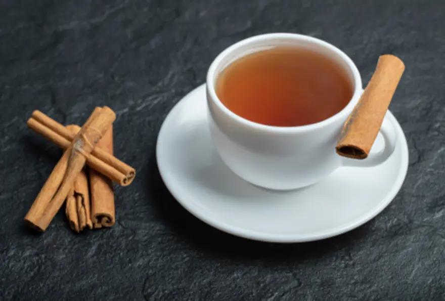
दालचिनीचे पाणी पिण्याचे फायदे: रोजच्या आहारात ते समाविष्ट करण्याचे 9 सोपे मार्ग
दालचिनीचे पाणी पिण्याचे फायदे त्याच्या छान चव आणि सुगंधापलीकडे जातात. हे साधे पेय दालचिनीतील सक्रिय घटकांची, विशेषतः सिनॅमल्डिहाइड आणि पॉलीफेनॉल्सची, ताकद वापरते; दालचिनीच्या बहुतेक आरोग्यदायी गुणधर्मांसाठी हे घटक जबाबदार असतात. दालचिनी पाण्यात भिजवल्यावर हे पाण्यात विरघळणारे घटक बाहेर पडतात आणि त्यामुळे उपचारात्मक तसेच आनंददायी असे पेय तयार होते. दालचिनीचे पाणी विशेष आकर्षक बनवणारी गोष्ट म्हणजे ते सहज उपलब्ध आणि अनेक प्रकारे वापरता येणारे आहे. महागड्या सप्लिमेंट्स किंवा गुंतागुंतीच्या आरोग्य पद्धतींच्या तुलनेत, दालचिनीचे पाणी तयार करण्यासाठी फक्त दोन घटक लागतात, जे बहुधा तुमच्या घरी आधीच उपलब्ध असतात. दालचिनीच्या पाण्याचे फायदे पारंपरिक वैद्यकीय पद्धतींमध्ये शतकानुशतके ओळखले गेले आहेत, आणि आधुनिक संशोधन आता या पारंपरिक उपयोगांपैकी अनेकांना मान्यता देत आहे. दालचिनीच्या पाण्याचे 9 फायदे 1. पचनाचे आरोग्य सुधारते दालचिनीतील जंतुरोधक गुणधर्म तुमच्या आतड्यांमधील उपयुक्त जिवाणूंचे संतुलन राखण्यास मदत करतात, तसेच हानिकारक सूक्ष्मजीवांशी लढतात. दालचिनीचा उष्ण गुणधर्म पचन एन्झाइम्सना चालना देतो, ज्यामुळे तुमचे शरीर अन्न अधिक कार्यक्षमतेने पचवू शकते. यामुळे पोषक तत्त्वांचे शोषण चांगले होऊ शकते आणि जेवणानंतर होणारी पचनाशी संबंधित अस्वस्थता कमी होऊ शकते. 2. रक्तातील साखर नियंत्रित करते यूएसडीएच्या संशोधनानुसार, दालचिनी इन्सुलिन संवेदनशीलता सुधारू शकते आणि रक्तातील ग्लुकोजची पातळी कमी करू शकते, जे टाइप 2 मधुमेह असलेल्या लोकांसाठी विशेषतः फायदेशीर आहे. दालचिनीचे पाणी नियमितपणे घेतल्याने रक्तातील साखरेची अचानक वाढ आणि घट टाळण्यास मदत होऊ शकते, ज्यामुळे अनेकदा ऊर्जा कमी होणे आणि खाण्याची तीव्र इच्छा निर्माण होते. 3. चयापचय वाढवते दालचिनीचे पाणी चयापचयाची कार्यक्षमता आणि इन्सुलिन संवेदनशीलता सुधारून वजन व्यवस्थापनास काही प्रमाणात मदत करू शकते. इन्सुलिनची कार्यक्षमता सुधारून आणि रक्तातील साखरेची पातळी स्थिर ठेवून, दालचिनी तुमच्या शरीराला पोषक तत्त्वे अधिक प्रभावीपणे प्रक्रिया करण्यास मदत करते. चयापचयाची ही सुधारलेली कार्यक्षमता ऊर्जेचा चांगला वापर आणि चरबी साठवण कमी करण्यात मदत करू शकते, ज्यामुळे तुमच्या एकूण आरोग्याच्या उद्दिष्टांना आधार मिळतो. 4. सूज कमी करते दालचिनीतील सूजरोधक घटक शरीरभरातील सूज कमी करण्यास मदत करू शकतात. नियमित सेवनामुळे सांधेदुखी कमी होणे, स्नायूंचा त्रास कमी होणे आणि पेशींचे एकूण आरोग्य सुधारण्यास मदत होऊ शकते. या सूजरोधक परिणामांमुळे दालचिनीचे पाणी सूज-संबंधित स्थितींना सामोरे जाणाऱ्या लोकांसाठी किंवा वयानुसार होणारे सूज-संबंधित आजार टाळू इच्छिणाऱ्यांसाठी विशेष फायदेशीर ठरू शकते. 5. हृदयाचे आरोग्य सुधारते संशोधनानुसार, दालचिनी एलडीएल (‘वाईट’) कोलेस्ट्रॉल आणि ट्रायग्लिसराइड्स कमी करण्यास मदत करू शकते, तसेच एचडीएल (‘चांगले’) कोलेस्ट्रॉलची पातळी कायम ठेवू शकते किंवा किंचित वाढवू शकते. तुमच्या लिपिड प्रोफाइलमधील हे बदल हृदयविकाराचा धोका लक्षणीयरीत्या कमी करू शकतात. याशिवाय, दालचिनीतील अँटिऑक्सिडंट्स तुमच्या रक्तवाहिन्यांचे ऑक्सिडेटिव्ह नुकसानापासून संरक्षण करतात, एकूण हृदयवाहिन्यासंबंधी आरोग्यास आधार देतात आणि रक्तदाब कमी करण्यासही मदत करू शकतात. 6. रोगप्रतिकारक शक्तीला आधार देते दालचिनीचे पाणी पचनासाठी फायदेशीर ठरणारे जंतुरोधक गुणधर्म तुमच्या रोगप्रतिकारक शक्तीलाही आधार देतात. दालचिनीमध्ये जीवाणूरोधक, बुरशीरोधक आणि विषाणूरोधक गुणधर्म असलेले घटक असतात, जे तुमच्या शरीराला विविध रोगजंतूंविरुद्ध संरक्षण करण्यास मदत करतात. सर्दी आणि फ्लूच्या काळात कोमट दालचिनीचे पाणी पिणे विशेषतः आरामदायी आणि संरक्षणात्मक ठरू शकते. 7. वजन कमी करण्यास मदत करते रक्तातील साखरेची पातळी नियंत्रित करण्यात मदत करून, दालचिनी खाण्याची तीव्र इच्छा कमी करू शकते आणि भूक नियंत्रणास आधार देऊ शकते. रक्तातील साखर स्थिर असताना, अचानक भूक लागणे आणि त्यामुळे आरोग्यास अपायकारक स्नॅक्स खाण्याची शक्यता कमी होते. काही अभ्यासांनुसार, दालचिनीचे पाणी चयापचय प्रक्रियांना चालना देऊन थेट चरबी कमी करण्यासही मदत करू शकते. 8. अँटिऑक्सिडंट्सने समृद्ध दालचिनीच्या पाण्यातील पॉलीफेनॉल्स आणि इतर अँटिऑक्सिडंट्स तुमच्या शरीरातील हानिकारक फ्री रॅडिकल्स निष्क्रिय करण्यास मदत करतात. हे फ्री रॅडिकल्स पेशींना नुकसान पोहोचवू शकतात आणि अकाली वृद्धत्व तसेच दीर्घकालीन आजारांना हातभार लावू शकतात. हे अँटिऑक्सिडंट संरक्षण तुमच्या त्वचेपासून मेंदूपर्यंत सर्व शरीर प्रणालींना लाभ देते, एकूण आरोग्यास आधार देते आणि वृद्धत्वाची प्रक्रिया मंदावण्यास मदत करू शकते. 9. मेंदूचे आरोग्य सुधारते उदयोन्मुख संशोधनानुसार दालचिनीच्या पाण्याचे फायदे मेंदूच्या आरोग्यापर्यंतही पोहोचू शकतात. प्रीक्लिनिकल अभ्यास, जे बहुतेक प्राण्यांवर झाले आहेत, असे सूचित करतात की दालचिनीतील घटक मज्जापेशींचे संरक्षण करण्यास आणि हालचालींच्या कार्याला आधार देण्यास मदत करू शकतात. मानवी संशोधन अजून अधिक आवश्यक असले तरी, हे निष्कर्ष अल्झायमर आणि पार्किन्सनसारख्या न्यूरोडीजेनेरेटिव्ह आजारांना प्रतिबंध करण्याच्या संभाव्य फायद्यांकडे इशारा करतात. दालचिनीचे पाणी कसे बनवावे: एक सोपी रेसिपी एका ग्लासमध्ये किंवा बरणीत दालचिनीची एक काडी (किंवा ½–1 टीस्पून दालचिनी पावडर) ठेवा दालचिनीवर 1–2 कप गरम (उकळते नाही) पाणी ओता हलकी चव हवी असल्यास 10–15 मिनिटे भिजू द्या किंवा अधिक तीव्र अर्कासाठी रात्रभर ठेवा दालचिनीची काडी काढून टाका किंवा दालचिनी पावडर वापरली असल्यास पाणी गाळून घ्या अतिरिक्त चव आणि फायद्यांसाठी लिंबाचा थोडा रस किंवा एक टीस्पून मध घाला तुमच्या आवडीनुसार दालचिनीचे पाणी कोमट किंवा थंड प्या रोजच्या आहारात दालचिनीचे पाणी समाविष्ट करण्याचे 9 सोपे मार्ग सकाळची सवय: चयापचय सुरू करण्यासाठी दिवसाची सुरुवात रिकाम्या पोटी एका ग्लास कोमट दालचिनीच्या पाण्याने करा स्मूदीचा आधार: सकाळची स्मूदी बनवताना साध्या पाण्याऐवजी दालचिनीचे पाणी वापरा, ज्यामुळे चव आणि फायदे वाढतात जेवणासोबतचे पेय: पचनास मदत करण्यासाठी जेवताना साध्या पाण्याऐवजी दालचिनीचे पाणी घ्या चहाची चव वाढवा: मसालेदार चवेसाठी तुमच्या आवडत्या हर्बल चहात दालचिनीचे पाणी मिसळा दुपारचे ताजेतवाने पेय: दुपारच्या वेळी ताजेतवाने वाटण्यासाठी थंड दालचिनीचे पाणी फ्रिजमध्ये ठेवा नाश्त्याला चालना: ओट्स किंवा नाश्त्याचे सीरिअल्स तयार करताना दालचिनीचे पाणी वापरा स्वयंपाकातील द्रव: सौम्य चवीसाठी सूप, स्ट्यू किंवा भाताच्या पदार्थांमध्ये दालचिनीचे पाणी वापरा बर्फाचे तुकडे: चवदार बर्फासाठी दालचिनीचे पाणी आइस क्यूब ट्रेमध्ये गोठवा फळांसोबत अर्क: चविष्ट अर्कयुक्त पेयासाठी दालचिनीचे पाणी सफरचंद किंवा संत्र्याच्या फोडींशी मिसळा दालचिनीचे पाणी रिकाम्या पोटी घेता येते का? बहुतेक लोकांसाठी रिकाम्या पोटी दालचिनीचे पाणी घेणे सुरक्षित असते आणि उपयुक्त घटकांचे शोषण वाढवू शकते. अन्नापूर्वी घेतल्यास, दालचिनीतील घटक अधिक सहजपणे शोषले जाऊ शकतात, ज्यामुळे रक्तातील साखर नियंत्रण आणि चयापचयासाठी दालचिनीचे पाणी पिण्याचा फायदा जास्तीत जास्त मिळू शकतो. मात्र, तुमचे पोट संवेदनशील असल्यास किंवा पचनाशी संबंधित समस्या असल्यास, कमी प्रमाणापासून सुरुवात करणे योग्य ठरेल. काही लोकांना अन्नाशिवाय दालचिनीचे पाणी घेतल्यास पोटात सौम्य त्रास होऊ शकतो. असे झाल्यास, ते हलक्या स्नॅकसोबत किंवा नाश्त्यानंतर पिण्याचा प्रयत्न करा. जास्तीत जास्त फायद्यांसाठी दालचिनीचे पाणी पिण्याची सर्वोत्तम वेळ तुम्हाला मिळणाऱ्या दालचिनीच्या पाण्याच्या फायद्यांवर वेळेचा मोठा परिणाम होऊ शकतो. दालचिनीचे पाणी पिण्याची योग्य वेळ साधारणतः सकाळी, नाश्त्याच्या सुमारे 20-30 मिनिटे आधी असते. या वेळेमुळे उपयुक्त घटक शरीरात काम सुरू करू शकतात आणि तुम्ही खाण्यापूर्वी रक्तातील साखरेची पातळी नियंत्रित करण्यास मदत होते. वजन कमी करण्यासाठी दालचिनीचे पाणी वापरणाऱ्यांसाठी ही सकाळची सवय दिवसभर भूक नियंत्रित करण्यात मदत करू शकते. दुसरी फायदेशीर वेळ म्हणजे जेवणापूर्वी, विशेषतः दुपारच्या आणि रात्रीच्या जेवणापूर्वी. जेवण्याच्या 15-20 मिनिटे आधी दालचिनीचे पाणी पिणे पचनास मदत करू शकते आणि जेवणानंतर रक्तातील साखरेची अचानक वाढ टाळण्यास मदत करू शकते. काही लोकांना झोपण्यापूर्वी कोमट दालचिनीचे पाणी घेतल्यानेही फायदा होतो, कारण ते रात्रीभर रक्तातील साखरेची पातळी स्थिर ठेवण्यास आणि झोपेची गुणवत्ता सुधारण्यास मदत करू शकते. दालचिनीचे पाणी पिण्याचे काही दुष्परिणाम आहेत का? दालचिनीचे पाणी पिण्याचे फायदे अनेक असले तरी, संभाव्य दुष्परिणामांची जाणीव असणे महत्त्वाचे आहे. मध्यम प्रमाणात घेतल्यास (दररोज सुमारे ½ टीस्पून दालचिनी पावडर किंवा एका काडीचा अर्क), दालचिनीचे पाणी निरोगी प्रौढांसाठी सर्वसाधारणपणे सुरक्षित असते. मात्र, जास्त प्रमाणात सेवन केल्यास कौमरिन नावाच्या घटकामुळे समस्या उद्भवू शकतात, विशेषतः कॅसिया दालचिनीमध्ये, जी सर्वात सामान्य प्रकार आहे. दालचिनीचे पाणी खूप जास्त प्रमाणात घेतल्यास संभाव्य दुष्परिणामांमध्ये पुढील गोष्टींचा समावेश होतो: कौमरिनचे जास्त सेवन झाल्यामुळे यकृतावर विषारी परिणाम तोंडात जखमा किंवा जळजळ संवेदनशील व्यक्तींमध्ये ॲलर्जिक प्रतिक्रिया रक्त पातळ करणाऱ्या औषधांशी परस्परसंवाद मधुमेहाच्या औषधांसोबत घेतल्यास रक्तातील साखर कमी होणे (हायपोग्लायसीमिया). गर्भवती महिला, यकृताचा आजार असलेले लोक आणि काही विशिष्ट औषधे घेत असलेल्यांनी दालचिनीचे पाणी नियमित दिनक्रमाचा भाग करण्यापूर्वी त्यांच्या डॉक्टरांचा सल्ला घ्यावा. मेट्रोपोलिस हेल्थकेअरमध्ये, आम्हाला समजते की आपल्या आरोग्याची जबाबदारी घेण्यासाठी ज्ञान आणि विश्वासार्ह निदान सहाय्य दोन्ही आवश्यक असतात. 4,000 हून अधिक चाचण्या आणि प्रोफाइल्सच्या आमच्या व्यापक पोर्टफोलिओद्वारे, आम्ही तुम्हाला रक्तातील साखर, कोलेस्ट्रॉल आणि सूज-संबंधित निर्देशकांसारखे महत्त्वाचे आरोग्य मार्कर्स तपासण्यात मदत करू शकतो. भारतभर 10,000 हून अधिक टचपॉइंट्सवर उपलब्ध असलेली आमची सोयीची घरून नमुना संकलन सेवा तुमचा दैनंदिन दिनक्रम न बिघडवता आरोग्याची प्रगती तपासणे शक्य करते. आमचे अचूक अहवाल आणि जलद टर्नअराउंड वेळा तुम्हाला तुमच्या आरोग्य प्रवासाबद्दल माहितीपूर्ण निर्णय घेण्यास सक्षम करतात. वारंवार विचारले जाणारे प्रश्न दालचिनीच्या पाण्यासाठी सर्वोत्तम दालचिनी कोणती? सिलोन दालचिनी, ज्याला अनेकदा "खरी दालचिनी" म्हटले जाते, नियमितपणे दालचिनीचे पाणी पिण्यासाठी प्राधान्य दिले जाते, कारण त्यात कौमरिनचे प्रमाण लक्षणीयरीत्या कमी असते; त्यामुळे सामान्यपणे आढळणाऱ्या कॅसिया दालचिनीपेक्षा ती रोजच्या वापरासाठी अधिक सुरक्षित ठरते. दालचिनीचे पाणी वजन कमी करण्यास मदत करू शकते का? होय, वजन कमी करण्यासाठी दालचिनीचे पाणी रक्तातील साखरेची पातळी स्थिर ठेवणे, खाण्याची तीव्र इच्छा कमी करणे आणि चयापचयाला संभाव्य चालना देणे यामुळे प्रभावी ठरू शकते; मात्र निरोगी आहार आणि नियमित व्यायामासोबत घेतल्यास परिणाम अधिक चांगले मिळतात. दालचिनीचे पाणी त्वचेच्या आरोग्यासाठी चांगले आहे का? दालचिनीच्या पाण्यातील अँटिऑक्सिडंट्स आणि सूजरोधक घटक त्वचेच्या पेशींचे संरक्षण करण्यास आणि सूज कमी करण्यास मदत करू शकतात, त्यामुळे त्वचेचे आरोग्य सुधारू शकते; मात्र या विशिष्ट फायद्यावर थेट संशोधन अद्याप मर्यादित आहे. दिवसातून किती दालचिनीचे पाणी प्यावे? बहुतेक प्रौढांसाठी, दररोज एक ते दोन कप दालचिनीचे पाणी, ज्यात ½ टीस्पूनपेक्षा जास्त दालचिनी पावडर किंवा एक काडी वापरलेली नसते, सुरक्षित आणि चांगल्या दालचिनीच्या पाण्याचे फायदे मिळवण्यासाठी उपयुक्त मानले जाते.


