


Latest Blogs
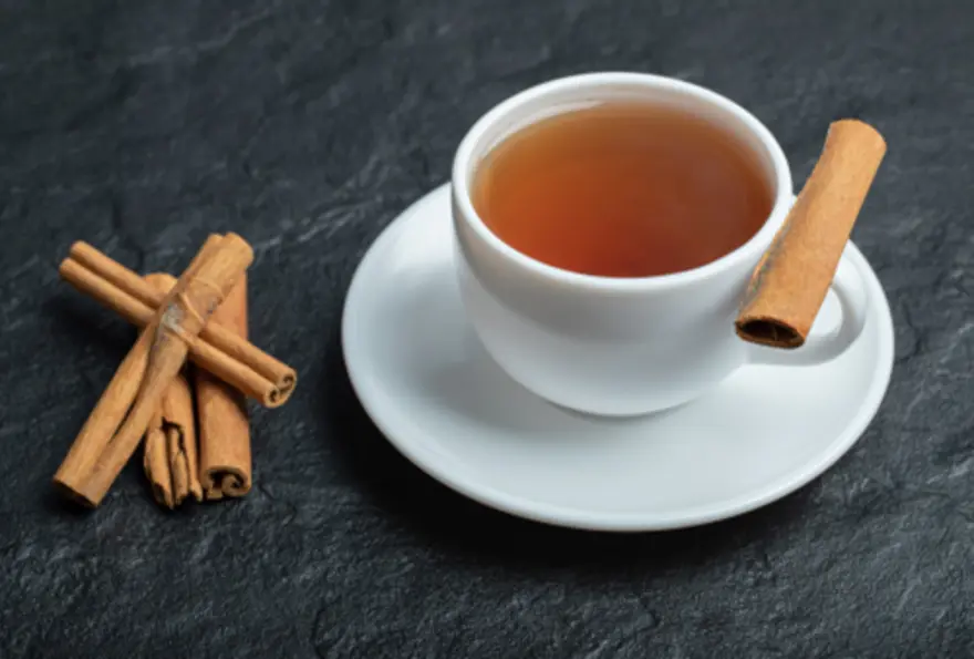
તજના પાણીના ફાયદા: તેને તમારા દૈનિક આહારમાં ઉમેરવાની 9 સરળ રીતો
તજનું પાણી પીવાના ફાયદા તેના સરસ સ્વાદ અને સુગંધથી ઘણા આગળ જાય છે. આ સરળ પીણું તજના સક્રિય સંયોજનો, ખાસ કરીને સિનામાલ્ડિહાઇડ અને પોલિફેનોલ્સની શક્તિનો ઉપયોગ કરે છે, જે તેના મોટાભાગના આરોગ્યવર્ધક ગુણધર્મો માટે જવાબદાર છે. જ્યારે તમે તજને પાણીમાં પલાળો છો, ત્યારે આ પાણીમાં દ્રાવ્ય સંયોજનો છૂટા પડે છે, જેના કારણે એવું પીણું બને છે જે ઉપચારાત્મક પણ છે અને પીવામાં આનંદદાયક પણ છે. તજનું પાણી ખાસ આકર્ષક બનવાનું કારણ તેની સરળ ઉપલબ્ધતા અને વિવિધ રીતે ઉપયોગ કરી શકાય તેવી પ્રકૃતિ છે. મોંઘા સપ્લિમેન્ટ્સ અથવા જટિલ આરોગ્ય રૂટિનથી વિપરીત, તજનું પાણી બનાવવા માટે ફક્ત બે ઘટકોની જરૂર પડે છે, જે શક્ય છે કે તમારા ઘરમાં પહેલેથી જ ઉપલબ્ધ હોય. દાલચિનીના પાણીના ફાયદા પરંપરાગત ચિકિત્સા પદ્ધતિઓમાં સદીઓથી માન્ય રહ્યા છે, અને આધુનિક સંશોધન હવે આ પરંપરાગત ઉપયોગોમાંથી ઘણાને માન્યતા આપી રહ્યું છે. તજના પાણીના 9 ફાયદા 1. પાચન આરોગ્ય સુધારે છે તજના એન્ટીમાઇક્રોબિયલ ગુણધર્મો તમારા આંતરડામાં રહેલા ફાયદાકારક બેક્ટેરિયાનું સંતુલન જાળવવામાં મદદ કરે છે અને સાથે સાથે નુકસાનકારક સૂક્ષ્મજીવો સામે લડે છે. તજની ઉષ્ણ પ્રકૃતિ પાચન એન્ઝાઇમ્સને ઉત્તેજિત કરે છે, જેના કારણે તમારું શરીર ખોરાકને વધુ અસરકારક રીતે તોડી શકે છે. આથી પોષક તત્ત્વોનું શોષણ વધુ સારું થઈ શકે છે અને ભોજન પછી પાચન સંબંધિત અસ્વસ્થતા ઘટી શકે છે. 2. બ્લડ શુગરને નિયંત્રિત કરે છે યુએસડીએના સંશોધને દર્શાવ્યું છે કે તજ ઇન્સ્યુલિન સંવેદનશીલતા સુધારી શકે છે અને બ્લડ ગ્લુકોઝનું સ્તર ઘટાડી શકે છે, જે ખાસ કરીને ટાઇપ 2 ડાયાબિટીસ ધરાવતા લોકો માટે લાભદાયક છે. તજનું પાણી નિયમિત રીતે પીવાથી બ્લડ શુગરમાં અચાનક થનારા તીવ્ર વધારો અને ઘટાડાને રોકવામાં મદદ મળી શકે છે, જે ઘણીવાર ઊર્જામાં ઘટાડો અને ખાવાની ઇચ્છા તરફ દોરી જાય છે. 3. મેટાબોલિઝમને પ્રોત્સાહન આપે છે તજનું પાણી મેટાબોલિક કાર્યક્ષમતા અને ઇન્સ્યુલિન સંવેદનશીલતા સુધારીને વજન વ્યવસ્થાપનમાં આંશિક રીતે મદદ કરી શકે છે. ઇન્સ્યુલિનની કાર્યક્ષમતા સુધારીને અને બ્લડ શુગરનું સ્તર સ્થિર રાખીને, તજ તમારા શરીરને પોષક તત્ત્વો વધુ અસરકારક રીતે પ્રોસેસ કરવામાં મદદ કરે છે. આ સુધારેલું મેટાબોલિક કાર્ય ઊર્જાનો વધુ સારો ઉપયોગ અને ચરબીના ઓછા સંગ્રહ તરફ દોરી શકે છે, જે તમારા સમગ્ર આરોગ્ય લક્ષ્યોને સમર્થન આપે છે. 4. સોજો ઘટાડે છે તજમાં રહેલા એન્ટી-ઇન્ફ્લેમેટરી સંયોજનો શરીરમાં સોજો ઘટાડવામાં મદદ કરી શકે છે. તેનું નિયમિત સેવન સાંધાના દુખાવાને ઘટાડવામાં, સ્નાયુઓના દુખાવામાં રાહત આપવામાં અને સમગ્ર કોષીય આરોગ્યને ટેકો આપવા માટે મદદરૂપ થઈ શકે છે. આ એન્ટી-ઇન્ફ્લેમેટરી અસરો તજના પાણીને ખાસ કરીને સોજા સંબંધિત પરિસ્થિતિઓનો સામનો કરતા લોકો અથવા વય સંબંધિત સોજાવાળા રોગોથી બચવા ઇચ્છતા લોકો માટે લાભદાયક બનાવે છે. 5. હૃદયના આરોગ્યને વધારે છે સંશોધન સૂચવે છે કે તજ એલડીએલ (‘ખરાબ’) કોલેસ્ટ્રોલ અને ટ્રાઇગ્લિસરાઇડ્સ ઘટાડવામાં મદદ કરી શકે છે, જ્યારે એચડીએલ (‘સારું’) કોલેસ્ટ્રોલનું સ્તર જાળવી રાખે છે અથવા થોડું વધારી શકે છે. તમારા લિપિડ પ્રોફાઇલમાં આ ફેરફારો હૃદયરોગનું જોખમ નોંધપાત્ર રીતે ઘટાડી શકે છે. ઉપરાંત, તજમાં રહેલા એન્ટીઓક્સિડન્ટ્સ તમારી રક્તવાહિનીઓને ઓક્સિડેટિવ નુકસાનથી બચાવવામાં મદદ કરે છે, સમગ્ર કાર્ડિયોવેસ્ક્યુલર આરોગ્યને ટેકો આપે છે અને શક્ય છે કે બ્લડ પ્રેશર ઘટાડવામાં મદદ કરે. 6. રોગપ્રતિકારક શક્તિને ટેકો આપે છે તજના પાણીના જે એન્ટીમાઇક્રોબિયલ ગુણધર્મો પાચન માટે ફાયદાકારક છે, તે તમારી રોગપ્રતિકારક શક્તિને પણ ટેકો આપે છે. તજમાં એન્ટીબેક્ટેરિયલ, એન્ટીફંગલ અને એન્ટીવાયરલ ગુણધર્મો ધરાવતા સંયોજનો હોય છે, જે તમારા શરીરને વિવિધ રોગકારકો સામે રક્ષણ આપવામાં મદદ કરે છે. શરદી અને ફ્લૂની ઋતુ દરમિયાન ગરમ તજનું પાણી પીવું ખાસ કરીને આરામદાયક અને રક્ષણાત્મક હોઈ શકે છે. 7. વજન ઘટાડવામાં મદદ કરે છે બ્લડ શુગરનું સ્તર નિયંત્રિત કરવામાં મદદ કરીને, તજ ખાવાની ઇચ્છા ઘટાડવામાં અને ભૂખના નિયંત્રણમાં મદદ કરી શકે છે. જ્યારે તમારું બ્લડ શુગર સ્થિર હોય છે, ત્યારે તમને અચાનક ભૂખ લાગવાની શક્યતા ઓછી રહે છે, જે અસ્વસ્થ નાસ્તા તરફ દોરી જાય છે. કેટલાક અભ્યાસો સૂચવે છે કે તજનું પાણી મેટાબોલિક પ્રક્રિયાઓને વધારીને સીધું ચરબી ઘટાડવામાં પણ મદદ કરી શકે છે. 8. એન્ટીઓક્સિડન્ટ્સથી સમૃદ્ધ છે તજના પાણીમાં રહેલા પોલિફેનોલ્સ અને અન્ય એન્ટીઓક્સિડન્ટ્સ તમારા શરીરમાં નુકસાનકારક મુક્ત રેડિકલ્સને નિષ્ક્રિય કરવામાં મદદ કરે છે. આ મુક્ત રેડિકલ્સ કોષોને નુકસાન પહોંચાડી શકે છે અને સમય પહેલાં વૃદ્ધત્વ તથા લાંબા ગાળાના રોગોમાં યોગદાન આપી શકે છે. આ એન્ટીઓક્સિડન્ટ સુરક્ષા તમારી ત્વચાથી લઈને મગજ સુધી, શરીરની તમામ પ્રણાલીઓ સુધી વિસ્તરે છે, સમગ્ર આરોગ્યને ટેકો આપે છે અને શક્ય છે કે વૃદ્ધત્વની પ્રક્રિયાને ધીમી કરે. 9. મગજના આરોગ્યમાં સુધારો કરે છે ઉભરતું સંશોધન સૂચવે છે કે દાલચિનીના પાણીના ફાયદા મગજના આરોગ્ય સુધી વિસ્તરી શકે છે. મોટાભાગે પ્રાણીઓમાં કરાયેલા પ્રીક્લિનિકલ અભ્યાસો સૂચવે છે કે તજના સંયોજનો નર્વ કોષોને સુરક્ષિત રાખવામાં અને મોટર કાર્યને ટેકો આપવા માટે મદદ કરી શકે છે. જોકે માનવોમાં વધુ સંશોધનની જરૂર છે, આ તારણો અલ્ઝાઇમર અને પાર્કિન્સન જેવા ન્યુરોડિજનરેટિવ રોગો અટકાવવા માટે સંભવિત ફાયદા સૂચવે છે. તજનું પાણી કેવી રીતે બનાવવું: એક સરળ રેસીપી એક તજની સ્ટિક (અથવા પીસેલા તજનો ½–1 ચમચી) એક ગ્લાસ અથવા બરણીમાં મૂકો તજ ઉપર 1–2 કપ ગરમ (ઉકળતું નહીં) પાણી રેડો હળવા સ્વાદ માટે તેને 10–15 મિનિટ સુધી પલળવા દો, અથવા વધુ ગાઢ અર્ક માટે આખી રાત રાખો તજની સ્ટિક કાઢી નાખો અથવા જો પીસેલો તજ વાપર્યો હોય તો પાણી ગાળી લો વધારાના સ્વાદ અને ફાયદા માટે લીંબુનો થોડો રસ અથવા એક ચમચી મધ ઉમેરો તમારી પસંદ મુજબ તજનું પાણી ગરમ અથવા ઠંડું પીવો તજનું પાણી તમારા દૈનિક આહારમાં ઉમેરવાની 9 સરળ રીતો સવારની રીત: તમારા મેટાબોલિઝમને શરૂ કરવા માટે દિવસની શરૂઆત ખાલી પેટે એક ગ્લાસ ગરમ તજના પાણીથી કરો સ્મૂધી બેઝ: વધારાના સ્વાદ અને ફાયદા માટે સવારે સ્મૂધી બનાવતી વખતે સાદા પાણીની જગ્યાએ તજનું પાણી વાપરો ભોજન સાથેનું પીણું: પાચનમાં મદદ માટે ભોજન દરમિયાન સાદા પાણીની જગ્યાએ તજનું પાણી પીવો ચામાં વધારો: મસાલેદાર ટ્વિસ્ટ માટે તજનું પાણી તમારી મનપસંદ હર્બલ ચા સાથે મિક્સ કરો બપોર પછી તાજગી આપતું પીણું: બપોર પછી તાજગીભર્યા પીણાં માટે ઠંડું તજનું પાણી ફ્રિજમાં રાખો નાસ્તામાં વધારો: ઓટમીલ અથવા નાસ્તાના સીરિયલ્સ બનાવતી વખતે તજનું પાણી વાપરો રાંધવાનું પ્રવાહી: હળવા સ્વાદ માટે તજનું પાણી સૂપ, સ્ટ્યૂ અથવા ભાતની વાનગીઓમાં ઉમેરો આઇસ ક્યુબનો નવો ઉપયોગ: સ્વાદવાળા બરફ માટે તજનું પાણી આઇસ ક્યુબ ટ્રેમાં જમાવો ફળો સાથે ઇન્ફ્યુઝન: સ્વાદિષ્ટ ઇન્ફ્યુઝ્ડ પીણાં માટે તજના પાણીમાં સફરજન અથવા નારંગીની સ્લાઇસ ઉમેરો શું તજનું પાણી ખાલી પેટે પી શકાય? મોટાભાગના લોકો માટે, ખાલી પેટે તજનું પાણી પીવું સલામત છે અને ફાયદાકારક સંયોજનોનું શોષણ વધારી શકે છે. ખોરાક પહેલાં લેવામાં આવે ત્યારે, તજમાં રહેલા સંયોજનો વધુ સરળતાથી શોષાઈ શકે છે, જેના કારણે બ્લડ શુગર નિયંત્રણ અને મેટાબોલિઝમ માટે તજનું પાણી પીવાનો લાભ શક્ય તેટલો વધી શકે છે. પરંતુ, જો તમારું પેટ સંવેદનશીલ હોય અથવા પાચન સમસ્યાઓ હોય, તો નાની માત્રાથી શરૂઆત કરવી સમજદારીભર્યું છે. કેટલાક લોકોને ખોરાક વિના તજનું પાણી પીવાથી પેટમાં હળવી બળતરા થઈ શકે છે. જો આવું થાય, તો તેને હળવા નાસ્તા સાથે અથવા નાસ્તા પછી પીવાનો પ્રયાસ કરો. મહત્તમ ફાયદા માટે તજનું પાણી પીવાનો શ્રેષ્ઠ સમય સમય તમારા અનુભવાતા તજના પાણીના ફાયદા પર નોંધપાત્ર અસર કરી શકે છે. તજનું પાણી પીવાનો શ્રેષ્ઠ સમય સામાન્ય રીતે સવારે, નાસ્તા પહેલાં લગભગ 20-30 મિનિટનો હોય છે. આ સમય ફાયદાકારક સંયોજનોને તમારી સિસ્ટમમાં કામ શરૂ કરવાની તક આપે છે, જે તમે ખાઓ તે પહેલાં બ્લડ શુગરનું સ્તર નિયંત્રિત કરવામાં મદદ કરે છે. વજન ઘટાડવા માટે તજનું પાણી વાપરતા લોકો માટે, આ સવારની રીત આખા દિવસ દરમિયાન ભૂખ નિયંત્રિત કરવામાં મદદ કરી શકે છે. બીજો લાભદાયક સમય ભોજન પહેલાંનો છે, ખાસ કરીને બપોરના ભોજન અને રાત્રિના ભોજન પહેલાં. ખાવાથી 15-20 મિનિટ પહેલાં તજનું પાણી પીવાથી પાચનમાં મદદ મળી શકે છે અને ભોજન પછી બ્લડ શુગરમાં અચાનક વધારો અટકાવવામાં મદદ મળી શકે છે. કેટલાક લોકોને સુતા પહેલાં એક કપ ગરમ તજનું પાણી પીવાથી પણ લાભ મળે છે, કારણ કે તે રાત દરમિયાન બ્લડ શુગરનું સ્તર સ્થિર રાખવામાં અને ઊંઘની ગુણવત્તા સુધારવામાં મદદ કરી શકે છે. શું તજનું પાણી પીવાથી કોઈ આડઅસરો થાય છે? તજનું પાણી પીવાના ફાયદા ઘણા છે, છતાં સંભવિત આડઅસરો વિશે જાણવું મહત્વપૂર્ણ છે. મધ્યમ માત્રામાં પીવામાં આવે ત્યારે (દૈનિક લગભગ ½ ચમચી પીસેલો તજ અથવા એક સ્ટિકનું અર્ક), તજનું પાણી સામાન્ય રીતે સ્વસ્થ પુખ્તો માટે સલામત છે. પરંતુ, વધુ પડતું સેવન કુમારિન નામના સંયોજનને કારણે સમસ્યાઓ તરફ દોરી શકે છે, ખાસ કરીને કેશિયા તજમાં, જે સૌથી સામાન્ય જાત છે. અતિશય તજનું પાણી પીવાથી થતી સંભવિત આડઅસરોમાં શામેલ છે: કુમારિનના અતિશય સેવનથી યકૃતની ઝેરી અસર મોઢામાં ઘા અથવા બળતરા સંવેદનશીલ વ્યક્તિઓમાં એલર્જિક પ્રતિક્રિયાઓ લોહી પાતળું કરતી દવાઓ સાથે પરસ્પર અસર જો ડાયાબિટીસની દવાઓ સાથે લેવામાં આવે તો લો બ્લડ શુગર (હાઇપોગ્લાયસેમિયા). ગર્ભવતી સ્ત્રીઓ, યકૃત રોગ ધરાવતા લોકો અને ચોક્કસ દવાઓ લેતા લોકોએ તજના પાણીને પોતાના રૂટિનનો નિયમિત ભાગ બનાવતા પહેલાં પોતાના ડોક્ટરની સલાહ લેવી જોઈએ. મેટ્રોપોલિસ હેલ્થકેરમાં, અમે સમજીએ છીએ કે તમારા આરોગ્યની જવાબદારી લેવી માટે જ્ઞાન અને વિશ્વસનીય નિદાન સહાય બંનેની જરૂર પડે છે. 4,000થી વધુ ટેસ્ટ અને પ્રોફાઇલ્સના અમારા વ્યાપક પોર્ટફોલિયોથી, અમે બ્લડ શુગર, કોલેસ્ટ્રોલ અને સોજા સંબંધિત સૂચકો જેવા મહત્વપૂર્ણ આરોગ્ય માર્કર્સનું નિરીક્ષણ કરવામાં તમારી મદદ કરી શકીએ છીએ. ભારતભરમાં 10,000થી વધુ ટચપોઇન્ટ્સને આવરી લેતી અમારી અનુકૂળ હોમ સેમ્પલ કલેક્શન સેવા સુનિશ્ચિત કરે છે કે તમે તમારા દૈનિક રૂટિનમાં વિક્ષેપ કર્યા વિના તમારા આરોગ્યની પ્રગતિનું નિરીક્ષણ કરી શકો. અમારા સચોટ રિપોર્ટ્સ અને ઝડપી ટર્નઅરાઉન્ડ સમય તમને તમારા આરોગ્યપ્રવાસ વિશે માહિતગાર નિર્ણયો લેવા સક્ષમ બનાવે છે. વારંવાર પૂછાતા પ્રશ્નો તજના પાણી માટે શ્રેષ્ઠ તજ કયું છે? સિલોન તજ, જેને ઘણીવાર "ટ્રુ સિનામન" કહેવામાં આવે છે, નિયમિત તજના પાણીના સેવન માટે પસંદગીનો વિકલ્પ છે, કારણ કે તેમાં કુમારિનનું પ્રમાણ નોંધપાત્ર રીતે ઓછું હોય છે, જે તેને વધુ સામાન્ય કેશિયા તજની જાત કરતાં દૈનિક ઉપયોગ માટે વધુ સલામત બનાવે છે. શું તજનું પાણી વજન ઘટાડવામાં મદદ કરી શકે છે? હા, વજન ઘટાડવા માટે તજનું પાણી બ્લડ શુગરનું સ્તર સ્થિર રાખીને, ખાવાની ઇચ્છા ઘટાડીને અને સંભવિત રીતે મેટાબોલિઝમ વધારીને અસરકારક થઈ શકે છે, જોકે તેના પરિણામો સ્વસ્થ આહાર અને નિયમિત વ્યાયામ સાથે જોડવામાં આવે ત્યારે શ્રેષ્ઠ રહે છે. શું તજનું પાણી ત્વચાના આરોગ્ય માટે સારું છે? તજના પાણીમાં રહેલા એન્ટીઓક્સિડન્ટ્સ અને એન્ટી-ઇન્ફ્લેમેટરી સંયોજનો ત્વચાના કોષોને સુરક્ષિત રાખવામાં અને સોજો ઘટાડવામાં મદદ કરી શકે છે, જેના કારણે ત્વચાના આરોગ્યમાં સંભવિત સુધારો થઈ શકે છે, જોકે આ ચોક્કસ ફાયદા અંગે સીધું સંશોધન હજુ મર્યાદિત છે. દિવસમાં કેટલું તજનું પાણી પીવું જોઈએ? મોટાભાગના પુખ્તો માટે, પીસેલા તજના ½ ચમચીથી વધુ નહીં અથવા એક સ્ટિકથી બનાવેલા તજના પાણીના દૈનિક એકથી બે કપ, શ્રેષ્ઠ તજના પાણીના ફાયદાનો અનુભવ કરવા માટે સલામત અને લાભદાયક માનવામાં આવે છે.
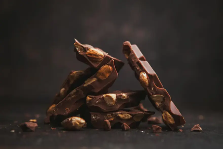
આરોગ્ય માટે ડાર્ક ચોકલેટના 10 અદ્ભુત ફાયદા
વિચારતા હો કે ડાર્ક ચોકલેટ ખરેખર તમારા આરોગ્ય માટે સારી છે કે નહીં? આ યોગ્ય પ્રશ્ન છે—ખાસ કરીને જ્યારે કંઈક એટલું સ્વાદિષ્ટ હોય કે તે સાચું હોવું મુશ્કેલ લાગે. પરંતુ મર્યાદામાં ખાવામાં આવે ત્યારે, ડાર્ક ચોકલેટ તેના સમૃદ્ધ સ્વાદથી આગળ વધીને પુરાવા આધારિત ફાયદા આપી શકે છે. તે તમારો મૂડ સુધારવામાં, હૃદયના આરોગ્યને ટેકો આપવામાં અને તણાવપૂર્ણ સમયમાં હળવો આરામ આપવામાં મદદ કરી શકે છે. જો તમે તમારા સુખાકારીને ટેકો આપવા માટે નાની અને આનંદદાયક રીત શોધી રહ્યા છો, તો ડાર્ક ચોકલેટ મૂલ્યવાન ઉમેરો બની શકે છે. થોડું જ પ્રમાણ તમારા દિવસમાં આનંદ અને હેતુ બંને આપી શકે છે, જે તેને વિચારપૂર્વક માણવા યોગ્ય નાસ્તો બનાવે છે. ડાર્ક ચોકલેટ શું છે? ડાર્ક ચોકલેટ કોકો સોલિડ્સ, કોકો બટર અને ઓછી માત્રામાં ખાંડથી બનાવવામાં આવે છે. મિલ્ક ચોકલેટથી વિપરીત, તેમાં દૂધનું પ્રમાણ ઓછું અથવા નથી હોતું, જેના કારણે તેને વધુ ગાઢ કોકો સ્વાદ અને ફાયદાકારક સંયોજનોનું વધુ પ્રમાણ મળે છે. ઓછામાં ઓછા 70% કોકો ધરાવતી ડાર્ક ચોકલેટ ખાસ કરીને ફ્લેવોનોઇડ્સ જેવા એન્ટીઓક્સિડન્ટ્સથી સમૃદ્ધ હોય છે, જે હૃદયના આરોગ્ય, મગજના કાર્ય અને ત્વચાની સુખાકારીને ટેકો આપી શકે છે. તેનો અનોખો સ્વાદ અને કુદરતી આરોગ્યને ટેકો આપતા ગુણધર્મો તેને વધુ વિચારપૂર્વકના નાસ્તાની શોધ કરતા લોકો માટે પસંદગીનો વિકલ્પ બનાવે છે. તેને હજુ પણ મીઠી વસ્તુ માનવામાં આવે છે, છતાં ડાર્ક ચોકલેટ માત્ર સ્વાદિષ્ટ આનંદથી વધુ આપે છે—મર્યાદામાં ખાવામાં આવે ત્યારે તે તમારી જીવનશૈલીનો હેતુપૂર્ણ ભાગ બની શકે છે. ઓછા ઉમેરણો ધરાવતી સારી ગુણવત્તાની ડાર્ક ચોકલેટ પસંદ કરવાથી તમે તેનો સમૃદ્ધ સ્વાદ અને સંભવિત સુખાકારી ફાયદા બંને માણી શકો છો. ડાર્ક ચોકલેટની પોષક માહિતી ડાર્ક ચોકલેટ વિવિધ પોષક તત્ત્વો આપે છે, જે તમારા સુખાકારીમાં યોગદાન આપી શકે છે. જો તમે 70–85% કોકો ધરાવતી બાર પસંદ કરી રહ્યા છો, તો તેમાં તમને મહત્વપૂર્ણ ખનિજો, એન્ટીઓક્સિડન્ટ્સ અને થોડું આહાર ફાઇબર મળવાની શક્યતા છે. અહીં ડાર્ક ચોકલેટ (70–85% કોકો)ના 30g સર્વિંગ માટે સામાન્ય વિગતો આપવામાં આવી છે: પોષક તત્ત્વ 30g દીઠ માત્રા કેલરી 170–190 kcal પ્રોટીન 2–3 g કુલ ચરબી 12–14 g સેચ્યુરેટેડ ચરબી 7–8 g કાર્બોહાઇડ્રેટ્સ 13–15 g ખાંડ 6–8 g ફાઇબર 3–4 g આયર્ન 3–4 mg (20–25% DV) મેગ્નેશિયમ 60–70 mg (15–18% DV) ઝિંક 0.8–1 mg કોપર 0.5–0.6 mg આ આંકડા બ્રાન્ડ મુજબ થોડા બદલાઈ શકે છે, પરંતુ તે ડાર્ક ચોકલેટની પોષક કિંમત દર્શાવે છે, જે તેના સંભવિત આરોગ્ય ફાયદાઓને ટેકો આપે છે. ડાર્ક ચોકલેટના 10 ફાયદા ડાર્ક ચોકલેટ માત્ર સ્વાદિષ્ટ નથી—તે અર્થપૂર્ણ રીતે તમારા આરોગ્યને પણ ટેકો આપી શકે છે. મર્યાદામાં ખાવામાં આવે ત્યારે, આ નાસ્તો માનસિક અને શારીરિક રીતે શક્તિશાળી ફાયદા આપી શકે છે. અહીં જાણવાની લાયક દસ સંભવિત ડાર્ક ચોકલેટના ફાયદા આપેલા છે: 1. હૃદયના આરોગ્યને ટેકો આપે છે ડાર્ક ચોકલેટમાં રહેલા ફ્લેવાનોોલ્સ રક્તપ્રવાહ સુધારવામાં, બ્લડ પ્રેશર ઘટાડવામાં અને હૃદયના કાર્યને ટેકો આપવામાં મદદ કરી શકે છે. આ સંયોજનો તમારી ધમનીઓને લવચીક રાખવામાં સહાય કરી શકે છે, જેના કારણે હૃદય માટે અસરકારક રીતે લોહી પંપ કરવું સરળ બને છે. 2. મગજના કાર્યને પ્રોત્સાહન આપે છે જે ફ્લેવાનોોલ્સ તમારા હૃદયને ટેકો આપે છે, તે જ તમારા મગજ માટે પણ ફાયદાકારક હોઈ શકે છે. મગજમાં વધુ સારો રક્તપ્રવાહ જ્ઞાનાત્મક કાર્ય, ધ્યાન કેન્દ્રિત કરવાની ક્ષમતા અને યાદશક્તિ સુધારી શકે છે—ખાસ કરીને વધુ માંગવાળા અથવા તણાવપૂર્ણ સમયમાં. 3. મૂડ સુધારે છે ડાર્ક ચોકલેટમાં સેરોટોનિન અને થિયોબ્રોમિન જેવા સંયોજનોની ઓછી માત્રા હોય છે, જે તમારો મૂડ સુધારવામાં મદદ કરી શકે છે. ઘણા લોકોને લાગે છે કે તે હળવો આરામ અને શાંતિનો અનુભવ આપે છે. 4. એન્ટીઓક્સિડન્ટ્સથી સમૃદ્ધ છે ડાર્ક ચોકલેટ એન્ટીઓક્સિડન્ટ્સથી ભરપૂર હોય છે, જેમાં પોલિફેનોલ્સ અને ફ્લેવોનોઇડ્સનો સમાવેશ થાય છે. આ તમારા શરીરને ઓક્સિડેટિવ તણાવ સામે લડવામાં મદદ કરે છે, જે વૃદ્ધત્વ અને લાંબા ગાળાની પરિસ્થિતિઓમાં ભૂમિકા ભજવે છે. 5. ત્વચાના આરોગ્યમાં સુધારો કરે છે કેટલાક સંશોધન સૂચવે છે કે ફ્લેવાનોોલ્સ તમારી ત્વચાને સૂર્યના નુકસાનથી બચાવી શકે છે, ભેજ સુધારી શકે છે અને ત્વચામાં રક્તપ્રવાહ વધારી શકે છે. આથી સમય જતાં તમારી ત્વચા વધુ સારી લાગે અને દેખાય શકે છે. 6. બ્લડ શુગર નિયંત્રણને ટેકો આપી શકે છે આ આશ્ચર્યજનક લાગે, છતાં કેટલાક અભ્યાસો સૂચવે છે કે ડાર્ક ચોકલેટનું મધ્યમ સેવન ઇન્સ્યુલિન સંવેદનશીલતા સુધારવામાં મદદ કરી શકે છે. જોકે, ઓછી ખાંડવાળી જાતો પસંદ કરવી અને વધુ પડતું સેવન ટાળવું મહત્વપૂર્ણ છે. 7. આવશ્યક ખનિજો આપે છે ડાર્ક ચોકલેટમાં આયર્ન, મેગ્નેશિયમ અને કોપર સારી માત્રામાં જોવા મળે છે. આ ખનિજો તમારા ઊર્જા સ્તરથી લઈને રોગપ્રતિકારક શક્તિ અને હાડકાંની મજબૂતાઈ સુધીની બાબતોને ટેકો આપે છે. 8. સોજો ઘટાડવામાં મદદ કરી શકે છે લાંબા ગાળાનો સોજો ઘણી આરોગ્ય ચિંતાઓમાં એક પરિબળ છે. ડાર્ક ચોકલેટમાં રહેલા એન્ટીઓક્સિડન્ટ અને એન્ટી-ઇન્ફ્લેમેટરી ગુણધર્મો સોજો ઘટાડવામાં મદદ કરી શકે છે, ખાસ કરીને જ્યારે તે સંતુલિત આહારનો ભાગ હોય. 9. પુરુષોના સમગ્ર આરોગ્યને ટેકો આપે છે પુરુષો માટે, ડાર્ક ચોકલેટ રક્તપ્રવાહ સુધારવામાં અને બ્લડ પ્રેશર ઘટાડવામાં મદદ કરી શકે છે, જે હૃદયના આરોગ્ય અને શારીરિક કાર્યક્ષમતાને લાભ આપી શકે છે. ડાર્ક ચોકલેટમાં રહેલા એન્ટીઓક્સિડન્ટ્સ તણાવ ઘટાડવામાં અને ભારે શારીરિક પ્રવૃત્તિ પછી પુનઃપ્રાપ્તિમાં મદદ કરી શકે છે. સંતુલિત જીવનશૈલીમાં શામેલ કરવામાં આવે ત્યારે આ પુરુષો માટે ડાર્ક ચોકલેટના ફાયદામાંથી થોડા છે. 10. સ્ત્રીઓની સુખાકારીને ટેકો આપે છે સ્ત્રીઓ માટે, ડાર્ક ચોકલેટ તેના મેગ્નેશિયમ સામગ્રીને કારણે માસિક દરમિયાન આરામ આપી શકે છે, જે કડકાવો અને થાક ઘટાડવામાં મદદ કરી શકે છે. તેના મૂડ સુધારતા ગુણધર્મો હોર્મોનલ ફેરફારો દરમિયાન ભાવનાત્મક સુખાકારીને પણ ટેકો આપી શકે છે. આ હળવા પ્રભાવો સ્ત્રીઓ માટે ડાર્ક ચોકલેટના ફાયદા અને તમારી દૈનિક રૂટિનમાં નાનો પરંતુ મદદરૂપ ઉમેરો બનવાની તેની સંભાવનાને દર્શાવે છે. આ બ્લેક ચોકલેટના ફાયદા સાથે, તમે એવો સ્વાદિષ્ટ નાસ્તો માણી શકો છો જે તમારી સમગ્ર સુખાકારીમાં સકારાત્મક યોગદાન આપે છે. સમજદારીથી પસંદ કરવામાં આવે અને ધ્યાનપૂર્વક ખાવામાં આવે ત્યારે, ડાર્ક ચોકલેટ ખાવાના ફાયદા આરોગ્યપ્રત્યે જાગૃત જીવનશૈલીનો ભાગ બની શકે છે. જોખમો અને ધ્યાનમાં રાખવાના મુદ્દા ડાર્ક ચોકલેટના ઘણા ફાયદા હોવા છતાં, થોડા મુદ્દા ધ્યાનમાં રાખવા જરૂરી છે. બધી ડાર્ક ચોકલેટ સમાન રીતે બનાવાતી નથી, અને તેને તમારી રૂટિનમાં શામેલ કરતી વખતે તમારી વ્યક્તિગત આરોગ્ય પરિસ્થિતિઓ પણ મહત્વપૂર્ણ છે. 1. કેલરી અને ચરબી વધારે હોય છે: ડાર્ક ચોકલેટ ઊર્જા-ઘન હોય છે. નાનાં ભાગોમાં પણ નોંધપાત્ર કેલરી અને સેચ્યુરેટેડ ચરબી હોય છે. જો તેને તમારા બાકીના આહાર સાથે સંતુલિત ન કરવામાં આવે, તો વધુ ખાવાથી સમય જતાં વજન વધી શકે છે. 2. ઉમેરેલી ખાંડ હોઈ શકે છે: જોકે ડાર્ક ચોકલેટમાં સામાન્ય રીતે મિલ્ક ચોકલેટ કરતાં ઓછી ખાંડ હોય છે, કેટલીક બ્રાન્ડ્સ તમારી અપેક્ષા કરતાં વધુ ખાંડ ઉમેરે છે. તમે ઓછા ઉમેરણોવાળા વિકલ્પો પસંદ કરી રહ્યા છો તેની ખાતરી કરવા માટે લેબલ્સ તપાસો. 3. કેફીન હોય છે: ડાર્ક ચોકલેટમાં કેફીન અને થિયોબ્રોમિન સહિત કુદરતી ઉત્તેજકો હોય છે. તેની માત્રા પ્રમાણમાં ઓછી હોવા છતાં, જો તમે ઉત્તેજકો પ્રત્યે સંવેદનશીલ હો અથવા સૂવાના સમયની નજીક તેને ખાતા હો, તો તે તમને અસર કરી શકે છે. 4. માઇગ્રેન શરૂ કરી શકે છે: કેટલાક લોકોને લાગે છે કે ડાર્ક ચોકલેટ માથાનો દુખાવો અથવા માઇગ્રેન શરૂ કરી શકે છે. જો તમને આ પેટર્ન દેખાય, તો તેનું સેવન મર્યાદિત કરવા અથવા તમારા હેલ્થકેર પ્રદાતા સાથે ચર્ચા કરવાનો વિચાર કરો. 5. ભારે ધાતુઓનું સંભવિત પ્રમાણ: તાજેતરના અહેવાલોએ કેટલાક કોકો ઉત્પાદનોમાં કેડમિયમ અને સીસાના સૂક્ષ્મ પ્રમાણ અંગે ચિંતાઓ ઉભી કરી છે, ખાસ કરીને ચોક્કસ પ્રદેશોમાંથી મેળવેલા ઉત્પાદનોમાં. પારદર્શક સોર્સિંગ ધરાવતી વિશ્વસનીય બ્રાન્ડ્સ પસંદ કરવાથી આ જોખમ ઘટાડવામાં મદદ મળી શકે છે. 6. બધા આહાર માટે યોગ્ય નથી: ચોક્કસ આહાર નિયંત્રણો ધરાવતા લોકો (ઉદાહરણ તરીકે, લો-હિસ્ટામિન આહારનું પાલન કરતા લોકો અથવા ચોક્કસ એલર્જી ધરાવતા લોકો)એ ડાર્ક ચોકલેટ ટાળવાની અથવા મર્યાદિત કરવાની જરૂર પડી શકે છે. જો તમને ચિંતા હોય, તો હેલ્થકેર પ્રદાતા સાથે તપાસ કરવી હંમેશાં સારી વિચારણા છે. 7. વધુ ખાવાથી ફાયદા ઘટી શકે છે: વધારે ડાર્ક ચોકલેટ ખાવાથી તેના સકારાત્મક પ્રભાવ ઓછા થઈ શકે છે. વધારે કેલરી, વધારે ખાંડ અને વધારે ચરબી તમારા આરોગ્યને ટેકો આપવાને બદલે તેના પર ભાર મૂકી શકે છે. આ મુદ્દાઓ સમજવાથી તમે માહિતગાર પસંદગીઓ કરી શકો છો. વાત ડરવાની નથી—વાત સંતુલનની છે. ધ્યાનપૂર્વક રહેતા અને સારી ગુણવત્તાની ચોકલેટ પસંદ કરતા, તમે સંભવિત ખામીઓ ટાળીને તેના ફાયદા માણી શકો છો. કેટલું ખાવું? મર્યાદા મહત્વપૂર્ણ છે. સારી ગુણવત્તાની ડાર્ક ચોકલેટનો રોજનો 20 થી 30 ગ્રામનો નાનો ભાગ (લગભગ એક કે બે સ્ક્વેર) અતિશયતા વિના ડાર્ક ચોકલેટના ફાયદા માણવા માટે પૂરતો છે. ઓછામાં ઓછા 70% કોકો ધરાવતી બાર શોધો, જેથી તમને વધુ માત્રામાં એન્ટીઓક્સિડન્ટ્સ અને ઓછી ખાંડ મળે તેની ખાતરી થાય. તેને વારંવારની આદત કરતાં વિચારપૂર્વક માણવાના નાસ્તા તરીકે રાખવું, તમારા સમગ્ર આહાર અને લક્ષ્યો પર આધાર રાખીને, સંતુલન જાળવવામાં અને અનાવશ્યક જોખમો ટાળવામાં મદદ કરી શકે છે. ઓછી માત્રામાં ડાર્ક ચોકલેટ માણવી તમારી સુખાકારી રૂટિનનો આરામદાયક અને લાભદાયક ભાગ બની શકે છે. નિષ્કર્ષ ડાર્ક ચોકલેટ તમારા સુખાકારી પ્રવાસમાં નાનો પરંતુ અર્થપૂર્ણ ઉમેરો બની શકે છે. મર્યાદામાં માણવામાં આવે ત્યારે તેના કુદરતી સંયોજનો તમારા હૃદય, મૂડ અને સમગ્ર સુખાકારીને ટેકો આપી શકે છે. પરંતુ તમામ આરોગ્ય પસંદગીઓની જેમ, વ્યક્તિગત સંભાળ મહત્વપૂર્ણ છે. તમારી પોષક જરૂરિયાતોને વધુ સારી રીતે સમજવા અથવા મહત્વપૂર્ણ આરોગ્ય માર્કર્સનું નિરીક્ષણ કરવા માટે, નિયમિત હેલ્થ ચેક-અપ્સનો વિચાર કરો. મેટ્રોપોલિસ હેલ્થકેર, તેની વિશ્વસનીય નિદાન સેવાઓ અને હોમ સેમ્પલ કલેક્શન સાથે, તમારા આરોગ્ય લક્ષ્યો માટે વિશ્વસનીય ટેકો આપે છે—તમને આત્મવિશ્વાસ સાથે માહિતગાર નિર્ણયો લેવામાં મદદ કરે છે. વારંવાર પૂછાતા પ્રશ્નો શું ડાર્ક ચોકલેટ ત્વચા માટે સારી છે? હા, ડાર્ક ચોકલેટ તમારી ત્વચા માટે સારી હોઈ શકે છે. તેમાં ફ્લેવોનોઇડ્સ જેવા એન્ટીઓક્સિડન્ટ્સ હોય છે, જે ત્વચાની ભેજ સુધારવામાં, સૂર્યના નુકસાનથી બચાવવામાં અને વધુ સારા રક્તપ્રવાહને ટેકો આપવા માટે મદદ કરી શકે છે. સંતુલિત આહારના ભાગરૂપે મર્યાદામાં ખાવાથી સમય જતાં તમારી ત્વચાના આરોગ્ય અને દેખાવને હળવો ટેકો મળી શકે છે, ખાસ કરીને જો તેને પૂરતું પાણી પીવું અને સૂર્યથી રક્ષણ જેવી ત્વચાને ટેકો આપતી અન્ય આદતો સાથે જોડવામાં આવે. ડાર્ક ચોકલેટના ગેરફાયદા શું છે? ડાર્ક ચોકલેટના ફાયદા હોવા છતાં, વધારે ખાવાથી સમસ્યાઓ થઈ શકે છે. તેમાં કેલરી, ચરબી અને ક્યારેક ખાંડ વધુ હોય છે. વધુ પડતું ખાવાથી કેટલાક લોકોમાં વજન વધવું, કેફીનને કારણે ઊંઘમાં ખલેલ અથવા માથાનો દુખાવો થઈ શકે છે. ઉપરાંત, કેટલીક બ્રાન્ડ્સમાં ભારે ધાતુઓના સૂક્ષ્મ અંશો હોઈ શકે છે. હંમેશાં લેબલ્સ તપાસો અને તેને મર્યાદામાં માણો. શું ડાર્ક ચોકલેટ પિરિયડ્સ માટે સારી છે? હા, ડાર્ક ચોકલેટ પિરિયડ્સ દરમિયાન મદદ કરી શકે છે. તેમાં રહેલું મેગ્નેશિયમ કડકાવો અને સ્નાયુ તણાવ ઘટાડવામાં મદદ કરી શકે છે, જ્યારે તેના મૂડ વધારતા સંયોજનો હોર્મોનલ ફેરફારો દરમિયાન ચીડિયાપણું અને અસ્વસ્થતા ઘટાડવામાં મદદ કરી શકે છે. ઓછી માત્રામાં ડાર્ક ચોકલેટ ખાવાથી શારીરિક અને ભાવનાત્મક બંને રીતે આરામ મળી શકે છે. શ્રેષ્ઠ અસર માટે તે ઓછી ખાંડવાળી હોય અને મર્યાદામાં ખવાય તેની ખાતરી કરો. ડાર્ક ચોકલેટમાં શું ટાળવું? ડાર્ક ચોકલેટ પસંદ કરતી વખતે, વધુ ખાંડ, કૃત્રિમ ઉમેરણો અથવા ઓછું કોકો પ્રમાણ (70%થી ઓછું) ધરાવતા ઉત્પાદનો ટાળો. હાઇડ્રોજનેટેડ તેલ અથવા અનાવશ્યક ફિલર્સથી પણ દૂર રહો. ગુણવત્તાની ખાતરી કરવા માટે પ્રમાણપત્રો અને ઘટકોની પારદર્શિતા તપાસો. ડાર્ક ચોકલેટનો યોગ્ય પ્રકાર પસંદ કરવાથી તેના આરોગ્ય ફાયદા માણવામાં મોટો ફરક પડે છે.
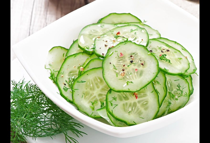
કાકડીના ફાયદા સમજાવ્યા: પોષક મૂલ્ય, હાઇડ્રેશન અને વધુ
શું કાકડી તમારા માટે સારી છે? કાકડી એક તાજગી આપતું, ઓછી કેલરીવાળું શાક છે, જે પ્રભાવશાળી આરોગ્ય ફાયદાઓથી ભરપૂર છે. 95% પાણીથી બનેલી કાકડી હાઇડ્રેશન માટે ઉત્તમ છે અને હળવો, તાજગી આપતો નાસ્તો બને છે. તેમાં વિટામિન C અને K, પોટેશિયમ અને એન્ટીઓક્સિડન્ટ્સ જેવા આવશ્યક પોષક તત્ત્વો હોય છે, જે શરીરના વિવિધ કાર્યોને ટેકો આપે છે. સંશોધન સૂચવે છે કે કાકડી સંભવિત એન્ટી-ઇન્ફ્લેમેટરી, એન્ટીઓક્સિડન્ટ અને એન્ટી-ડાયાબિટિક અસરો આપી શકે છે. કાકડીના ફાયદા કાર્ડિયોવેસ્ક્યુલર આરોગ્ય, કિડનીના કાર્ય, ત્વચાના આરોગ્ય અને કેન્સર નિવારણ સુધી વિસ્તરે છે. તેના શાંતિકારક ગુણધર્મો માટે જાણીતી કાકડી પાચન અને સમગ્ર સુખાકારી માટે ઉત્તમ છે. કાકડીનું પોષક મૂલ્ય પોષક તત્ત્વ 100g દીઠ માત્રા આરોગ્યમાં ભૂમિકા પાણી 95g હાઇડ્રેશન, શરીરના કાર્યોને ટેકો આપે છે કેલરી 16 kcal વજન વ્યવસ્થાપન માટે ઓછી કેલરીવાળો ખોરાક વિટામિન C 2.8 mg એન્ટીઓક્સિડન્ટ, રોગપ્રતિકારક આરોગ્યને ટેકો આપે છે વિટામિન K 16.4 mcg લોહી ગંઠાવા અને હાડકાંના આરોગ્ય માટે મહત્વપૂર્ણ પોટેશિયમ 147 mg બ્લડ પ્રેશર અને હૃદયના કાર્યને નિયંત્રિત કરે છે મેગ્નેશિયમ 13 mg સ્નાયુ અને નર્વ કાર્યને ટેકો આપે છે ફાઇબર 0.5 g પાચનમાં મદદ કરે છે અને આંતરડાના આરોગ્યને પ્રોત્સાહન આપે છે એન્ટીઓક્સિડન્ટ્સ ક્યુકરબિટાસિન્સ, ફ્લેવોનોઇડ્સનો સમાવેશ થાય છે કોષોને નુકસાનથી બચાવે છે અને સોજો ઘટાડે છે કાકડીના 12 આરોગ્ય ફાયદા આ સરળ શાકને તમારા આહારમાં સામેલ કરવાથી તમારા આરોગ્યને નોંધપાત્ર રીતે કેવી રીતે પ્રોત્સાહન મળી શકે છે તે જાણો. તમને હાઇડ્રેટેડ રાખે છે કાકડી ખાવાના ફાયદામાંથી એક મુખ્ય ફાયદો તેની અદ્ભુત હાઇડ્રેટિંગ શક્તિ છે. લગભગ 95% પાણી ધરાવતી કાકડી શરીરમાં ઘટેલા પ્રવાહીને ફરી ભરવા અને સમગ્ર હાઇડ્રેશનને ટેકો આપવા માટે ઉત્તમ છે. યોગ્ય હાઇડ્રેશન આ માટે મહત્વપૂર્ણ છે: શરીરનું યોગ્ય તાપમાન જાળવવું સાંધાઓને સ્નિગ્ધ રાખવું કોષીય કાર્યોને ટેકો આપવો કાકડીનો નાસ્તો કરવો, ખાસ કરીને ગરમ હવામાનમાં અથવા વર્કઆઉટ પછી, તમારી પ્રવાહી જરૂરિયાતો પૂરી કરવામાં મદદ કરી શકે છે. પાણીનું પ્રમાણ ઝેરી પદાર્થોને બહાર કાઢવામાં પણ મદદ કરે છે, જે કિડની અને મૂત્રમાર્ગના આરોગ્યને પ્રોત્સાહન આપે છે. પોટેશિયમ જેવા ઇલેક્ટ્રોલાઇટ્સ સાથે, કાકડી પ્રવાહી સંતુલનને ટેકો આપે છે અને ડિહાઇડ્રેશન અટકાવે છે. વજન ઘટાડવામાં મદદ કરે છે થોડું વજન ઘટાડવા માંગો છો? કાકડીના આરોગ્ય ફાયદા તેને તમારા વજન ઘટાડવાના આયોજનમાં સમજદાર ઉમેરો બનાવે છે. કેલરીમાં ઓછી અને પાણી તથા ફાઇબરથી સમૃદ્ધ કાકડી તમને લાંબા સમય સુધી પેટ ભરેલું અને સંતોષદાયક અનુભવવામાં મદદ કરે છે. ફાઇબર તૃપ્તિને પ્રોત્સાહન આપે છે, જે ભૂખ નિયંત્રિત કરવામાં અને કુલ કેલરી સેવન ઘટાડવામાં મદદ કરે છે. પાણીનું ઊંચું પ્રમાણ વધારાની કેલરી વિના તમારા ભોજનમાં માત્રા ઉમેરે છે. આ કાકડીને આરોગ્યપ્રદ નાસ્તા, સલાડમાં વધુ માત્રા ઉમેરવા અને સ્મૂધીમાં ઉમેરવા માટે ઉત્તમ બનાવે છે. ઉપરાંત, કેટલાક અભ્યાસો સૂચવે છે કે એન્ટીઓક્સિડન્ટ્સ મેટાબોલિઝમ અને સોજાને અસર કરી શકે છે, જે મેટાબોલિઝમ અને સોજાના નિયંત્રણ દ્વારા પેટની ચરબી સાથે જોડાયેલા છે. તેથી, તમારા વજન ઘટાડવાના લક્ષ્યોને ટેકો આપવા માટે આ તાજગી આપતું શાક ખાઓ. એન્ટીઓક્સિડન્ટ્સથી સમૃદ્ધ છે કાકડી એન્ટીઓક્સિડન્ટ્સનો ઉત્તમ સ્ત્રોત છે, જેમાં ક્યુકરબિટાસિન્સ, વિટેક્સિન, ઓરિએન્ટિન અને આઇસોસ્કોપેરિન જેવા ફ્લેવોનોઇડ્સ અને ટેનિન્સનો સમાવેશ થાય છે. આ શક્તિશાળી સંયોજનો નુકસાનકારક મુક્ત રેડિકલ્સને નિષ્ક્રિય કરવામાં મદદ કરે છે, જે ઓક્સિડેટિવ તણાવમાં યોગદાન આપે છે – કેન્સર, હૃદયરોગ અને ડાયાબિટીસ જેવા લાંબા ગાળાના રોગોમાં એક મુખ્ય પરિબળ છે. મુક્ત રેડિકલ્સના નુકસાન સામે લડીને, કાકડીમાં રહેલા એન્ટીઓક્સિડન્ટ્સ તમારા કોષો અને તંતુઓને સુરક્ષિત રાખવામાં મદદ કરે છે અને શરીરમાં સોજો ઘટાડે છે. સ્વસ્થ ત્વચાને પ્રોત્સાહન આપે છે ઇચ્છિત સ્વસ્થ ચમક મેળવવા માંગો છો? ત્વચા માટે કાકડીના ફાયદા તેના હાઇડ્રેટિંગ અને એન્ટીઓક્સિડન્ટ ગુણધર્મોને કારણે પ્રભાવશાળી છે. પાણીનું ઊંચું પ્રમાણ તમારી ત્વચાને ભેજયુક્ત અને નરમ રાખવામાં મદદ કરે છે. કાકડીનો પરંપરાગત રીતે ત્વચાની બળતરા શાંત કરવા, સોજો ઘટાડવા અને સનબર્નનો દુખાવો હળવો કરવા માટે ઉપયોગ થયો છે. કાકડીમાં રહેલા ત્વચા માટે ફાયદાકારક વિટામિન C અને K કોલેજન ઉત્પાદનને ટેકો આપે છે અને આંખોની આસપાસના ડાર્ક સર્કલ્સ અને સોજો ઘટાડવામાં મદદ કરી શકે છે. તેજસ્વી, યુવાન દેખાતી ત્વચા માટે તમારા આહારમાં કાકડી માણો અને તેની સ્લાઇસ ઉપરથી લગાવવાનો પણ પ્રયાસ કરો. બ્લડ શુગર ઘટાડવામાં મદદ કરે છે કાકડીના ફાયદા સંભવિત રીતે બ્લડ શુગર વ્યવસ્થાપનમાં પણ મદદ કરી શકે છે. અભ્યાસો સૂચવે છે કે કાકડીના બીજ અને ગૂદામાં રહેલા કેટલાક બાયોએક્ટિવ સંયોજનો ઇન્સ્યુલિન સંવેદનશીલતા સુધારી શકે છે અને બ્લડ ગ્લુકોઝનું સ્તર ઘટાડવામાં મદદ કરી શકે છે. આ ખાસ કરીને ટાઇપ 2 ડાયાબિટીસ ધરાવતા લોકો માટે બ્લડ શુગર નિયંત્રિત કરવામાં અને ડાયાબિટીસ સંબંધિત જટિલતાઓનું જોખમ ઘટાડવામાં મદદરૂપ થઈ શકે છે. કાકડીમાં કાર્બોહાઇડ્રેટનું ઓછું પ્રમાણ તેને બ્લડ શુગરનું ધ્યાન રાખતા લોકો માટે પણ સમજદાર પસંદગી બનાવે છે. તમારા સ્તરને નિયંત્રણમાં રાખવામાં મદદ કરવા માટે ભોજન અને નાસ્તામાં કાકડી ઉમેરવાનો પ્રયાસ કરો. પાચનમાં મદદ કરે છે કાકડીના આરોગ્ય ફાયદા તમારી પાચન પ્રણાલી સુધી પણ વિસ્તરે છે. કાકડીમાં સારી માત્રામાં ફાઇબર હોય છે, ખાસ કરીને તેની છાલમાં, જે પાચનને ઉત્તેજિત કરવામાં અને નિયમિત આંતરડાની ગતિને પ્રોત્સાહન આપવામાં મદદ કરે છે. પાણીનું ઊંચું પ્રમાણ મળને નરમ બનાવીને અને કબજિયાત અટકાવીને પાચન આરોગ્યને વધુ ટેકો આપે છે. ઉપરાંત, કાકડીના બીજમાં હળવી લેક્સેટિવ અસર હોય છે, જે પેટ ફૂલવું અને અસ્વસ્થતા ઘટાડવામાં મદદ કરે છે. આ શાકના શાંતિકારક ગુણધર્મો ગેસ્ટ્રિક સોજો અને એસિડિટી શાંત કરવામાં પણ મદદ કરી શકે છે. તમારા આંતરડાને ખુશ અને સ્વસ્થ રાખવા માટે કાકડી નિયમિત રીતે માણો. હૃદયના આરોગ્યને ટેકો આપે છે તમારા હૃદયને પણ કાકડી ગમે છે! કાકડીમાં રહેલું પોટેશિયમ શરીરમાં સોડિયમનું સ્તર સંતુલિત કરીને બ્લડ પ્રેશરને નિયંત્રિત કરવામાં મહત્વપૂર્ણ ભૂમિકા ભજવે છે. આ તમારી કાર્ડિયોવેસ્ક્યુલર સિસ્ટમ પરનો ભાર ઘટાડવામાં મદદ કરે છે, જેના કારણે હાઇપરટેન્શનનું જોખમ અને સ્ટ્રોકનું જોખમ ઘટે છે. કાકડીમાં રહેલા એન્ટીઓક્સિડન્ટ્સ તમારી રક્તવાહિનીઓને ઓક્સિડેટિવ નુકસાનથી બચાવવામાં પણ મદદ કરે છે, જે વધુ સારા વાસ્ક્યુલર આરોગ્યને પ્રોત્સાહન આપે છે. ઉપરાંત, ફાઇબરનું પ્રમાણ એલડીએલ અથવા "ખરાબ" કોલેસ્ટ્રોલ ઘટાડવામાં યોગદાન આપે છે – જે હૃદયરોગ અટકાવવા માટેનું બીજું મુખ્ય પરિબળ છે. વધારાના ટેકા માટે તમારા હૃદય-સ્વસ્થ આહાર આયોજનમાં કાકડી ઉમેરો. શ્વાસને તાજું કરે છે ખરાબ શ્વાસ શરમજનક હોઈ શકે છે, પરંતુ કાકડી મદદ કરી શકે છે! આ શાકમાં પાણીનું ઊંચું પ્રમાણ તમારા મોંમાંથી દુર્ગંધ પેદા કરતા બેક્ટેરિયાને દૂર કરવામાં મદદ કરે છે. ઉપરાંત, કાકડીમાં ફાઇટોકેમિકલ્સ હોય છે, જે ખરાબ શ્વાસ માટે જવાબદાર સૂક્ષ્મજીવોને મારી શકે છે. ભોજન પછી શ્વાસને કુદરતી રીતે તાજું કરવા માટે કાકડીની સ્લાઇસ ખાવાનો પ્રયાસ કરો. દુર્ગંધ સામે લડવામાં મદદ કરવા માટે તમે કાકડીની સ્લાઇસ દાંત અને મસૂડા પર ઘસી શકો છો અથવા તેના બીજ ચાવી શકો છો. મોંને સ્વચ્છ અનુભવતું રાખવાની આ સરળ અને તાજગી આપતી રીત છે. સોજા સામે લડવામાં મદદ કરી શકે છે સોજો ઘણા લાંબા ગાળાના રોગો પાછળનું સામાન્ય કારણ છે. સદનસીબે, કાકડીના આરોગ્ય ફાયદામાં સંભવિત એન્ટી-ઇન્ફ્લેમેટરી અસરોનો સમાવેશ થાય છે. કાકડીમાં ક્યુકરબિટાસિન્સ અને ફ્લેવોનોઇડ્સ જેવા સંયોજનો હોય છે, જે શરીરમાં સોજો ઘટાડવામાં મદદ કરી શકે છે. અભ્યાસો સૂચવે છે કે આ સંયોજનો COX-2 જેવા પ્રો-ઇન્ફ્લેમેટરી એન્ઝાઇમ્સની પ્રવૃત્તિને અટકાવી શકે છે. કોષીય સ્તરે સોજા સામે લડીને, કાકડી આર્થ્રાઇટિસ, ઇન્ફ્લેમેટરી બાઉલ ડિસીઝ અને કેટલાક કેન્સર જેવી પરિસ્થિતિઓનું જોખમ ઘટાડવામાં મદદ કરી શકે છે. હાડકાંના આરોગ્ય માટે સારી છે કાકડીમાં રહેલું વિટામિન K મજબૂત અને સ્વસ્થ હાડકાં જાળવવા માટે આવશ્યક છે. આ પોષક તત્ત્વ હાડકાંના મેટાબોલિઝમમાં મહત્વપૂર્ણ ભૂમિકા ભજવે છે, હાડકાંની રચનાને પ્રોત્સાહન આપવામાં અને હાડકાંની ક્ષતિ અટકાવવામાં મદદ કરે છે. એક કપ કાકડી તમારી દૈનિક વિટામિન K જરૂરિયાતનો લગભગ 19% ભાગ આપે છે. કાકડીમાં કેલ્શિયમ અને મેગ્નેશિયમ જેવા ખનિજો પણ હોય છે, જે હાડકાંના આરોગ્ય માટે ખૂબ મહત્વપૂર્ણ છે. કાકડી નિયમિત રીતે ખાવાથી હાડકાંની ઘનતાને ટેકો મળી શકે છે અને ખાસ કરીને ઉંમર વધે તેમ ઓસ્ટિયોપોરોસિસનું જોખમ ઘટી શકે છે. ડિટોક્સિફિકેશનને ટેકો આપે છે કાકડી તમારા શરીરની કુદરતી ડિટોક્સિફિકેશન પ્રક્રિયાને ટેકો આપવા માટે સારી સાથી બની શકે છે. પાણીનું ઊંચું પ્રમાણ તમારી સિસ્ટમમાંથી ઝેરી પદાર્થોને બહાર કાઢવામાં મદદ કરે છે, જ્યારે એન્ટીઓક્સિડન્ટ્સ ઓક્સિડેટિવ નુકસાન સામે લડવામાં મદદ કરે છે. કાકડીમાં રહેલા કેટલાક સંયોજનો, જેમ કે ક્યુકરબિટાસિન્સ, યકૃતમાં ડિટોક્સિફિકેશન એન્ઝાઇમ્સને ઉત્તેજિત કરવામાં પણ મદદ કરી શકે છે. તમારા યકૃત અને કિડનીને ટેકો આપીને, કાકડી નુકસાનકારક પદાર્થોને વધુ અસરકારક રીતે દૂર કરવામાં મદદ કરે છે. તણાવ ઘટાડવામાં મદદ કરે છે તણાવ અનુભવો છો? કાકડી ખાવાના ફાયદા તમને આરામ કરવામાં મદદ કરી શકે છે. કાકડીમાં અનેક B વિટામિન્સની ઓછી માત્રા હોય છે, જે નર્વસ સિસ્ટમના આરોગ્યને ટેકો આપે છે, જેમાં વિટામિન B1, વિટામિન B5 અને વિટામિન B7 (બાયોટિન)નો સમાવેશ થાય છે. આ પોષક તત્ત્વો જી એ બી એ (GABA)ના ઉત્પાદનમાં સામેલ છે, જે નર્વસ સિસ્ટમને શાંત કરવામાં મદદ કરતું ન્યુરોટ્રાન્સમિટર છે. કાકડી મેગ્નેશિયમથી પણ સમૃદ્ધ છે, જે તણાવ ઘટાડવામાં અને સારી ઊંઘને પ્રોત્સાહન આપવામાં મદદ કરી શકે તેવું ખનિજ છે. કાકડીના હાઇડ્રેટિંગ ગુણધર્મો તણાવ સંબંધિત માથાનો દુખાવો હળવો કરવામાં પણ મદદ કરી શકે છે. તેથી, આવતા વખતે જ્યારે તમે તણાવ અનુભવો, ત્યારે આરામ અને નવી ઊર્જા મેળવવામાં મદદ માટે કાકડીની સ્લાઇસ લો. કાકડીનું સેવન કરવાના સંભવિત આડઅસરો કાકડીના આરોગ્ય ફાયદા ઘણા હોવા છતાં, સંભવિત આડઅસરો વિશે જાણવું મહત્વપૂર્ણ છે: રેગવીડ પરાગકણ પ્રત્યે સંવેદનશીલ લોકોમાં એલર્જિક પ્રતિક્રિયાઓ બીજ અને છાલને કારણે ગેસ અને પેટ ફૂલવું જેવી પાચન સમસ્યાઓ વિટામિન Kની સામગ્રીને કારણે લોહી પાતળું કરતી દવાઓમાં વિક્ષેપ કડવી લાગતી કાકડી મોટી માત્રામાં ખાવાથી ક્યુકરબિટાસિન્સ નામના સંયોજનોને કારણે પેટમાં અસ્વસ્થતા પણ થઈ શકે છે. જોકે, સામાન્ય આહારમાં કાકડી ખાવાથી આડઅસરો દુર્લભ છે. તમારા આહારમાં કાકડી કેવી રીતે સામેલ કરવી કાકડીના આરોગ્ય ફાયદામાંથી એક શ્રેષ્ઠ બાબત એ છે કે તેને તમારા આહારમાં ઉમેરવી કેટલી સરળ છે! તાજગી આપતા, હાઇડ્રેટિંગ પીણાં માટે પાણીમાં તેની સ્લાઇસ ઉમેરો અથવા સંતોષદાયક કરકરાપણું મેળવવા માટે તેને સલાડમાં કાપીને ઉમેરો. તમે તેને તમારા મનપસંદ ફળો સાથે સ્મૂધી અને જ્યૂસમાં પણ બ્લેન્ડ કરી શકો છો, સેન્ડવિચ અને રેપ્સમાં પાતળી સ્લાઇસ વાપરી શકો છો, ઠંડો કાકડીનો સૂપ બનાવી શકો છો અથવા પ્રોબાયોટિકથી સમૃદ્ધ અથાણા તરીકે ફર્મેન્ટ કરી શકો છો. નિષ્કર્ષ જેમ આપણે જોયું, કાકડી ખાવાના ફાયદા પ્રભાવશાળી છે. ત્વચા માટે કાકડીના ફાયદા અને સમગ્ર આરોગ્ય માટે મહત્તમ લાભ મેળવવા માટે, વધારાના ફાઇબર અને પોષક તત્ત્વો માટે તેની છાલ રાખો. તેની વિવિધ રીતે ઉપયોગ કરી શકાય તેવી પ્રકૃતિ અને તાજગી આપતા સ્વાદને કારણે, કાકડીને દૈનિક આદત બનાવવી સરળ છે. ઉત્તમ આહાર ઉપરાંત, જો તમે નિદાન પરીક્ષણો દ્વારા તમારા આરોગ્યની જવાબદારી લેવા માંગતા હો, તો મેટ્રોપોલિસ હેલ્થકેરનો વિચાર કરો. ભારતભરમાં અનુકૂળ ઘેરબેઠાં બ્લડ કલેક્શન, અદ્યતન લેબ્સમાં વિશ્લેષિત સેમ્પલ્સ અને ઝડપી ટેસ્ટ પરિણામો માટે, વધુ સ્વસ્થ તમારા માટે મેટ્રોપોલિસ પસંદ કરો! વારંવાર પૂછાતા પ્રશ્નો શું કાકડી ઝુકીની જેવી જ છે? ના, બંને કુકરબિટેસી કુળના હોવા છતાં, કાકડી અને ઝુકીની અલગ છોડ છે. કાકડી સામાન્ય રીતે કાચી ખાવામાં આવે છે અને રસદાર કરકરાપણું ધરાવે છે, જ્યારે ઝુકીની ઘણીવાર રાંધીને ખવાય છે અને તેનો સ્વાદ થોડો મીઠો હોય છે. શું કાકડીના બીજ આરોગ્ય માટે ફાયદાકારક છે? હા, કાકડીના બીજમાં ફાઇબર અને ફાયદાકારક પોષક તત્ત્વો હોય છે. તેમાં ઠંડક આપતી અસર હોય છે અને તે કબજિયાત અટકાવવામાં મદદ કરી શકે છે. બીજ કાકડીના એન્ટીઓક્સિડન્ટ અને લિપિડ ઘટાડનારા ગુણધર્મોમાં પણ યોગદાન આપે છે. શું કાકડીમાં પૂરતું ફાઇબર હોય છે? કાકડીમાં ફાઇબરનું મધ્યમ પ્રમાણ હોય છે, ખાસ કરીને જ્યારે તેને છાલ અને બીજ સાથે ખવાય છે. કેટલીક શાકભાજીની તુલનામાં તેમાં ફાઇબર બહુ વધારે નથી, છતાં તે તમારી દૈનિક જરૂરિયાતોમાં યોગદાન આપી શકે છે. શું કાકડી એસિડિટી અથવા હાર્ટબર્નમાં મદદ કરી શકે છે? કાકડીની ક્ષારીયતા અને પાણીનું ઊંચું પ્રમાણ પેટના એસિડને નિષ્ક્રિય કરવામાં અને હાર્ટબર્નના લક્ષણોને શાંત કરવામાં મદદ કરી શકે છે. તેની ઠંડક આપતી અસર બળતરાગ્રસ્ત પાચન માર્ગને શાંત કરી શકે છે. શું કાકડી કીટો આહાર માટે યોગ્ય છે? હા, ચોક્કસ! બહુ ઓછી કેલરી અને કાર્બોહાઇડ્રેટ્સ સાથે, કાકડી કીટોજેનિક આહારમાં સંપૂર્ણ રીતે ફિટ થાય છે. તે કીટોસિસમાં વિક્ષેપ કર્યા વિના પાણી અને પોષક તત્ત્વો આપે છે. શું કાકડી ખાવાથી શરદી થાય છે? ના, આ એક માન્યતા છે. કાકડીમાં ઠંડક આપતો ગુણ હોવા છતાં, તે શરદી અથવા ચેપનું કારણ બનતી નથી. શું કાકડી ત્વચાનો ટેન દૂર કરવામાં મદદ કરી શકે છે? કાકડીનો રસ તેના એન્ટી-ઇન્ફ્લેમેટરી સંયોજનોને કારણે સનબર્ન થયેલી ત્વચાને શાંત કરવામાં અને ટેન હળવો કરવામાં મદદ કરી શકે છે. ટેન થયેલા ભાગો પર તાજી કાકડીની સ્લાઇસ અથવા રસ લગાવવાથી સમય જતાં હળવી લાઇટનિંગ અસર મળી શકે છે.
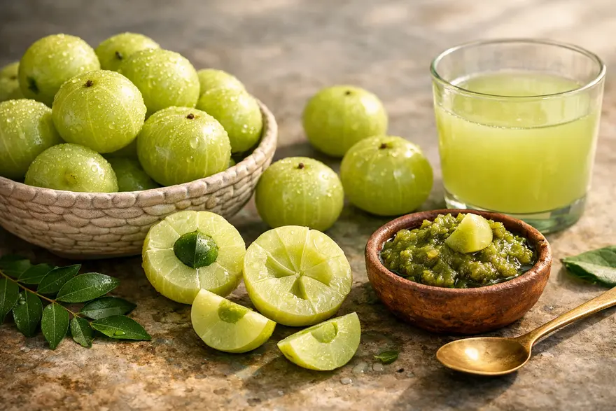
ગૂસબેરી (આમળા)ના 10 અદ્ભુત ફાયદા જે તમારે જાણવા જોઈએ
જો તમે ભારતમાં મોટા થયા હો, તો શક્ય છે કે તમે સાંભળ્યું હશે કે આમળા તમારા આરોગ્ય માટે સારા છે. ઇન્ડિયન ગૂસબેરી તરીકે પણ ઓળખાતું આ નાનું, ખાટું ફળ રોજિંદા આહાર અને પરંપરાગત સુખાકારીની પદ્ધતિઓમાં લાંબા સમયથી મૂલ્યવાન માનવામાં આવે છે. આજે પણ તેનો વ્યાપક ઉપયોગ થાય છે, કારણ કે તે વિટામિન C, એન્ટીઓક્સિડન્ટ્સ, ફાઇબર અને સમગ્ર સુખાકારી સાથે જોડાયેલા અન્ય વનસ્પતિ સંયોજનોથી સમૃદ્ધ છે. આમળા કોઈ ચમત્કારિક ઈલાજ નથી. પરંતુ જ્યારે તમે તેને સંતુલિત આહારમાં સામેલ કરો છો, ત્યારે તે તમારી રોગપ્રતિકારક શક્તિ, પાચન, હૃદયનું આરોગ્ય, ત્વચા અને વધુને ટેકો આપી શકે છે. આ લેખમાં, તમે જાણશો કે આમળા શું છે, તેમાં શું હોય છે, તેના સંભવિત આરોગ્ય ફાયદા, તેને કેવી રીતે ખાવું અને ક્યારે સાવચેત રહેવું જોઈએ. ગૂસબેરી (આમળા) શું છે? વિશ્વના અલગ અલગ ભાગોમાં ગૂસબેરી શબ્દ અલગ ફળો માટે વપરાઈ શકે છે. ભારતીય સંદર્ભમાં, ગૂસબેરીનો અર્થ સામાન્ય રીતે આમળા, અથવા ઇન્ડિયન ગૂસબેરી થાય છે. આમળા નાનું, ગોળ ફળ છે, જેનો રંગ હળવો લીલો હોય છે અને તેનો સ્વાદ સ્પષ્ટ રીતે ખાટો અને તીખો હોય છે. તેને સામાન્ય રીતે તાજું ખવાય છે, તેનો રસ બનાવવામાં આવે છે, અથાણું બનાવવામાં આવે છે, મીઠાઈરૂપે તૈયાર કરવામાં આવે છે અથવા પાઉડર અને પ્રિઝર્વ્સમાં ઉપયોગ થાય છે. આયુર્વેદમાં પણ તેનો લાંબો ઇતિહાસ છે અને સામાન્ય જીવનશક્તિ માટેની પરંપરાગત વાનગીઓમાં તેનો સમાવેશ ઘણીવાર થાય છે. આમળાને ખાસ બનાવે છે તેનો ઘન પોષક પ્રોફાઇલ. તે નાનું હોવા છતાં, તેમાં એવા ઘણા સંયોજનો હોય છે જે વિવિધ આહારના ભાગરૂપે ખાવામાં આવે ત્યારે તમારા આરોગ્યને ટેકો આપી શકે છે. ગૂસબેરી (આમળા)નું પોષક મૂલ્ય આમળામાં રહેલા ચોક્કસ પોષક તત્ત્વો તેની જાત, ઉગાડવાની પરિસ્થિતિઓ અને પ્રોસેસિંગ પદ્ધતિ મુજબ બદલાઈ શકે છે. તેમ છતાં, આમળા પોષક તત્ત્વોથી સમૃદ્ધ ફળ તરીકે વ્યાપક રીતે માન્ય છે. પોષક તત્ત્વ અથવા સંયોજન તમારા આરોગ્ય માટે તે કેમ મહત્વનું છે વિટામિન C રોગપ્રતિકારક શક્તિ, કોલેજન બનવું અને એન્ટીઓક્સિડન્ટ રક્ષણને ટેકો આપે છે ફાઇબર પાચન અને પેટ ભરાયેલું લાગવામાં મદદ કરે છે પોલિફેનોલ્સ અને ટેનિન્સ તમારા કોષોને ઓક્સિડેટિવ તણાવથી બચાવવામાં મદદ કરે છે કેલ્શિયમ, આયર્ન અને ફોસ્ફરસ હાડકાં અને મેટાબોલિક આરોગ્ય સહિત શરીરના સામાન્ય કાર્યોને ટેકો આપે છે કેરોટીનોઇડ્સ અને અન્ય વનસ્પતિ સંયોજનો આંખો અને સમગ્ર કોષીય આરોગ્યને ટેકો આપી શકે છે આમળા ખાસ કરીને તેના ઊંચા વિટામિન C અને એન્ટીઓક્સિડન્ટ સામગ્રી માટે જાણીતા છે, જે એક કારણ છે કે તેને ઘણીવાર સુખાકારીને ટેકો આપતું ફળ કહેવામાં આવે છે. તમારા આરોગ્ય માટે ગૂસબેરી (આમળા)ના ટોચના 10 ફાયદા 1. તમારી રોગપ્રતિકારક શક્તિને ટેકો આપે છે આમળા તેના વિટામિન C સામગ્રી માટે સૌથી વધુ જાણીતા છે. વિટામિન C તમારી રોગપ્રતિકારક શક્તિને ટેકો આપવામાં મદદ કરે છે અને એન્ટીઓક્સિડન્ટ તરીકે પણ કામ કરે છે, એટલે કે તે તમારા કોષોને ઓક્સિડેટિવ તણાવથી બચાવવામાં મદદ કરે છે. આ મહત્વનું છે કારણ કે બીમારી, પ્રદૂષણનો સંપર્ક, ઊંઘની અછત અને શરીરના રોજિંદા ઘસારા દરમિયાન ઓક્સિડેટિવ તણાવ વધી શકે છે. આમળા જેવા વિટામિન Cથી સમૃદ્ધ ખોરાકને તમારા આહારમાં ઉમેરવાથી તમારા શરીરની કુદરતી રક્ષણ પ્રણાલીને ટેકો મળી શકે છે. 2. વધુ સારા પાચનને પ્રોત્સાહન આપે છે આમળામાં ફાઇબર હોય છે, જે સ્વસ્થ પાચન અને નિયમિત આંતરડાની ગતિને ટેકો આપવા માટે મદદ કરી શકે છે. જો તમારા આહારમાં ફાઇબર ઓછું હોય, તો આમળા જેવા ફળોને સામેલ કરવાથી પાચન સંબંધિત આરામમાં સુધારો થઈ શકે છે. પરંપરાગત ઉપયોગ આમળાને પાચન સુખાકારી સાથે પણ જોડે છે. જોકે કેટલાક ક્ષેત્રોમાં હજુ વધુ મજબૂત માનવીય સંશોધનની જરૂર છે, હાલના પુરાવા સૂચવે છે કે તેના વનસ્પતિ સંયોજનો અને ફાઇબર આંતરડાના આરોગ્યને ટેકો આપી શકે છે. 3. સ્વસ્થ ત્વચાને ટેકો આપવામાં મદદ કરે છે તમારી ત્વચાને કોલેજન બનાવવા માટે વિટામિન Cની જરૂર હોય છે, જે એક પ્રોટીન છે અને ત્વચાને મજબૂત તથા સ્વસ્થ રાખવામાં મદદ કરે છે. આમળા વિટામિન C અને એન્ટીઓક્સિડન્ટ્સથી સમૃદ્ધ હોવાથી, તે અંદરથી ત્વચાના આરોગ્યને ટેકો આપવામાં મદદ કરી શકે છે. આનો અર્થ એ નથી કે આમળા પોતે જ ત્વચાની સમસ્યાઓ દૂર કરી શકે છે. પરંતુ સંતુલિત આહારના ભાગરૂપે, તે તમારા શરીરને ઓક્સિડેટિવ તણાવ સંભાળવામાં અને સામાન્ય કોલેજન ઉત્પાદન જાળવવામાં મદદ કરીને તમારી ત્વચાને ટેકો આપી શકે છે. 4. તમારા વાળને મજબૂત બનાવી શકે છે આમળાનો ઉપયોગ વર્ષોથી પરંપરાગત વાળ સંભાળમાં થયો છે, અને આધુનિક સંશોધન હવે આ બાબતને વધુ નજીકથી સમજવાનો પ્રયાસ કરી રહ્યું છે. તેની એન્ટીઓક્સિડન્ટ સામગ્રી માથાની ત્વચાના આરોગ્યને ટેકો આપી શકે છે, જ્યારે તેની પોષક પ્રોફાઇલ વધુ મજબૂત દેખાતા વાળમાં યોગદાન આપી શકે છે. તમે ઘણીવાર વાળના તેલ, માસ્ક અને સપ્લિમેન્ટ્સમાં આમળા જોશો. શરૂઆતના અભ્યાસો આશાજનક હોવા છતાં, વાળ ખરવા માટે આમળાને એકમાત્ર ઉકેલ તરીકે નહીં પરંતુ સહાયક સંભાળ તરીકે જોવું શ્રેષ્ઠ છે. 5. ઓક્સિડેટિવ તણાવ સંભાળવામાં મદદ કરે છે ઓક્સિડેટિવ તણાવ ત્યારે થાય છે જ્યારે મુક્ત રેડિકલ્સ કહેવાતા નુકસાનકારક અણુઓ તમારા શરીર દ્વારા સંભાળી શકાય તે કરતાં ઝડપથી એકઠા થાય છે. સમય જતાં, આ વૃદ્ધત્વ અને લાંબા ગાળાના રોગોમાં યોગદાન આપી શકે છે. આમળામાં વિટામિન C અને પોલિફેનોલ્સ જેવા એન્ટીઓક્સિડન્ટ્સ હોય છે, જે શરીરમાં ઓક્સિડેટિવ તણાવ ઘટાડવામાં મદદ કરી શકે છે. આ એક કારણ છે કે આમળાને ઘણીવાર સામાન્ય સુખાકારી અને સ્વસ્થ વૃદ્ધત્વ સાથે જોડવામાં આવે છે. 6. હૃદયના આરોગ્યને ટેકો આપી શકે છે કેટલાક અભ્યાસો સૂચવે છે કે આમળા કેટલાક બ્લડ લિપિડ માર્કર્સ સુધારવામાં અને ઓક્સિડેટિવ તણાવ ઘટાડવામાં મદદ કરીને હૃદયના આરોગ્યને ટેકો આપી શકે છે. આ ઉપયોગી હોઈ શકે છે કારણ કે કોલેસ્ટ્રોલનું અસંતુલન અને સોજો સમય જતાં તમારા હૃદય અને રક્તવાહિનીઓને અસર કરી શકે છે. તેમ છતાં, આમળા તબીબી સંભાળ, વ્યાયામ અથવા નિર્ધારિત સારવારનો વિકલ્પ નથી. તેને હૃદયપ્રત્યે જાગૃત જીવનશૈલીમાં એક સ્વસ્થ ઉમેરા તરીકે જોવું શ્રેષ્ઠ છે. 7. બ્લડ શુગર સંતુલનમાં મદદ કરી શકે છે આમળામાં ફાઇબર અને વનસ્પતિ સંયોજનો હોય છે, જે બ્લડ શુગર સંતુલનને ટેકો આપવા માટે મદદ કરી શકે છે. કેટલાક માનવીય અભ્યાસો અને સમીક્ષાઓ સૂચવે છે કે તે ફાસ્ટિંગ બ્લડ શુગર અને લિપિડ સ્તરો પર સકારાત્મક અસર કરી શકે છે. તેમ છતાં, તમારે તેને ડાયાબિટીસની સારવારનો વિકલ્પ માનવામાં સાવચેત રહેવું જોઈએ. જો તમને ડાયાબિટીસ અથવા પ્રિડાયાબિટીસ હોય, તો ખોરાકની પસંદગીઓ મહત્વપૂર્ણ છે, પરંતુ યોગ્ય નિદાન, મોનિટરિંગ અને તબીબી માર્ગદર્શન પણ એટલું જ મહત્વપૂર્ણ છે. 8. આંખોના આરોગ્યને ટેકો આપે છે આમળામાં વિટામિન C અને અન્ય વનસ્પતિ સંયોજનો હોય છે, જે સમગ્ર કોષીય આરોગ્યને ટેકો આપે છે. તેમાં આંખોના આરોગ્ય સાથે જોડાયેલા કેરોટીનોઇડ સંબંધિત સંયોજનો પણ હોય છે. જોકે માત્ર આમળા આંખોના રોગને અટકાવી શકતા નથી, સંતુલિત આહારના ભાગરૂપે પોષક તત્ત્વોથી ભરપૂર ફળો ખાવાથી લાંબા ગાળાની આંખોની સુખાકારીને ટેકો મળી શકે છે. 9. મગજના આરોગ્યને ટેકો આપી શકે છે એન્ટીઓક્સિડન્ટ્સ કોષોને ઓક્સિડેટિવ નુકસાનથી બચાવવામાં મદદ કરે છે, અને તેમાં મગજના કોષો પણ આવે છે. શરૂઆતના પુરાવા સૂચવે છે કે આમળાના બાયોએક્ટિવ સંયોજનોમાં ન્યુરોપ્રોટેક્ટિવ સંભાવના હોઈ શકે છે. આ ક્ષેત્રમાં હજુ માનવોમાં વધુ સંશોધનની જરૂર છે, તેથી અપેક્ષાઓ વાસ્તવિક રાખવી શ્રેષ્ઠ છે. તેમ છતાં, ફળો અને વનસ્પતિ ખોરાકથી સમૃદ્ધ આહાર લાંબા ગાળાના મગજના આરોગ્યને ટેકો આપવાની સૌથી વ્યવહારુ રીતોમાંથી એક છે. 10. વજન વ્યવસ્થાપનને ટેકો આપી શકે છે આમળા ઓછી કેલરીવાળા છે અને તેમાં ફાઇબર હોય છે, જે તમને વધુ સમય સુધી પેટ ભરેલું અનુભવવામાં અને સારી ખાવાની આદતોને ટેકો આપવા માટે મદદ કરી શકે છે. જો તમે વજન સંભાળવાનો પ્રયાસ કરી રહ્યા હો, તો અતિ-પ્રક્રિયિત નાસ્તાની જગ્યાએ આખા ફળો પસંદ કરવું ઘણીવાર સમજદાર પગલું છે. આમળા પોતે જ વજન ઘટાડશે નહીં. પરંતુ તે તમારા સમગ્ર આરોગ્ય લક્ષ્યોને ટેકો આપતા સંતુલિત આહારમાં સારી રીતે ફિટ થઈ શકે છે. તમારા આહારમાં ગૂસબેરી (આમળા) કેવી રીતે ઉમેરવા તમે આમળાનો આનંદ ઘણી સરળ રીતોથી લઈ શકો છો: જો તમને ગમે તો થોડું મીઠું ઉમેરીને તેને તાજું ખાઓ નાની માત્રામાં આમળાનો રસ પીવો આમળા પાઉડર નવશેકા પાણી, સ્મૂધી અથવા દહીંમાં ઉમેરો તેનો ચટણી અથવા ઘરે બનાવેલા સ્વાદવર્ધક પદાર્થોમાં ઉપયોગ કરો ક્યારેક આમળાનું અથાણું અજમાવો આમળાનો મુરબ્બો મર્યાદામાં ખાઓ સમારેલા આમળા સલાડ અથવા રોજિંદી વાનગીઓમાં ઉમેરો જો તમે તમારી બ્લડ શુગર અથવા કેલરીના સેવન પર ધ્યાન રાખી રહ્યા હો, તો કેન્ડી, સિરપ અને મુરબ્બા જેવા મીઠાશવાળા ઉત્પાદનો સાથે સાવચેત રહો. શું ગૂસબેરી (આમળા) દરેક માટે સલામત છે? મોટાભાગના લોકો માટે, ખોરાક તરીકે આમળા ખાવા સામાન્ય રીતે સલામત માનવામાં આવે છે. પરંતુ કોઈપણ ખોરાક અથવા સપ્લિમેન્ટની જેમ, તે દરેકને સમાન રીતે અનુકૂળ ન પણ આવે. ગૂસબેરીની સંભવિત આડઅસરો આમળા ખૂબ ખાટા અને સક્રિય સંયોજનોથી ભરપૂર હોવાથી, મોટી માત્રા કેટલાક લોકોમાં પેટની અસ્વસ્થતા પેદા કરી શકે છે. કેટલાક લોકોને એ પણ લાગી શકે છે કે ખાલી પેટે લેવાથી એસિડિટી અથવા પાચન સંબંધિત બળતરા વધી જાય છે. જો તમે પહેલી વાર આમળાનો રસ, પાઉડર અથવા સપ્લિમેન્ટ્સ અજમાવી રહ્યા હો, તો નાની માત્રાથી શરૂઆત કરો અને તમારું શરીર કેવી પ્રતિક્રિયા આપે છે તે જુઓ. કોણે સાવચેત રહેવું જોઈએ? જો નીચેની પરિસ્થિતિઓ હોય, તો નિયમિત આમળા સપ્લિમેન્ટ્સ સાથે તમારે વધુ સાવચેત રહેવું જોઈએ: તમને ડાયાબિટીસ અથવા લો બ્લડ શુગર હોય તમે લોહી પાતળું કરતી અથવા અન્ય લાંબા ગાળાની દવાઓ લેતા હો તમારું પેટ સંવેદનશીલ હોય તમે સર્જરીનું આયોજન કરી રહ્યા હો તમને લાંબા ગાળાની આરોગ્ય પરિસ્થિતિ હોય અને તમે તેને દરરોજ સપ્લિમેન્ટ સ્વરૂપે વાપરવા માંગતા હો ખોરાક તરીકે ખવાતા આખા આમળા સંકેન્દ્રિત પાઉડર, કેપ્સ્યુલ્સ અથવા એક્સ્ટ્રેક્ટ્સથી અલગ હોય છે. સપ્લિમેન્ટ્સ અલગ રીતે વર્તી શકે છે અને દવાઓ સાથે પરસ્પર અસર કરી શકે છે. શું ગર્ભાવસ્થા દરમિયાન ગૂસબેરી ખાઈ શકાય? ઘણા લોકો ગર્ભાવસ્થા દરમિયાન સામાન્ય ખોરાકના ભાગરૂપે આમળા ખાય છે. જોકે, જો તમે ગર્ભવતી હો, સ્તનપાન કરાવતા હો અથવા આમળા પાઉડર, કેપ્સ્યુલ્સ અથવા હર્બલ ફોર્મ્યુલેશન્સનો નિયમિત ઉપયોગ કરવાની યોજના ધરાવતા હો, તો પહેલાં તમારા ડોક્ટરને પૂછવું શ્રેષ્ઠ છે. આ ખાસ કરીને મહત્વપૂર્ણ છે કારણ કે હર્બલ સપ્લિમેન્ટ્સનો અભ્યાસ હંમેશાં દવાઓ જેટલો વિગતવાર કરવામાં આવતો નથી, ખાસ કરીને ગર્ભાવસ્થામાં. ક્યારે આરોગ્ય તપાસનો વિચાર કરવો જોઈએ? સ્વસ્થ ખોરાક તમારી સુખાકારીને ટેકો આપી શકે છે, પરંતુ તે યોગ્ય તબીબી મૂલ્યાંકનનો વિકલ્પ નથી. જો તમને સતત થાક, પાચન સમસ્યાઓ, વાળ ખરવા, અસ્પષ્ટ વજનમાં ફેરફાર, ઊંચું બ્લડ શુગર, કોલેસ્ટ્રોલની ચિંતાઓ અથવા કોઈપણ સતત લક્ષણો હોય, તો તપાસ કરાવવી સમજદારીભર્યું છે. સમયસર પરીક્ષણ તમારા શરીરને શું જોઈએ છે તે સમજવામાં અને સંભાળમાં વિલંબ અટકાવવામાં મદદ કરી શકે છે. પ્રિવેન્ટિવ હેલ્થ ચેક-અપ્સ અને યોગ્ય નિદાન પરીક્ષણો તમને સ્પષ્ટતા આપી શકે છે, ખાસ કરીને જ્યારે લક્ષણો હળવા, ગૂંચવણભર્યા અથવા અવગણવા સરળ હોય. મુખ્ય નિષ્કર્ષ આમળા, અથવા ઇન્ડિયન ગૂસબેરી, પોષક તત્ત્વોથી સમૃદ્ધ ફળ છે, જે તમારી રોગપ્રતિકારક શક્તિ, પાચન, ત્વચા, વાળ, હૃદયનું આરોગ્ય અને સમગ્ર સુખાકારીને ટેકો આપી શકે છે. તેનું વિટામિન C, ફાઇબર અને એન્ટીઓક્સિડન્ટ સંયોજનો તેને સંતુલિત આહારમાં ઉપયોગી ઉમેરો બનાવે છે. મુખ્ય બાબત એ છે કે તમારી અપેક્ષાઓ વાસ્તવિક રાખવી. આમળા આરોગ્યને ટેકો આપી શકે છે, પરંતુ તે તબીબી સલાહ, સારવાર અથવા યોગ્ય નિદાનનો વિકલ્પ બની શકતા નથી. જો તમે તમારા આરોગ્ય વિશે સક્રિય રહેવા માંગતા હો, તો સ્વસ્થ ખોરાકની પસંદગીઓને સમયસર સ્ક્રીનિંગ સાથે જોડવું મદદરૂપ બને છે. મેટ્રોપોલિસ હેલ્થકેર તમારી સુખાકારી વિશે માહિતગાર નિર્ણયો લેવામાં મદદ કરવા માટે વિવિધ પ્રકારના નિદાન પરીક્ષણો, હેલ્થ ચેક-અપ્સ અને અનુકૂળ હોમ સેમ્પલ કલેક્શન આપે છે. રોજિંદી સુખાકારી અંગે વ્યવહારુ માર્ગદર્શન માટે તમે મેટ્રોપોલિસ હેલ્થકેર વેબસાઇટ પર વધુ નિષ્ણાત-આધારિત આરોગ્ય લેખો પણ જોઈ શકો છો. ગૂસબેરી (આમળા)ના ફાયદા અંગે વારંવાર પૂછાતા પ્રશ્નો ગૂસબેરીની આડઅસરો શું છે? ખોરાક તરીકે ખવાય ત્યારે આમળા સામાન્ય રીતે સલામત હોય છે, પરંતુ વધુ પડતું ખાવાથી કેટલાક લોકોમાં પેટની અસ્વસ્થતા થઈ શકે છે. જો તમે દવાઓ લેતા હો અથવા આમળા સપ્લિમેન્ટ્સનો નિયમિત ઉપયોગ કરવા માંગતા હો, તો તમારા ડોક્ટર સાથે તપાસ કરવી શ્રેષ્ઠ છે. શું ગૂસબેરી વજન ઘટાડવામાં મદદ કરી શકે છે? આમળા વજન વ્યવસ્થાપનને ટેકો આપી શકે છે, કારણ કે તે ઓછી કેલરીવાળા છે અને તેમાં ફાઇબર હોય છે. પરંતુ તે પોતે જ વજન ઘટાડતા નથી. તે સંતુલિત આહાર અને સક્રિય જીવનશૈલીના ભાગરૂપે શ્રેષ્ઠ રીતે કામ કરે છે. શું ગૂસબેરી ત્વચા માટે સારી છે? આમળા ત્વચાના આરોગ્યને ટેકો આપી શકે છે, કારણ કે તેમાં વિટામિન C અને એન્ટીઓક્સિડન્ટ્સ હોય છે. આ પોષક તત્ત્વો કોલેજન ઉત્પાદનને ટેકો આપવા અને તમારા કોષોને ઓક્સિડેટિવ તણાવથી બચાવવામાં મદદ કરે છે. શું ગર્ભાવસ્થા દરમિયાન ગૂસબેરી ખાઈ શકાય? સામાન્ય ખોરાકની માત્રામાં ખવાતા આમળાનો સેવન સામાન્ય રીતે થાય છે. પરંતુ જો તમે ગર્ભાવસ્થા દરમિયાન તેને પાઉડર, રસના સંકેન્દ્રિત સ્વરૂપ અથવા સપ્લિમેન્ટ સ્વરૂપે નિયમિત લેવા માંગતા હો, તો પહેલાં તમારા ડોક્ટરને પૂછવું વધુ સલામત છે. આમળા કાચા ખાવા સારા કે રસ તરીકે પીવા? જો તમને તેનો સ્વાદ ગમે, તો કાચા આમળા સારી પસંદગી છે. રસ પણ ચાલે, પરંતુ શક્ય હોય ત્યાં સુધી બિનમીઠાશવાળા વિકલ્પો પસંદ કરો. શ્રેષ્ઠ સ્વરૂપ એ છે જે તમે વધુ ખાંડ ઉમેર્યા વગર નિયમિત લઈ શકો. શું દરરોજ આમળા ખાઈ શકાય? ઘણા લોકો મધ્યમ માત્રામાં આમળાને પોતાની રૂટિનમાં સામેલ કરે છે. દૈનિક ઉપયોગ કેટલાક લોકોને અનુકૂળ આવી શકે છે, પરંતુ જો તમને કોઈ તબીબી પરિસ્થિતિ હોય અથવા તમે નિયમિત દવાઓ લેતા હો, તો દરરોજ સંકેન્દ્રિત સ્વરૂપો વાપરતા પહેલાં તમારા ડોક્ટર સાથે વાત કરો. શું આમળા ડાયાબિટીસ ધરાવતા લોકો માટે સારા છે? આમળા બ્લડ શુગર સંતુલનને ટેકો આપી શકે છે, પરંતુ તે ડાયાબિટીસની સારવાર નથી. જો તમને ડાયાબિટીસ હોય, તો તેને માત્ર તેના પર આધાર રાખવાને બદલે ડોક્ટરના માર્ગદર્શન હેઠળની આહાર યોજનાના ભાગરૂપે વાપરો.
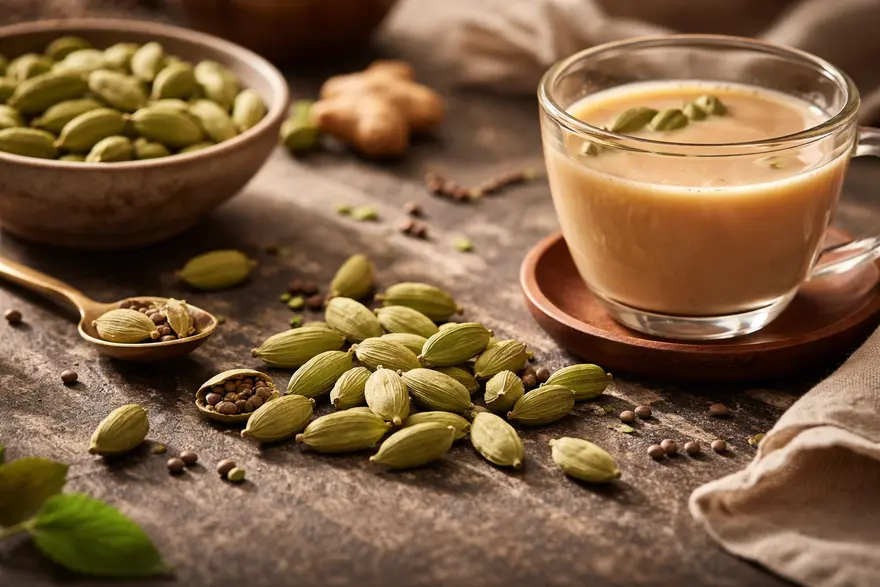
એલચી (કાર્ડમમ)ના 10 ફાયદા અને તેનો ઉપયોગ કરવાની શ્રેષ્ઠ રીતો
એલચી, જેને કાર્ડમમ તરીકે પણ ઓળખવામાં આવે છે, ભારતીય રસોડામાં સૌથી વધુ ગમતા મસાલાઓમાંથી એક છે. તમે તેને તેની ગરમ સુગંધ અને થોડા મીઠા સ્વાદ માટે ચા, ખીર, કરી અથવા મીઠાઈઓમાં ઉમેરી શકો છો. સ્વાદ ઉપરાંત, એલચીનો ઉપયોગ પેઢીઓથી રોજિંદી સુખાકારીની રૂટિનમાં પણ કરવામાં આવે છે. આધુનિક સંશોધન સૂચવે છે કે એલચી પાચનને ટેકો આપી શકે છે, શ્વાસને તાજું કરી શકે છે અને હૃદય તથા મેટાબોલિક આરોગ્યના કેટલાક પાસાઓમાં મદદ કરી શકે છે. તે જ સમયે, અપેક્ષાઓ વાસ્તવિક રાખવી શ્રેષ્ઠ છે. એલચી એક મદદરૂપ મસાલો છે, ઈલાજ નથી. શ્રેષ્ઠ પરિણામો ત્યારે મળે છે જ્યારે તમે તેનો ઉપયોગ સંતુલિત આહાર અને સ્વસ્થ જીવનશૈલીના ભાગરૂપે કરો છો. એલચી (કાર્ડમમ) શું છે? એલચી આદુ કુળના છોડની ફળીની અંદરના બીજમાંથી બનેલો મસાલો છે. લીલી એલચી એ એવી જાત છે જેનો મોટાભાગના લોકો રોજિંદા રસોઈમાં ઉપયોગ કરે છે, જ્યારે કાળી એલચીનો સ્વાદ વધુ સ્મોકી હોય છે અને તેનો ઉપયોગ ઘણીવાર મસાલેદાર વાનગીઓમાં થાય છે. આ લેખમાં, મુખ્ય ધ્યાન લીલી એલચી પર છે. તે તેના કુદરતી એસેન્શિયલ ઓઇલ્સ અને વનસ્પતિ સંયોજનોને કારણે સુગંધથી સમૃદ્ધ છે. તેથી જ તેની નાની માત્રા પણ તમારા ખોરાક અને પીણાંમાં ઘણો સ્વાદ ઉમેરી શકે છે. એલચી (કાર્ડમમ)નું પોષક મૂલ્ય એલચી સામાન્ય રીતે ઓછી માત્રામાં ખવાય છે, તેથી તમે એક સમયે તેના પોષક તત્ત્વો મોટી માત્રામાં લેતા નથી. તેમ છતાં, તેમાં ફાઇબર, ખનિજો અને એન્ટીઓક્સિડન્ટ વનસ્પતિ સંયોજનો હોય છે, જે મસાલા તરીકે તેના મૂલ્યમાં યોગદાન આપે છે. કાર્ડમમનું પોષક વિભાજન (100g દીઠ) પોષક તત્ત્વ 100g દીઠ અંદાજિત માત્રા ઊર્જા 311 kcal કાર્બોહાઇડ્રેટ્સ 68.47 g ફાઇબર 28 g પ્રોટીન 10.76 g ચરબી 6.70 g કેલ્શિયમ 383 mg આયર્ન 13.97 mg મેગ્નેશિયમ 229 mg પોટેશિયમ 1119 mg વિટામિન C 21 mg કાર્ડમમમાં રહેલા મુખ્ય સક્રિય સંયોજનો 1,8-cineole Alpha-terpinyl acetate Linalool અન્ય વોલેટાઇલ ઓઇલ્સ અને એન્ટીઓક્સિડન્ટ સંયોજનો એલચી (કાર્ડમમ)ના 10 ફાયદા 1. પાચનને ટેકો આપી શકે છે ભોજન પછી એલચીનો વ્યાપક ઉપયોગ થાય છે કારણ કે તે પેટ ફૂલવું, ગેસ અને ભારેપણાની લાગણી ઘટાડવામાં મદદ કરી શકે છે. પરંપરાગત ઉપયોગ અને શરૂઆતનું સંશોધન સૂચવે છે કે તે પાચન આરામને ટેકો આપી શકે છે અને પાચનને ઉત્તેજિત કરી શકે છે. આ એક કારણ છે કે ઘણા લોકો ભોજન પછી એલચીની ફળી ચાવવી અથવા તેને ગરમ પીણાંમાં ઉમેરવી પસંદ કરે છે. 2. શ્વાસને તાજું કરી શકે છે અને મૌખિક આરોગ્યને ટેકો આપી શકે છે એલચીનો ઉપયોગ ઘણીવાર કુદરતી માઉથ ફ્રેશનર તરીકે થાય છે. તેના સુગંધિત તેલ ભોજન પછી તમારા મોંને વધુ સ્વચ્છ અને તાજું અનુભવવામાં મદદ કરી શકે છે. કેટલાક પ્રયોગશાળા અને સમીક્ષા આધારિત પુરાવા પણ સૂચવે છે કે કાર્ડમમના સંયોજનો કેટલાક મૌખિક બેક્ટેરિયા સામે કાર્ય કરી શકે છે. આ એલચીને તમારી મૌખિક સંભાળની રૂટિનમાં એક સરળ અને આનંદદાયક ઉમેરો બનાવે છે, જોકે તે બ્રશિંગ અને ફ્લોસિંગનો વિકલ્પ નથી. 3. સ્વસ્થ બ્લડ પ્રેશરને ટેકો આપવા મદદ કરી શકે છે કાર્ડમમમાં એન્ટીઓક્સિડન્ટ સંયોજનો હોય છે, અને એક નાના ક્લિનિકલ અભ્યાસમાં જોવા મળ્યું કે કાર્ડમમ પાઉડર નવા નિદાન થયેલા ઊંચા બ્લડ પ્રેશર ધરાવતા પુખ્તોમાં બ્લડ પ્રેશર ઘટાડવામાં મદદ કરી શકે છે. આનો અર્થ એ નથી કે એલચી તમારી દવાઓનો વિકલ્પ બનવી જોઈએ. પરંતુ તેને તમારા આહારમાં ઉમેરવાથી હૃદયને અનુકૂળ ખાવાની આદતોને ટેકો મળી શકે છે. 4. હૃદયના આરોગ્યને ટેકો આપી શકે છે કારણ કે કાર્ડમમ બ્લડ પ્રેશર અને ઓક્સિડેટિવ તણાવમાં મદદ કરી શકે છે, તે સમગ્ર કાર્ડિયોવેસ્ક્યુલર આરોગ્યને પણ ટેકો આપી શકે છે. આ ફાયદો મોટા ચિત્રમાં વધુ સારી રીતે સમજાય છે. જો તમે સંતુલિત આહાર લો છો, સક્રિય રહો છો, સારી ઊંઘ લો છો અને તણાવ સંભાળો છો, તો એલચી તે રૂટિનનો નાનો સહાયક ભાગ બની શકે છે. 5. સોજો ઘટાડવામાં મદદ કરી શકે છે કાર્ડમમ એન્ટીઓક્સિડન્ટ વનસ્પતિ સંયોજનોથી સમૃદ્ધ છે. આ સંયોજનો તમારા કોષોને ઓક્સિડેટિવ તણાવથી બચાવવામાં મદદ કરી શકે છે અને સોજા પ્રત્યે શરીરની પ્રતિક્રિયાને ટેકો આપી શકે છે. માનવીય પુરાવા હજુ મર્યાદિત છે, પરંતુ કેટલીક સમીક્ષાઓ અને ટ્રાયલ્સ સૂચવે છે કે કાર્ડમમ સોજાના કેટલાક માર્કર્સમાં સુધારો કરી શકે છે. 6. શ્વસન આરામને ટેકો આપી શકે છે ગળામાં અસ્વસ્થતા અથવા છાતીમાં ભારેપણું લાગે ત્યારે એલચી સામાન્ય રીતે ગરમ ચા અને ઘરેલુ પીણાંમાં ઉમેરવામાં આવે છે. પરંપરાગત ઉપયોગ સૂચવે છે કે તે હળવી ઉધરસ અથવા કફ દરમિયાન તમને વધુ આરામદાયક અનુભવવામાં મદદ કરી શકે છે. કેટલાક સંશોધન સંભવિત વાયુમાર્ગ-શિથિલતા અને એન્ટીમાઇક્રોબિયલ અસરો તરફ પણ ઈશારો કરે છે, જોકે વધુ મજબૂત માનવીય અભ્યાસોની હજુ જરૂર છે. 7. મેટાબોલિક આરોગ્યમાં મદદ કરી શકે છે કેટલાક નાના અભ્યાસો સૂચવે છે કે કાર્ડમમ બ્લડ શુગર નિયંત્રણ, ટ્રાઇગ્લિસરાઇડ્સ અને ઇન્સ્યુલિન રેઝિસ્ટન્સ સાથે જોડાયેલા કેટલાક માર્કર્સ સુધારવામાં મદદ કરી શકે છે. તેમ છતાં, પુરાવા એટલા મજબૂત નથી કે તેને ડાયાબિટીસના ઈલાજ તરીકે ગણવામાં આવે. તમારે તેને એક મદદરૂપ મસાલા તરીકે વિચારવો જોઈએ, જે સ્વસ્થ ખાવાની પદ્ધતિમાં ફિટ થઈ શકે છે, તબીબી સંભાળના વિકલ્પ તરીકે નહીં. 8. પ્રવાહી સંતુલનને ટેકો આપી શકે છે કેટલાક અભ્યાસોમાં કાર્ડમમને હળવી ડાય્યુરેટિક અસર સાથે જોડવામાં આવ્યું છે. તેનો અર્થ એ છે કે તે તમારા શરીરને પ્રવાહી સંતુલન વધુ અસરકારક રીતે સંભાળવામાં મદદ કરી શકે છે. આ એક કારણ છે કે લોકો ક્યારેક તેને હળવાશ અથવા પેટ ઓછું ફૂલેલું લાગવા સાથે જોડે છે. તેને ડિટોક્સ ઈલાજ તરીકે નહીં, પરંતુ સહાયક તરીકે જોવું વધુ યોગ્ય છે. 9. એન્ટીઓક્સિડન્ટ રક્ષણ આપી શકે છે એલચીની સૌથી સ્પષ્ટ શક્તિઓમાંથી એક તેની એન્ટીઓક્સિડન્ટ સામગ્રી છે. એન્ટીઓક્સિડન્ટ્સ તમારા કોષોને મુક્ત રેડિકલ્સથી થતા નુકસાનથી બચાવવામાં મદદ કરે છે. સમય જતાં, એન્ટીઓક્સિડન્ટથી સમૃદ્ધ ખોરાક અને મસાલા સમગ્ર સુખાકારીને ટેકો આપી શકે છે અને તમને વધુ રક્ષણાત્મક દૈનિક આહાર બનાવવામાં મદદ કરી શકે છે. 10. રોજિંદી સુખાકારીની લાગણીને પ્રોત્સાહન આપી શકે છે ઉપયોગી થવા માટે એલચીનો પ્રભાવ નાટકીય હોવો જરૂરી નથી. તેની સુગંધ, સ્વાદ અને ગરમાશ સરળ ખોરાકને વધુ સંતોષદાયક બનાવી શકે છે. આ મહત્વનું છે કારણ કે જ્યારે સ્વસ્થ ખોરાક વધુ સ્વાદિષ્ટ લાગે છે, ત્યારે તમે તેને નિયમિત માણવાની શક્યતા વધુ હોય છે. આ કાર્ડમમના સૌથી સરળ અને વ્યવહારુ ફાયદાઓમાંથી એક છે. પુરુષો માટે એલચીના ફાયદા કાર્ડમમની એવી કોઈ સાબિત અસર નથી જે માત્ર પુરુષો માટે જ હોય, પરંતુ એલચી થોડા વ્યવહારુ રીતે હજુ પણ ઉપયોગી થઈ શકે છે. તે ભારે અથવા સમૃદ્ધ ભોજન પછી પાચનને ટેકો આપી શકે છે. તે ચા, કોફી અથવા ભોજન પછી કુદરતી રીતે શ્વાસને તાજું કરવામાં મદદ કરી શકે છે. તે હૃદયને અનુકૂળ અને મેટાબોલિક રીતે અનુકૂળ ખાવાની આદતોને ટેકો આપી શકે છે. તે પીણાં અથવા મીઠાઈઓમાં વધારાની ખાંડની જરૂરિયાત વિના સ્વાદ ઉમેરી શકે છે. મીઠા સ્વાદવર્ધકોની જગ્યાએ ઉપયોગમાં લેવાય ત્યારે તે વજનપ્રત્યે જાગૃત રૂટિનમાં ફિટ થઈ શકે છે. જો તમે પુરુષ આરોગ્ય માટે એલચીના ફાયદા શોધી રહ્યા છો, તો સૌથી સલામત જવાબ એ છે કે એલચી સામાન્ય સુખાકારીને ટેકો આપી શકે છે, પરંતુ તેની કોઈ અનોખી પુરુષ-વિશિષ્ટ અસર હોય તેવા મજબૂત પુરાવા નથી. એલચીના પાણીના ફાયદા એલચીનું પાણી એટલે હળવેથી કચડેલી એલચીની ફળીઓથી ઇન્ફ્યુઝ કરેલું પાણી. ઘણા લોકો તેની હળવી સુગંધ અને શાંતિકારક સ્વાદ માટે તેને પીવે છે. એલચીના પાણીના મુખ્ય ફાયદા હાઇડ્રેશન, પાચન આરામ અને ભોજન પછી હળવી તાજગીની લાગણી સાથે જોડાયેલા છે. જો તમને સ્વાદવાળું પાણી ગમે છે, તો તે ખાંડવાળા પીણાં ઘટાડવામાં પણ મદદ કરી શકે છે. આ બાબતને વાસ્તવિક રીતે સમજવી મહત્વપૂર્ણ છે. એલચીના પાણીના ફાયદા સ્વસ્થ રૂટિનનો ઉપયોગી ભાગ બની શકે છે, પરંતુ તે કોઈ ચમત્કારિક પીણું નથી. એલચીનું પાણી કેવી રીતે બનાવવું 1 થી 2 લીલી એલચીની ફળીઓને હળવેથી કચડો. તેને 1 થી 2 કપ ગરમ અથવા ઉકાળેલા પાણીમાં ઉમેરો. પાણીને 5 થી 10 મિનિટ સુધી પલળવા દો. ગાળી લો અને તેને ગરમ અથવા રૂમ તાપમાને પીવો. એલચી (કાર્ડમમ)નો ઉપયોગ કરવાની શ્રેષ્ઠ રીતો એલચીને તમારા દૈનિક ભોજનમાં સામેલ કરવી સરળ છે. તેને માણવા માટે તમને સપ્લિમેન્ટ્સની જરૂર નથી. હકીકતમાં, મોટાભાગના લોકો માટે રસોઈમાં તેનો ઉપયોગ સૌથી સરળ અને સલામત અભિગમ છે. રસોઈમાં એલચીનો ઉપયોગ કેવી રીતે કરવો ચા અથવા દૂધમાં કચડેલી ફળીઓ ઉમેરો. ખીર, ફિરની, કસ્ટર્ડ અને બેક કરેલી મીઠાઈઓમાં પીસેલી એલચી વાપરો. પોરિજ અથવા ઓટ્સમાં ચપટી ઉમેરો. સુગંધ અને ગરમાશ માટે તેને સ્મૂધીમાં ઉમેરો. તેનો ઉપયોગ પુલાવ, બિરયાની અને સમૃદ્ધ ગ્રેવીમાં કરો. ફળોના બાઉલ અથવા દહીંમાં થોડી છાંટો. આરોગ્ય ફાયદા માટે એલચીવાળી ચા કેવી રીતે બનાવવી 1 થી 2 કપ પાણી ઉકાળો. 2 થી 3 એલચીની ફળીઓને હળવેથી કચડો. તેને પાણીમાં ઉમેરો અને થોડા મિનિટ સુધી ધીમે ઉકાળો. જો તમને સામાન્ય ચા જોઈએ હોય, તો ચાની પત્તી ઉમેરો. ગાળી લો અને ગરમ પીવો. અરોમાથેરાપી માટે કાર્ડમમ ઓઇલ કાર્ડમમ ઓઇલમાં ગરમ, મસાલેદાર સુગંધ હોય છે. કેટલાક લોકો આરામદાયક વાતાવરણ માટે તેનો ડિફ્યુઝરમાં ઉપયોગ કરે છે. તે તમારી જગ્યા વધુ શાંત અને તાજી લાગવામાં મદદ કરી શકે છે. લાયકાત ધરાવતા ક્લિનિશિયન સલાહ આપે નહીં ત્યાં સુધી એસેન્શિયલ ઓઇલનું સેવન ન કરો. ત્વચા પર લગાવતા પહેલાં તેને હંમેશાં યોગ્ય રીતે પાતળું કરો. વજન ઘટાડવા માટે મસાલા તરીકે એલચી એલચી ખાંડ ઉમેર્યા વિના ખોરાકમાં સ્વાદ ઉમેરી શકે છે. તે તમને સરળ ખોરાક વધુ માણવામાં મદદ કરી શકે છે. જ્યારે તમે તેને ચા, ઓટ્સ અથવા ફળોમાં વાપરો છો, ત્યારે તે વજનપ્રત્યે જાગૃત આહારમાં ફિટ થઈ શકે છે. મોટા પ્રમાણમાં વજન ઘટાડા માટે સીધા પુરાવા મર્યાદિત છે, તેથી માત્ર તેના પર આધાર રાખશો નહીં. એલચી કેળાના ફાયદા કેળા અને એલચીનું સરળ સંયોજન સ્મૂધી, પોરિજ અથવા ઝડપી મૅશ કરેલા નાસ્તામાં સારી રીતે કામ કરી શકે છે. કેળું તમને કાર્બોહાઇડ્રેટ્સ, પોટેશિયમ અને ફાઇબર આપે છે, જ્યારે એલચી સ્વાદ અને હળવી પાચન લાગણી ઉમેરે છે. એલચી કેળાના સૌથી સરળ ફાયદા એ છે કે તે વધુ ઉમેરેલી ખાંડની જરૂરિયાત વિના પૌષ્ટિક નાસ્તાનો સ્વાદ વધુ સારો બનાવે છે. જો તમને ઘરે બનાવેલી સ્મૂધી ગમે છે, તો એલચી કેળાના ફાયદામાં વધુ સારો સ્વાદ, વધુ સંતોષ અને વધુ સંતુલિત નાસ્તાનો વિકલ્પ સામેલ થઈ શકે છે. સંભવિત આડઅસરો અને સાવચેતીઓ સામાન્ય ખોરાકની માત્રામાં એલચી સામાન્ય રીતે સલામત છે. તેમ છતાં, થોડા મુદ્દા ધ્યાનમાં રાખવા યોગ્ય છે. વધુ પડતી માત્રા કેટલાક લોકોમાં પેટની અસ્વસ્થતા કરી શકે છે. જો તમે ગર્ભવતી હો, તો સંકેન્દ્રિત સપ્લિમેન્ટ્સ કરતાં રસોઈમાં ઉપયોગ સામાન્ય રીતે પસંદ કરવો જોઈએ. જો તમને કોઈ તબીબી પરિસ્થિતિ હોય, તો કાર્ડમમ એક્સ્ટ્રેક્ટ્સ અથવા કેપ્સ્યુલ્સ લેતા પહેલાં તમારા ડોક્ટર સાથે વાત કરો. જો તમે નિયમિત દવાઓ લો છો, ખાસ કરીને બ્લડ પ્રેશર અથવા બ્લડ શુગર માટે, તો પોતે જ સપ્લિમેન્ટ્સ લેવાનું ટાળો. એસેન્શિયલ ઓઇલ્સનું અનૌપચારિક રીતે સેવન ન કરવું જોઈએ. દિવસમાં કેટલી એલચી લઈ શકાય? ખોરાકમાં ઉપયોગ થતી એલચી માટે કોઈ પ્રમાણભૂત દૈનિક જરૂરિયાત નથી. સામાન્ય રસોઈની માત્રામાં, તે સામાન્ય રીતે સારી રીતે સહન થાય છે. મોટાભાગના લોકો માટે, ચા અથવા ભોજનમાં 1 થી 2 ફળી, અથવા પાઉડરની નાની ચપટી, વ્યવહારુ માત્રા છે. સ્વાદ અથવા સંભવિત આરોગ્ય મૂલ્ય માણવા માટે તમને મોટી માત્રાની જરૂર નથી. જો તમે નિયમિતપણે એલચીના પાણીના ફાયદા અજમાવવા માંગતા હો, તો તેને ખૂબ સંકેન્દ્રિત બનાવવાને બદલે હળવી અને મધ્યમ તૈયારી રાખો. નિષ્કર્ષ એલચી સુગંધિત રસોડાના મસાલાથી ઘણું વધુ છે. તે પાચનને ટેકો આપી શકે છે, શ્વાસને તાજું કરી શકે છે, એન્ટીઓક્સિડન્ટ સેવનમાં યોગદાન આપી શકે છે અને હૃદય તથા મેટાબોલિક આરોગ્ય માટે હળવો ટેકો આપી શકે છે. પુરાવા આશાસ્પદ છે, પરંતુ કાર્ડમમને સારવાર કરતાં સહાયક ખોરાક ઘટક તરીકે વિચારવું હજુ પણ શ્રેષ્ઠ છે. તેનો ઉપયોગ કરવાની સૌથી સરળ રીત દૈનિક રસોઈ, ચા અથવા હળવા ઇન્ફ્યુઝ્ડ પાણીમાં છે. ગરમ પીણાં, ફળોના બાઉલ અથવા સ્વસ્થ નાસ્તામાં એલચી ઉમેરવા જેવી સરળ આદતો પૌષ્ટિક ખાવાનું વધુ આનંદદાયક બનાવી શકે છે. જો તમે પોષણ, નિવારણ, લક્ષણો અને પરીક્ષણ અંગે વધુ વિશ્વસનીય આરોગ્ય માર્ગદર્શન ઇચ્છતા હો, તો તમે મેટ્રોપોલિસ હેલ્થકેરના વધુ નિષ્ણાત-આધારિત લેખો જોઈ શકો છો. અદ્યતન નિદાન ટેકનોલોજી, નિષ્ણાત પેથોલોજિસ્ટ્સ, 4,000+ ટેસ્ટ અને અનુકૂળ હોમ સેમ્પલ કલેક્શન સાથે, મેટ્રોપોલિસ હેલ્થકેર તમને વધુ આત્મવિશ્વાસ સાથે માહિતગાર આરોગ્ય નિર્ણયો લેવામાં મદદ કરે છે. વારંવાર પૂછાતા પ્રશ્નો કાર્ડમમના આરોગ્ય ફાયદા શું છે? કાર્ડમમ પાચન, મૌખિક તાજગી, એન્ટીઓક્સિડન્ટ સેવન અને બ્લડ પ્રેશર તથા મેટાબોલિક આરોગ્યના કેટલાક પાસાઓને ટેકો આપી શકે છે. સૌથી મજબૂત પુરાવા હજુ મર્યાદિત છે, તેથી તેને એકલા ઈલાજ તરીકે નહીં પરંતુ સ્વસ્થ આહારના ભાગરૂપે વાપરવું શ્રેષ્ઠ છે. પુરુષો માટે એલચીના આરોગ્ય ફાયદા શું છે? એલચી પુરુષોમાં પાચન, શ્વાસની તાજગી અને હૃદયને અનુકૂળ ખાવાની આદતોને ટેકો આપી શકે છે, જેમ તે સ્ત્રીઓમાં પણ આપી શકે છે. માત્ર પુરુષો માટે અનોખી અસર અંગે મજબૂત પુરાવા નથી. શું કાર્ડમમ વજન ઘટાડવામાં મદદ કરી શકે છે? કાર્ડમમ વધારાની ખાંડ વિના સ્વસ્થ ખોરાક અને પીણાંને વધુ સ્વાદિષ્ટ બનાવીને પરોક્ષ રીતે મદદ કરી શકે છે. જોકે, કાર્ડમમ એકલું અર્થપૂર્ણ વજન ઘટાડે છે એવું કહેવા માટે પૂરતા પુરાવા નથી. શું કાર્ડમમ તમારા હૃદય માટે સારું છે? તે હોઈ શકે છે. કેટલાક નાના અભ્યાસો અને મેટા-એનાલિસિસ સૂચવે છે કે કાર્ડમમ બ્લડ પ્રેશર નિયંત્રણ અને સોજાના માર્કર્સને ટેકો આપી શકે છે. તેમ છતાં, તે તબીબી સંભાળ અને સ્વસ્થ જીવનશૈલીની આદતોનો પૂરક હોવો જોઈએ, વિકલ્પ નહીં. ત્વચા ફાયદા માટે કાર્ડમમનો ઉપયોગ કેવી રીતે કરવો? કાર્ડમમને ત્વચા સારવાર તરીકે ભલામણ કરવા માટે પૂરતા મજબૂત માનવીય પુરાવા નથી. એલચીને ખોરાકના મસાલા તરીકે વાપરવું અને ચોક્કસ ત્વચા ચિંતાઓ માટે યોગ્ય સ્કિનકેર પ્લાન અનુસરવું વધુ સલામત છે. એલચીના પાણીના ફાયદા શું છે? એલચીના પાણીના ફાયદામાં હળવો પાચન ટેકો, વધુ સારી હાઇડ્રેશન સપોર્ટ અને ખાંડવાળા પીણાંને હળવા વિકલ્પથી બદલવાની સરળ રીત સામેલ હોઈ શકે છે. તમારી અપેક્ષાઓ મધ્યમ રાખો અને તેનો ઉપયોગ સમગ્ર સ્વસ્થ રૂટિનના ભાગરૂપે કરો. શું દરરોજ એલચી લઈ શકાય? હા, મોટાભાગના લોકો રોજ નાની ખોરાકની માત્રામાં એલચી લઈ શકે છે. ચા, ભોજન અથવા મીઠાઈઓમાં મધ્યમ ઉપયોગ સામાન્ય રીતે સલામત માનવામાં આવે છે. શું ગર્ભાવસ્થા દરમિયાન એલચી સલામત છે? ગર્ભાવસ્થા દરમિયાન સામાન્ય રસોઈમાં વપરાતી એલચી સામાન્ય રીતે સંકેન્દ્રિત સપ્લિમેન્ટ્સ કરતાં વધુ પસંદનીય છે. જો તમે ગર્ભવતી હો અને કાર્ડમમ એક્સ્ટ્રેક્ટ્સ, ઓઇલ્સ અથવા કેપ્સ્યુલ્સ વાપરવા માંગતા હો, તો પહેલાં તમારા ડોક્ટર સાથે વાત કરો.
त्रिफळा चूर्ण: फायदे, उपयोग, मात्रा आणि दुष्परिणाम समजून घ्या
त्रिफळा चूर्ण म्हणजे काय? त्रिफळा चूर्ण, ज्याचा संस्कृतमध्ये अर्थ “तीन फळे” असा होतो, हे आवळा, हिरडा आणि बेहडा यांचे पारंपरिक आयुर्वेदिक मिश्रण आहे. या प्रत्येक घटकात वेगवेगळे उपचारात्मक गुणधर्म असल्याचे मानले जाते. आवळा व्हिटॅमिन C आणि अँटिऑक्सिडंट्समुळे रोगप्रतिकारशक्ती वाढवतो, हिरडा पचनास मदत करतो आणि बेहडा कोलेस्टेरॉल व श्वसन आरोग्यास आधार देतो. एकत्रितपणे, त्यांचा पारंपरिक वापर पचन, विषहरण आणि एकूण स्फूर्तीला आधार देण्यासाठी केला जातो, त्यामुळे त्रिफळा दीर्घायुष्य आणि स्फूर्तीसाठी एक पारंपरिक आयुर्वेदिक रसायन मानले जाते. विषहरणासाठी त्रिफळा चूर्ण कसे कार्य करते? नॅशनल इन्स्टिट्यूट्स ऑफ हेल्थ (एनआयएच) डेटाबेसमध्ये संदर्भित अभ्यासांनुसार, त्रिफळा पचन आणि यकृताच्या कार्यास आधार देणारे सौम्य विषहरणकारी आणि अँटिऑक्सिडंट घटक म्हणून कार्य करू शकते. हे पचनसंस्थेवर लक्ष केंद्रित करते, ज्याला आयुर्वेदात अनेकदा एकूण आरोग्याचे मूळ म्हटले जाते, आणि आतड्यांतील नैसर्गिक सूक्ष्मजीव संतुलन बिघडविल्याशिवाय साचलेले विषद्रव्य (आम) काढून टाकते. हे कसे कार्य करते ते येथे दिले आहे: आवळा यकृताच्या कार्यास आधार देतो आणि अँटिऑक्सिडंट एन्झाइमची क्रिया वाढवतो, ज्यामुळे नैसर्गिक चयापचय प्रक्रियांना मदत होते. हिरडा निरोगी मलविसर्जनाला चालना देतो आणि मोठे आतडे स्वच्छ करतो, ज्यामुळे विषद्रव्ये बाहेर टाकण्यास मदत होते. बेहडा लिपिड चयापचय नियंत्रित करतो आणि जड धातू व मुक्त रॅडिकल्स बाहेर टाकण्यास आधार देतो. नियमित, मध्यम वापरामुळे शरीरावर भार टाकणारे ऑक्सिडेटिव्ह ताणदायक घटक काढून टाकून त्वचेचे आरोग्य, ऊर्जा आणि रक्ताभिसरण संतुलनाला आधार मिळतो, असे आयुर्वेदात मानले जाते. वात, पित्त आणि कफ या तीन दोषांमध्ये संतुलन राखण्याचा हा आयुर्वेदिक मार्ग आहे. त्रिफळा चूर्णाचे पौष्टिक मूल्य फळ वैज्ञानिक नाव मुख्य पोषक घटक मुख्य फायदे आवळा एम्ब्लिका ऑफिसिनॅलिस व्हिटॅमिन C, पॉलीफेनॉल्स, फ्लॅव्होनॉइड्स रोगप्रतिकारशक्ती मजबूत करते, यकृत आणि हृदयाच्या आरोग्यास आधार देते हिरडा टर्मिनालिया चेब्युला टॅनिन्स, अमिनो आम्ले, गॅलिक आम्ल पचन सुधारते, चयापचय वाढवते आणि ऊतींचे विषहरण करते बेहडा टर्मिनालिया बेलिरिका लिग्नॅन्स, एलॅजिक आम्ल, अँटिऑक्सिडंट्स कोलेस्टेरॉल व्यवस्थापनास मदत करते, श्वसन आरोग्य सुधारते पचन आरोग्यासाठी त्रिफळा पचनाचे आरोग्य हा त्रिफळाचा सर्वात ओळखला जाणारा फायदा आहे. हे खालील बाबींसाठी ओळखले जाते: निरोगी आतड्यांची हालचाल आणि नियमित मलविसर्जनास आधार देणे पोटफुगी, वायू आणि आम्लता कमी करणे भूक आणि पोषक घटकांचे शोषण सुधारणे मोठ्या आतड्याच्या नैसर्गिक विषहरणास मदत करणे आतड्यांतील सूक्ष्मजीव संतुलन पुन्हा प्रस्थापित करणे तुमच्या आरोग्यासाठी त्रिफळा चूर्णाचे 10 फायदे पचन आरोग्य सुधारते: त्रिफळा नियमित मलविसर्जनाला चालना देऊन आणि बद्धकोष्ठता, आम्लता व पोटफुगी कमी करून निरोगी पचनास आधार देते. हे आतड्यांचे कार्य मजबूत करते आणि पोषक घटकांचे प्रभावी शोषण होण्यास मदत करते. रोगप्रतिकारशक्ती वाढवते: आवळ्यातील अँटिऑक्सिडंट्स आणि व्हिटॅमिन C मुबलक असल्याने, त्रिफळा रोगप्रतिकारक प्रणाली मजबूत करते, शरीराला संसर्गांशी लढण्यास आणि नैसर्गिकरीत्या ऑक्सिडेटिव्ह ताण कमी करण्यास मदत करते. वजन व्यवस्थापनास मदत करते: चयापचय सुधारून आणि पचनसंस्थेचे विषहरण करून, त्रिफळा भूक नियंत्रित करण्यास मदत करते आणि हळूहळू, निरोगी वजन कमी करण्यास आधार देते. यकृताचे कार्य सुधारते: याच्या विषहरणकारी गुणधर्मांमुळे यकृतातील एन्झाइम्स उत्तेजित होतात, विषद्रव्ये बाहेर टाकण्याची प्रक्रिया सुधारते आणि चरबी व पित्ताच्या चयापचयास मदत होते. त्वचेला नैसर्गिक तेज देते: नियमित सेवनामुळे रक्त शुद्ध होते, शरीरातील विषद्रव्ये कमी होतात आणि मुरूम किंवा रंगद्रव्याचे डाग कमी होतात, त्यामुळे त्वचेला नैसर्गिक आणि निरोगी तेज मिळते. डोळ्यांचे आरोग्य राखते त्रिफळातील आवळ्याचा पुनरुज्जीवित करणारा प्रभाव दृष्टीच्या आरोग्यास आधार देतो, डोळ्यांचा ताण कमी करतो आणि वयानुसार होणारे क्षीण होणे टाळण्यास मदत करतो. निरोगी रक्तशर्करेस आधार देते: प्राथमिक अभ्यासांनुसार, संतुलित आहारासोबत वापरल्यास त्रिफळा ग्लुकोज चयापचय सुधारण्यास मदत करू शकते आणि संतुलित आहारासोबत घेतल्यास कालांतराने रक्तशर्करेची पातळी स्थिर ठेवण्यास मदत होऊ शकते. कोलेस्टेरॉल व्यवस्थापित करते: एकूण जीवनशैली योजनेचा भाग म्हणून बेहडा निरोगी कोलेस्टेरॉल पातळीला आधार देण्यास मदत करू शकतो, ज्यामुळे अधिक निरोगी लिपिड प्रोफाइल आणि हृदय व रक्तवाहिन्यांचे कार्य प्रोत्साहित होते. तोंडाच्या आरोग्यास आधार देते: याच्या सूक्ष्मजीवरोधी गुणधर्मांमुळे, त्रिफळा तोंडातील अल्सर टाळण्यास, प्लाक साचणे कमी करण्यास आणि ताज्या श्वासासह निरोगी हिरड्यांना प्रोत्साहन देण्यास मदत करते. केस आणि टाळू मजबूत करते: पोषक घटकांनी समृद्ध हे मिश्रण केसांच्या मुळांना पोषण देते, अकाली केस पांढरे होणे टाळते आणि अधिक मजबूत, चमकदार केसांसाठी एकूण टाळूचे आरोग्य सुधारते. जास्तीत जास्त फायदे मिळवण्यासाठी त्रिफळा चूर्ण कसे वापरावे वैयक्तिक पसंतीनुसार त्रिफळा चूर्ण पावडर, कॅप्सूल किंवा गोळीच्या स्वरूपात घेतले जाऊ शकते. पारंपरिकपणे, ते कोमट पाणी किंवा मधासोबत मिसळून सकाळी लवकर रिकाम्या पोटी किंवा झोपण्यापूर्वी घेतले जाते. पचनास आधार देण्यासाठी: 1 टीस्पून कोमट पाण्यात मिसळा, 10 मिनिटे तसेच ठेवा आणि झोपण्यापूर्वी प्या. पचन आरोग्यासाठी: नाश्त्यापूर्वी कोमट पाण्यासोबत कमी प्रमाणात घ्या. त्वचा आणि रोगप्रतिकारशक्तीसाठी: शोषण आणि पौष्टिक समन्वय वाढवण्यासाठी मध किंवा तुपासोबत घ्या. त्रिफळा चूर्णाच्या मात्रेबाबत शिफारसी प्रौढ: 3–5 grams (about 1 teaspoon) दिवसातून एकदा किंवा दोनदा कोमट पाण्यासोबत. बद्धकोष्ठतेसाठी: झोपण्यापूर्वी कोमट पाणी किंवा दुधासोबत घ्या. वजन व्यवस्थापनासाठी: सकाळी लवकर जेवणापूर्वी घ्या. मुले: अर्धी मात्रा (फक्त देखरेखीखाली). कॅप्सूल स्वरूपात: साधारणपणे प्रत्येक मात्रेत 500–1000 mg. त्रिफळा चूर्ण इतर पूरक घटकांसोबत वापरता येते का? व्यावसायिक मार्गदर्शनाखाली त्रिफळा अश्वगंधा किंवा हळद यांसारख्या इतर आयुर्वेदिक औषधी वनस्पतींसोबत घेतले जाऊ शकते, ज्यामुळे अँटिऑक्सिडंट आणि पुनरुज्जीवित करणारे गुणधर्म वाढू शकतात. मात्र, डॉक्टरांच्या शिफारशीशिवाय ते इतर रेचक किंवा विषहरण करणाऱ्या औषधी वनस्पतींसोबत घेणे टाळावे, कारण त्यामुळे आतड्यांची हालचाल वाढू शकते किंवा औषधांचे शोषण बदलू शकते. जर तुम्ही मधुमेह, रक्तदाब किंवा थायरॉईडसाठी औषधे घेत असाल, तर त्रिफळा समाविष्ट करण्यापूर्वी तुमची दिनचर्या तुमच्या आरोग्यसेवा तज्ज्ञांशी चर्चा करा, कारण ते चयापचय दरांवर सूक्ष्म परिणाम करू शकते. त्रिफळा चूर्ण: दुष्परिणाम आणि काळजी शिफारस केलेल्या मात्रेत घेतल्यास त्रिफळा बहुतेक प्रौढांसाठी सुरक्षित मानले जाते. मात्र, अतिरेकी किंवा देखरेखीशिवाय सेवन केल्यास खालील त्रास होऊ शकतात: पोटात अस्वस्थता किंवा मुरडा सैल शौच किंवा निर्जलीकरण काही औषधांचे शोषण कमी होणे काळजी: गर्भवती किंवा स्तनपान करणाऱ्या महिलांनी त्रिफळा वापरण्यापूर्वी पात्र आरोग्यसेवा तज्ज्ञांचा सल्ला घ्यावा, कारण त्याचे स्वच्छ करणारे गुणधर्म योग्य असतीलच असे नाही. दीर्घकालीन अतिसार, आयबीएस किंवा कमी रक्तदाब असलेल्या व्यक्तींनी ते वापरण्यापूर्वी डॉक्टरांचा सल्ला घ्यावा. अचानक विषहरण प्रतिक्रिया टाळण्यासाठी कमी मात्रेपासून सुरुवात करून हळूहळू मात्रा वाढवणे उत्तम. त्रिफळा चूर्ण दीर्घकाळ वापरण्यास सुरक्षित आहे का? शिफारस केलेल्या मात्रेत घेतल्यास, त्रिफळा सर्वसाधारणपणे दीर्घकालीन वापरासाठी योग्य मानले जाते. रासायनिक रेचकांप्रमाणे, ते सवय लावणारे नाही आणि कालांतराने आतड्यांच्या आतील अस्तराला पोषण देते. मात्र, शरीराच्या नैसर्गिक प्रणालींना पुन्हा संतुलित होण्यासाठी काही महिन्यांच्या सतत वापरानंतर थोडा विराम (सुमारे 1–2 weeks) घेण्याची आयुर्वेदिक तज्ज्ञ शिफारस करतात. त्रिफळा वजन कमी करण्यात मदत करू शकते का? आहार आणि व्यायामासोबत घेतल्यास त्रिफळा निरोगी चयापचय आणि लिपिड संतुलनास आधार देऊ शकते. याचा सौम्य विषहरणकारी परिणाम पचनसंस्था स्वच्छ करतो, ज्यामुळे पोषक घटकांचे शोषण होण्यास मदत होते. तृप्ती सुधारते आणि रात्री उशिरा खाण्याची इच्छा नियंत्रित करण्यास मदत होते. संतुलित आहार आणि नियमित शारीरिक हालचालींसोबत घेतल्यास उत्तम परिणाम होतो. हे “त्वरित उपाय” नाही, तर टिकाऊ वजन व्यवस्थापन आणि चयापचय आरोग्यासाठी आधार देणारी औषधी वनस्पती आहे. त्रिफळा चूर्ण विरुद्ध इतर आयुर्वेदिक विषहरण उपाय संतुलित सूत्र: त्रिफळा तीन औषधी वनस्पती एकत्र करते: आवळा, हिरडा आणि बेहडा, जे वात, पित्त आणि कफ या तीनही दोषांचे संतुलन राखतात; तर एकाच औषधी वनस्पतीवर आधारित विषहरण उपाय फक्त एका पैलूवर लक्ष केंद्रित करतात. सौम्य पण प्रभावी: तीव्र रेचक स्वच्छीकरणांप्रमाणे अशक्तपणा किंवा निर्जलीकरण न घडवता हे पचनसंस्था हळूहळू स्वच्छ करते. पोषक घटकांनी समृद्ध विषहरण: त्रिफळा विषद्रव्ये बाहेर टाकताना ऊतींना पोषण देते आणि आवश्यक पोषक घटकांची भरपाई करते, त्यामुळे पोषक घटकांची हानी न होता विषहरण होते. संपूर्ण शरीरावर परिणाम: हे यकृत, आतडी आणि रक्तावर सर्वांगीणरीत्या कार्य करते, तर इतर विषहरण उपाय अनेकदा फक्त एका अवयवावर लक्ष केंद्रित करतात. नियमित वापरासाठी सुरक्षित: सवय न लावणारे आणि दीर्घकालीन आरोग्यासाठी योग्य असल्याने, हे आक्रमक किंवा अल्पकालीन आयुर्वेदिक स्वच्छीकरणांपेक्षा सुरक्षित ठरते. पुनरुज्जीवित करणारा प्रभाव: हे स्वच्छ करणारे आणि रसायन (पुनरुज्जीवक) अशा दोन्ही प्रकारे कार्य करते, साध्या विषहरणापलीकडे स्फूर्ती आणि दीर्घायुष्याला प्रोत्साहन देते. निष्कर्ष त्रिफळा चूर्ण हे पचन आरोग्य, चयापचय आणि एकूण संतुलनाला आधार देण्यासाठी पारंपरिकपणे वापरले जाणारे कालपरिक्षित आयुर्वेदिक सूत्र आहे, जे पचन, विषहरण, चयापचय आणि एकूण पुनरुज्जीवनास आधार देते. सकाळच्या दिनचर्येचा भाग म्हणून वापरले किंवा रात्रीच्या विषहरणासाठी घेतले तरी, याचे फायदे आतड्यांपासून त्वचेपर्यंत पोहोचतात आणि खरे अंतर्गत संतुलन दर्शवतात. मेट्रोपोलिस हेल्थकेअर निवडा — भारतातील विश्वासार्ह निदान ब्रँड, ज्यामध्ये 4,000+ चाचण्या, एनएबीएल आणि सीएपी-मान्यताप्राप्त प्रयोगशाळा आणि अचूकता व विश्वासार्हता सुनिश्चित करणारे तज्ज्ञ पॅथॉलॉजिस्ट आहेत. 10,000+ टचपॉइंट्सवर घरून नमुना संकलन, जलद रिपोर्ट टर्नअराउंड आणि वेबसाइट, कॉल, अॅप किंवा व्हॉट्सअॅपद्वारे सोपे बुकिंग यांमुळे सोयीचा अनुभव घ्या. नेहमी विचारले जाणारे प्रश्न त्रिफळा चूर्ण कशापासून बनते? त्रिफळा चूर्ण आवळा, हिरडा आणि बेहडा या तीन फळांपासून बनते; ती वाळवून, पूड करून समान प्रमाणात मिसळली जातात. हे 100% वनस्पती-आधारित असून नैसर्गिकरीत्या अँटिऑक्सिडंट्सने समृद्ध आहे. मी त्रिफळा चूर्ण किती वेळा घ्यावे? दिवसातून एकदा सामान्य आरोग्यासाठी दिवसातून दोनदा विशिष्ट विषहरण किंवा पचनाच्या उद्दिष्टांसाठी दीर्घकालीन वापरासाठी नेहमी व्यावसायिक देखरेखीखाली त्रिफळा चूर्ण बद्धकोष्ठतेसाठी मदत करू शकते का? होय. हे सौम्य नैसर्गिक रेचक म्हणून कार्य करते, मलविसर्जन सुधारते आणि कृत्रिम रेचकांप्रमाणे अवलंबित्व न निर्माण करता विषद्रव्ये बाहेर टाकते. त्रिफळा चूर्ण गर्भवती महिलांसाठी सुरक्षित आहे का? नाही. त्याच्या स्वच्छ करणाऱ्या गुणधर्मांमुळे, जे गर्भाशयाचे आकुंचन उत्तेजित करू शकतात, गर्भधारणेदरम्यान सामान्यतः याची शिफारस केली जात नाही. नेहमी प्रथम आरोग्यसेवा तज्ज्ञांचा सल्ला घ्या. त्रिफळा चूर्ण त्वचेचे आरोग्य सुधारू शकते का? नक्कीच. रक्त शुद्ध करून, पचन सुधारून आणि ऑक्सिडेटिव्ह ताण कमी करून, त्रिफळा अधिक स्वच्छ, उजळ आणि तरुण दिसणारी त्वचा मिळवण्यास मदत करते.
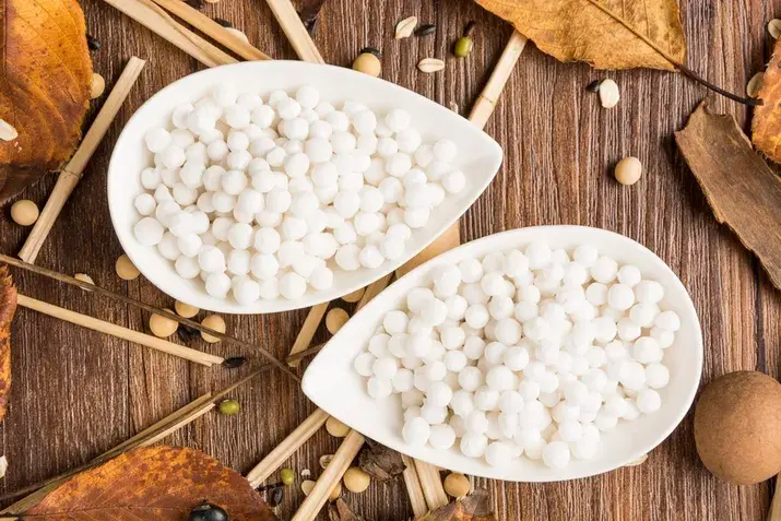
साबुदाण्याचे 10 आश्चर्यकारक फायदे, जे तुम्हाला कदाचित माहित नसतील
तुम्ही कधी उपवासादरम्यान किंवा प्रकृती सुधारताना साबुदाण्याकडे वळला असाल, तर तो फक्त झटपट ऊर्जा देण्यापेक्षा बरेच काही करू शकतो हे जाणून तुम्हाला आश्चर्य वाटू शकते. हा साधा घटक पोटासाठी सौम्य असतो आणि अनपेक्षित प्रकारे तुमच्या आरोग्याला आधार देऊ शकतो. तुम्ही पचन सांभाळत असाल, हलका पोषणयुक्त आहार शोधत असाल किंवा फक्त आरोग्यदायी पर्यायांचा विचार करत असाल, साबुदाण्याकडे बरेच काही देण्यासारखे आहे. या ब्लॉगमध्ये, तुम्हाला असे कमी परिचित फायदे कळतील, जे तुमच्या शरीरासाठी आणि आरोग्याच्या गरजांसाठी योग्य निवड करताना तुम्हाला अधिक आत्मविश्वास देऊ शकतात. साबुदाणा म्हणजे काय? साबुदाणा, ज्याला टॅपिओका पर्ल्स असेही म्हणतात, हा कसावा वनस्पतीच्या मुळापासून बनवलेला स्टार्चयुक्त अन्नपदार्थ आहे. हे छोटे, पांढरे दाणे शिजवल्यावर मऊ आणि अर्धपारदर्शक होतात, त्यामुळे ते पचायला सोपे आणि खायला आरामदायी असतात. उपवासाच्या काळात सामान्यतः वापरला जाणारा साबुदाणा पोटासाठी सौम्य आणि शरीराला ऊर्जा देणारा म्हणून ओळखला जातो. तो अनेक प्रकारच्या खिरी, नाश्त्याचे पदार्थ आणि चविष्ट पदार्थांमध्ये वापरला जातो. साधा दिसत असला, तरी साबुदाणा फक्त ऊर्जेचा स्रोत नाही—त्यात आरोग्याला आधार देणारे अनेक गुणधर्म आहेत, ज्याकडे अनेकदा दुर्लक्ष केले जाते. साबुदाण्याचे पौष्टिक मूल्य साबुदाणा मुख्यतः कार्बोहायड्रेट्सचा स्रोत असला, तरी संतुलित प्रमाणात सेवन केल्यास त्यात आरोग्यास आधार देणारे इतर काही पोषक घटक अल्प प्रमाणात असतात. साबुदाण्याचे पौष्टिक मूल्य समजून घेतल्याने तो तुमच्या आहारात समाविष्ट करण्याबाबत माहितीपूर्ण निर्णय घेता येतो. पोषक घटक प्रति 100g (न शिजवलेला साबुदाणा) ऊर्जा 358 kcal कार्बोहायड्रेट्स 88.7 g प्रथिने 0.2 g चरबी 0.2 g तंतुमय पदार्थ 0.9 g कॅल्शियम 20 mg लोह 1.58 mg पोटॅशियम 11 mg सोडियम 1 mg प्रथिने आणि चरबीचे प्रमाण कमी असले, तरी साबुदाणा झटपट ऊर्जा देतो, त्यामुळे प्रकृती सुधारताना आणि उपवासाच्या आहारात तो उपयुक्त ठरतो. दूध, शेंगदाणे किंवा भाज्या यांसारख्या घटकांसोबत घेतल्यास तो अधिक पौष्टिक होतो. साबुदाण्याचे 10 आरोग्य फायदे उपवासादरम्यान साबुदाण्याचा वापर तुम्हाला आधीच माहित असू शकतो, परंतु त्याबद्दल आणखी बरेच काही जाणून घेण्यासारखे आहे. तुमच्या आरोग्याला आणि एकूण स्वास्थ्याला आधार देऊ शकणारे दहा आश्चर्यकारक साबुदाण्याचे फायदे येथे दिले आहेत: 1. झटपट ऊर्जा वाढवतो साबुदाण्यात कार्बोहायड्रेट्स, विशेषतः स्टार्च भरपूर असतो, ज्यामुळे ऊर्जा पातळी लवकर भरून निघण्यास मदत होते. तुम्हाला अशक्तपणा जाणवत असेल किंवा आजारातून सावरत असाल, तर ताकद पुन्हा मिळवण्यासाठी साबुदाणा पचायला सोपा ऊर्जा स्रोत देऊ शकतो. 2. पचन आरोग्यास आधार देतो साबुदाण्याचा हलका आणि त्रास न देणारा स्वभाव तुमच्या पचनसंस्थेसाठी सौम्य ठरतो. संवेदनशील पोट असलेल्या लोकांसाठी त्याची अनेकदा शिफारस केली जाते, कारण तो आतड्यांवर जास्त ताण न आणता पचनातील अस्वस्थता कमी करण्यास मदत करतो. 3. ग्लूटेन-संवेदनशील व्यक्तींसाठी मुक्त पर्याय तुम्हाला ग्लूटेनची संवेदनशीलता असेल किंवा सेलिएक आजार असेल, तर साबुदाणा सुरक्षित पर्याय आहे. तो नैसर्गिकरीत्या ग्लूटेन-मुक्त असल्याने, पचनाशी संबंधित त्रास न होता गव्हावर आधारित धान्यांच्या ऐवजी वापरता येतो. 4. स्नायूंच्या दुरुस्तीस मदत करू शकतो स्वतःमध्ये प्रथिने कमी असली, तरी दूध किंवा शेंगदाण्यांसोबत शिजवल्यास साबुदाणा प्रकृती सुधारण्यासाठी चांगला अन्नपदार्थ ठरतो. या स्रोतांमधून मिळणारी अतिरिक्त प्रथिने शरीराला ऊती दुरुस्त करण्यास आणि शारीरिक क्रिया किंवा आजारानंतर सावरण्यास मदत करतात. 5. हाडांची मजबुती वाढवतो साबुदाण्यात कॅल्शियम आणि लोह अल्प प्रमाणात असते, जे हाडांच्या आरोग्यास आधार देतात. तो तुमचा एकमेव स्रोत नसावा, पण दुग्धजन्य पदार्थ किंवा पालेभाज्यांसोबत घेतल्यास आवश्यक खनिजांचे सेवन वाढू शकते. 6. चिंता कमी करण्यासाठी सौम्य शांत करणारा परिणाम काही पारंपरिक वापरांनुसार साबुदाण्याचा शांत करणारा परिणाम असू शकतो. वैज्ञानिक पुरावे मर्यादित असले, तरी त्याची सहज पचनक्षमता आणि ऊर्जा देणारा प्रभाव तुम्हाला अधिक स्थिर आणि कमी चिंताग्रस्त वाटण्यास मदत करू शकतो. 7. कमी वजनाच्या व्यक्तींमध्ये वजन वाढीस आधार देतो उच्च कॅलरी आणि कार्बोहायड्रेट्समुळे, निरोगी वजन वाढवण्याची गरज असलेल्या लोकांसाठी साबुदाणा योग्य आहे. पोषक घटकांनी समृद्ध अन्नपदार्थांसोबत शिजवल्यास तो टिकाऊ ऊर्जा आणि पोषण देतो. 8. गर्भधारणेदरम्यान उपयुक्त (मध्यम प्रमाणात) मध्यम प्रमाणात घेतल्यास, साबुदाणा गर्भधारणेदरम्यान ऊर्जा देण्यास आणि थकवा कमी करण्यास मदत करू शकतो. तो तुमच्या वैयक्तिक आहाराच्या गरजांना अनुरूप आहे की नाही हे सुनिश्चित करण्यासाठी नेहमी तुमच्या आरोग्यसेवा तज्ज्ञांशी बोला. 9. पुरुषांच्या आरोग्यासाठी फायदेशीर पुरुषांच्या आरोग्यासाठी कमी परिचित साबुदाण्याच्या फायद्यांपैकी एक म्हणजे हार्मोन्सचे संतुलन आणि सहनशक्तीला संभाव्य आधार, विशेषतः दूध आणि सुका मेवा यांसारख्या पोषक घटकांनी समृद्ध अन्नपदार्थांसोबत तयार केल्यास. 10. विषहरण आहारांसाठी उपयुक्त असू शकतो साबुदाणा पचायला सोपा आणि मसालेदार नसल्याने, तो हलक्या किंवा विषहरण करणाऱ्या आहारात अनेकदा समाविष्ट केला जातो. त्याचा साधेपणा तुमच्या पचनसंस्थेला विश्रांती देतो आणि सौम्य औषधी वनस्पती किंवा नैसर्गिक मसाल्यांसोबत वापरल्यास दाह कमी करण्यास मदत करू शकतो. हे साबुदाण्याचे फायदे त्याला तुमच्या आहार योजनेत बहुपयोगी भर बनवतात, विशेषतः इतर आरोग्यदायी अन्नपदार्थांसोबत घेतल्यास. फक्त लक्षात ठेवा—मध्यम प्रमाण महत्त्वाचे आहे. साबुदाण्यातील ग्लायसेमिक इंडेक्स साबुदाण्याचा ग्लायसेमिक इंडेक्स (GI) जास्त असतो, म्हणजे तो खाल्ल्यानंतर रक्तातील साखरेची पातळी वेगाने वाढते. तुमची ऊर्जा कमी असताना आणि तुम्हाला झटपट ऊर्जा हवी असताना हे उपयुक्त ठरू शकते. मात्र, तुम्ही मधुमेह, प्रीडायबिटीज किंवा इन्सुलिन रेझिस्टन्स सांभाळत असाल, तर त्याचे प्रमाण आणि तयारीची पद्धत याबाबत काळजी घेणे महत्त्वाचे आहे. साबुदाणा एकटाच खाल्ल्यास ग्लुकोजमध्ये अचानक वाढ होऊ शकते. हा परिणाम कमी करण्यासाठी, तो तंतुमय पदार्थांनी समृद्ध भाज्या, सुका मेवा किंवा दही किंवा शेंगदाणे यांसारख्या प्रथिनांच्या स्रोतांसोबत घ्या. हे मिश्रण कार्बोहायड्रेट्सचे पचन मंदावण्यास मदत करते आणि रक्तप्रवाहात साखरेचा अधिक स्थिर पुरवठा होतो. संतुलित जेवणात योग्य पद्धतीने समाविष्ट केल्यास, तुम्ही रक्तातील साखर लक्षात ठेवत असलात तरीही साबुदाणा तुमच्या आहाराचा भाग होऊ शकतो. नेहमी तुमच्या शरीराचे संकेत ऐका आणि शंका असल्यास तुमच्या आरोग्यसेवा तज्ज्ञांचा सल्ला घ्या. साबुदाणा खाल्ल्याने काही दुष्परिणाम होतात का? संतुलित जेवणाचा भाग म्हणून आणि मध्यम प्रमाणात घेतल्यास साबुदाणा बहुतेक लोकांसाठी सामान्यतः सुरक्षित असतो. मात्र, संभाव्य चिंता समजून घेतल्याने तुम्हाला तुमच्या शरीरासाठी आणि आरोग्यासाठी योग्य निवड करता येते. 1. रक्तातील साखरेची संभाव्य अचानक वाढ कार्बोहायड्रेट्सचे प्रमाण जास्त असल्यामुळे, साबुदाणा रक्तातील साखरेची पातळी लवकर वाढवू शकतो. तुम्ही मधुमेह किंवा प्री-डायबिटीज सांभाळत असाल, तर साबुदाण्याचे सेवन मर्यादित ठेवणे किंवा साखरेचे शोषण मंदावण्यासाठी तो प्रथिने आणि तंतुमय पदार्थांसोबत घेणे महत्त्वाचे आहे. 2. एकटाच खाल्ल्यास पोषक घटक कमी साबुदाणा स्वतः पुरेसे प्रथिने, तंतुमय पदार्थ किंवा आवश्यक जीवनसत्त्वे देत नाही. त्यामुळे त्याचे पौष्टिक मूल्य वाढवण्यासाठी दूध, तूप, सुका मेवा किंवा भाज्या यांसारख्या इतर घटकांसोबत वापरणे उत्तम. 3. जास्त खाल्ल्यास वजन वाढू शकते साबुदाणा कमी वजनाच्या व्यक्तींना मदत करू शकतो, परंतु मोठ्या प्रमाणात नियमित सेवन केल्यास अनावश्यक वजन वाढण्यास कारणीभूत ठरू शकतो. पुरेसे तंतुमय पदार्थ किंवा प्रथिने नसताना उच्च कॅलरीचे प्रमाण कालांतराने चरबी साठण्यास कारणीभूत ठरू शकते. 4. काही लोकांमध्ये पोटफुगी होऊ शकते काही प्रकरणांमध्ये, साबुदाण्यामुळे पोटफुगी किंवा वायू होऊ शकतो, विशेषतः तो नीट शिजवला नसेल किंवा मोठ्या प्रमाणात खाल्ला असेल तर. योग्य तयारी आणि प्रमाण नियंत्रण केल्यास हा त्रास कमी करण्यास मदत होऊ शकते. 5. योग्य प्रक्रिया न केल्यास दूषित होण्याचा धोका कच्च्या टॅपिओकातील सायनाइडसारखी नैसर्गिकरीत्या आढळणारी विषद्रव्ये काढून टाकण्यासाठी योग्य प्रक्रिया आवश्यक असते. सुरक्षितता सुनिश्चित करण्यासाठी नेहमी विश्वासार्ह स्रोतांमधून साबुदाणा खरेदी करा आणि तो नीट शिजवा. हे साबुदाण्याचे दुष्परिणाम समजून घेतल्याने तुम्ही त्याचे फायदे सजगपणे घेऊ शकता. नेहमी तुमच्या शरीराचे संकेत ऐका आणि तो खाल्ल्यानंतर कसे वाटते यानुसार प्रमाण समायोजित करा. घरी नक्की करून पाहाव्या अशा साबुदाण्याच्या पाककृती साबुदाणा स्वयंपाकघरात समाविष्ट करणे कंटाळवाणे असण्याची गरज नाही. त्याचे फायदे वेगवेगळ्या प्रकारे घेण्यासाठी येथे काही चविष्ट, आरोग्यदायी आणि सोप्या पाककृती दिल्या आहेत. 1. साबुदाणा खिचडी उपवासासाठी योग्य असा पदार्थ, साबुदाणा खिचडी साबुदाणा रात्रभर भिजवून आणि तो शेंगदाणे, जिरे आणि उकडलेले बटाटे यांसोबत शिजवून तयार केली जाते. चव आणि ताजेपणासाठी चिरलेली कोथिंबीर आणि लिंबाचा रस घाला. 2. साबुदाणा खीर या मलईदार गोड पदार्थात साबुदाणा दूध, वेलची आणि थोडा गूळ किंवा साखर घालून मंद आचेवर शिजवला जातो. पोत आणि अधिक पोषणासाठी चिरलेले बदाम किंवा काजू घाला. 3. साबुदाणा वडा हे कुरकुरीत वडे भिजवलेला साबुदाणा, मॅश केलेले बटाटे, हिरव्या मिरच्या आणि भाजलेले शेंगदाणे एकत्र करून बनवले जातात. अधिक आरोग्यदायी प्रकारासाठी ते कमी तेलात तव्यावर किंवा एअर-फ्रायरमध्ये तयार केले जातात, ज्यामुळे कुरकुरीतपणा आणि मऊपणाचा सुंदर समतोल मिळतो. 4. साबुदाणा थालीपीठ भिजवलेला साबुदाणा मॅश केलेले बटाटे, मसाले आणि राजगिरा किंवा तांदळाच्या पिठासारख्या पिठाच्या पर्यायांसोबत मिसळा. त्याचे थालीपीठ लाटून तव्यावर शिजवा. पोटभर नाश्त्यासाठी दह्यासोबत सर्व्ह करा. 5. साबुदाणा उपमा खिचडीसारखाच पण हलका, साबुदाणा उपम्यामध्ये कमी मसाले वापरले जातात आणि किसलेले खोबरे किंवा गाजरांसारख्या भाज्या घातल्या जातात. दिवसातील कोणत्याही वेळी शांत करणाऱ्या, पोषक जेवणासाठी तो उत्तम आहे. या पाककृती तुमच्या ताटात विविधता आणतातच, पण संतुलित आहाराला आधार देणाऱ्या पद्धतीने साबुदाण्याचे फायदे घेण्यासही मदत करतात. आतापर्यंत, तुम्हाला कळले असेल की साबुदाण्याचे फायदे तुमच्या आधीच्या समजुतींपेक्षा खूप पुढे जातात. तुम्ही वजन कमी करण्यासाठी साबुदाणा विचारात घेत असाल किंवा फक्त साबुदाणा शिजवण्याचे नवे मार्ग शोधत असाल, योग्य दृष्टिकोनाने हा साधा घटक तुमच्या स्वयंपाकघरातील नियमित भाग बनू शकतो. निष्कर्ष अनेकदा कमी लेखला जाणारा साबुदाणा फक्त ऊर्जा देण्यापेक्षा बरेच काही देतो—योग्य पद्धतीने वापरल्यास तो पचन, प्रकृती सुधारण्याची प्रक्रिया आणि पुरुषांच्या आरोग्यालाही आधार देतो. संतुलित जेवणात त्याचा समावेश करून, तुम्ही आरामदायी चव आणि पोषण दोन्हींचा आनंद घेऊ शकता. तुम्हाला आहाराबाबत अधिक माहितीपूर्ण निर्णय घ्यायचे असतील किंवा अशा अन्नपदार्थांचा तुमच्या आरोग्यावर कसा परिणाम होतो याबाबत स्पष्टता हवी असेल, तर व्यावसायिक निदान तपासणीचा विचार करा. मेट्रोपोलिस हेल्थकेअर तुमच्या आरोग्य प्रवासाला आधार देण्यासाठी विश्वासार्ह, अचूक चाचण्या आणि तज्ज्ञ वेलनेस पॅकेजेस देते. नेहमी विचारले जाणारे प्रश्न Q1. रोज साबुदाणा खाणे ठीक आहे का? होय, तुम्ही रोज कमी प्रमाणात साबुदाणा खाऊ शकता, पण तो तंतुमय पदार्थ, प्रथिने आणि इतर पोषक घटकांसोबत संतुलित ठेवा. Q2. साबुदाणा भातापेक्षा जास्त आरोग्यदायी आहे का? साबुदाणा झटपट ऊर्जा देतो पण त्यात पोषक घटक कमी असतात. वापरलेल्या प्रकारानुसार भातात अधिक तंतुमय पदार्थ आणि प्रथिने असू शकतात. Q3. साबुदाण्यात प्रथिने जास्त असतात का? नाही, साबुदाण्यात प्रथिने खूप कमी असतात. त्याचे पौष्टिक मूल्य वाढवण्यासाठी तो दूध, सुका मेवा किंवा कडधान्यांसोबत घ्या. Q4. साबुदाणा तंतुमय पदार्थांनी समृद्ध आहे का? साबुदाण्यात तंतुमय पदार्थ अत्यल्प असतात, त्यामुळे फक्त त्यावर पचन सुधारण्यासाठी अवलंबून राहणे योग्य नाही. चांगल्या परिणामांसाठी भाज्या किंवा बिया घाला. Q5. कोणाला साबुदाणा टाळावा? मधुमेह किंवा इन्सुलिन रेझिस्टन्स असलेल्या लोकांनी साबुदाणा मर्यादित प्रमाणात घ्यावा, कारण त्याचा ग्लायसेमिक इंडेक्स जास्त असतो आणि तो रक्तातील साखर वेगाने वाढवू शकतो. Q6. साबुदाणा मैद्यापासून बनतो का? नाही, साबुदाणा टॅपिओका (कसावा मूळ) पासून बनतो, मैद्यापासून नाही. तो नैसर्गिक, ग्लूटेन-मुक्त स्टार्चयुक्त उत्पादन आहे.


