


Web Stories


Latest Blogs

Pneumococcal Disease: Symptoms, Prevention & Vaccines
Pneumococcal disease affects millions worldwide, causing illnesses that range from mild ear infections to life-threatening meningitis and sepsis. This bacterial infection, caused by Streptococcus pneumoniae, can affect anyone but poses the greatest risk to young children, older adults, and people with weakened immune systems. What Is Pneumococcal Disease? Pneumococcal disease refers to any infection caused by Streptococcus pneumoniae bacteria, commonly called pneumococcus. This bacterial infection can affect various parts of your body, including the lungs, brain, blood, ears, and sinuses. The bacteria can live harmlessly in the nose and throat, but problems arise when they spread to other parts of the body. When pneumococcal infection invades normally sterile sites like your lungs, blood, or the fluid surrounding your brain and spinal cord, it becomes invasive pneumococcal disease. Invasive forms are typically more severe and require prompt medical attention. Non-invasive pneumococcal infections include ear infections and sinusitis, while invasive types encompass pneumonia, meningitis, and sepsis. The severity of pneumococcal disease varies significantly. Some people experience mild symptoms, while others develop life-threatening complications. Early recognition of pneumococcal disease symptoms and prompt treatment are crucial for preventing serious outcomes. Types of Pneumococcal Disease Understanding the different forms helps you recognise when to seek medical care: Pneumococcal pneumonia: A lung infection causing fever, cough, chest pain, and breathing difficulties Pneumococcal meningitis: Infection of brain and spinal cord membranes, potentially causing permanent damage Pneumococcal sepsis: Bloodstream infection that can lead to organ failure Pneumococcal otitis media: Middle ear infection, particularly common in children Pneumococcal sinusitis: Sinus cavity infection causing facial pain and congestion Other invasive infections: Including bone infections, joint infections, and heart valve infections What Causes Pneumococcal Disease? Pneumococcal disease causes centre around infection with Streptococcus pneumoniae bacteria. These bacteria have over 90 different strains, each with unique characteristics that affect how severe the infection becomes. The bacteria's outer capsule helps them evade your immune system, making infection more likely. Many healthy individuals carry pneumococcus bacteria in their nose and throat without developing symptoms; a condition called colonisation. Disease occurs when your immune defences are compromised or when the bacteria overcome your natural protective barriers. Viral respiratory infections like flu can weaken your defences, making pneumococcal infection more likely. Factors such as smoking, chronic illnesses, and weakened immunity significantly increase susceptibility to invasive pneumococcal disease Understanding these pneumococcal disease causes helps you take appropriate preventive measures. How Pneumococcal Disease Spreads Pneumococcal infection spreads through several pathways: Respiratory droplets released when infected people cough, sneeze, or talk Close contact in crowded environments like schools, nursing homes, or military facilities Direct contact with contaminated saliva or nasal secretions Asymptomatic carriers who harbour bacteria without showing symptoms but can still transmit infection Seasonal patterns with higher transmission rates during winter months Who Is at Highest Risk? Infants and children under five years, particularly those under two Adults aged 65 and older with highest mortality rates People with chronic conditions including heart disease, lung disease, diabetes, and kidney disease Immunocompromised individuals with HIV, cancer, or taking immunosuppressive medications Long-term care facility residents Cigarette smokers and those with alcohol use disorders Indigenous populations and certain ethnic minorities Symptoms of Pneumococcal Disease Recognising pneumococcal disease symptoms early can prevent serious complications: High fever and chills Persistent cough, often producing thick sputum Shortness of breath or rapid breathing Sharp chest pain worsening with deep breaths Severe headache and neck stiffness Confusion or altered mental state, particularly in older adults Extreme fatigue and weakness Nausea and vomiting Ear pain and irritability in children with middle ear infections Symptoms by Type (Pneumonia, Meningitis, Bacteraemia) Type Primary Symptoms Additional Signs Pneumococcal Pneumonia Fever, productive cough, chest pain, breathing difficulties Rapid breathing, confusion in elderly, bloody sputum Pneumococcal Meningitis Severe headache, high fever, stiff neck, light sensitivity Confusion, seizures, bulging fontanelle in infants Pneumococcal Sepsis High fever, rapid heart rate, low blood pressure Confusion, decreased urination, cold clammy skin Complications of Pneumococcal Disease Respiratory failure requiring mechanical ventilation Septic shock with potential organ failure and death Brain damage and learning difficulties following meningitis Permanent hearing loss after meningitis or recurrent ear infections Heart valve infections in invasive disease cases Bone and joint infections causing long-term mobility issues Death, particularly in vulnerable populations When to See a Doctor High fever above 38.5 °C (101.3 °F), especially in infants or older adults Difficulty breathing or chest pain Severe headache with neck stiffness Confusion or unusual drowsiness Persistent vomiting or inability to keep fluids down Symptoms worsening despite initial treatment How Pneumococcal Disease Is Diagnosed Medical history review including symptoms, risk factors, and vaccination status Physical examination checking vital signs, breathing, and neurological function Laboratory tests including blood cultures, sputum samples, and cerebrospinal fluid analysis Imaging studies such as chest X-rays or CT scans to identify infection sites Rapid diagnostic tests to detect pneumococcal antigens in body fluids Treatment Options for Pneumococcal Disease Treatment for pneumococcal disease depends on the type and severity of infection. Antibiotics remain the primary treatment, with penicillin traditionally being first-line therapy. However, treatment selection now considers bacterial resistance patterns and patient factors. Severe cases may require hospitalisation for intravenous antibiotics, oxygen therapy, or mechanical ventilation. Early pneumococcal disease treatment significantly improves outcomes and reduces complication risks. Supportive care includes pain management, fever reduction, and maintaining proper hydration. For meningitis cases, corticosteroids may help reduce inflammation and prevent neurological damage. Antibiotic Resistance Concerns Antibiotic resistance poses growing challenges in pneumococcal disease treatment. Some pneumococcal strains have developed resistance to penicillin and other commonly used antibiotics. This bacterial infection's resistance patterns vary geographically and continue evolving, making appropriate antibiotic selection crucial. Healthcare providers now rely on local resistance surveillance data to guide treatment decisions. Alternative antibiotics such as ceftriaxone, vancomycin, or fluoroquinolones may be required for resistant infections. This highlights the importance of completing prescribed antibiotic courses and avoiding unnecessary antibiotic use. Pneumococcal Vaccines: PCV & PPSV Two main vaccine types protect against pneumococcal disease: Pneumococcal conjugate vaccines (PCVs), which contain polysaccharides linked to carrier proteins, provide stronger and longer-lasting immunity Pneumococcal polysaccharide vaccine (PPSV23) covering 23 bacterial strains causing most adult infections Who Should Get the Pneumococcal Vaccine? All infants starting at 2 months with PCV13 Children under 5 who haven't completed the series Adults aged 65 years and older should receive both PCV13 and PPSV23, spaced appropriately as recommended High-risk individuals with chronic conditions or immunocompromise Healthcare workers and caregivers in high-risk settings How Effective Are the Vaccines? Pneumococcal vaccines demonstrate excellent effectiveness in preventing invasive disease. PCV13 shows approximately 75-85% effectiveness against vaccine-type pneumococcal disease in healthy adults. PPSV23 provides 60-70% protection against invasive disease in healthy older adults. Vaccination protects individuals and also provides wider community protection through herd immunity. Widespread childhood vaccination has dramatically reduced pneumococcal disease rates across all age groups. However, vaccine effectiveness may be lower in immunocompromised individuals, making other prevention strategies important. Prevention Tips Beyond Vaccination Good hygiene practices including frequent handwashing and covering coughs Avoiding close contact with sick individuals when possible Smoking cessation to improve lung health and immune function Managing chronic conditions like diabetes and heart disease effectively Maintaining a healthy lifestyle with adequate sleep, good nutrition, and regular exercise Pneumococcal Disease in Children Children under 5 face highest risks for pneumococcal infection, particularly those under 2 whose immune systems haven't fully matured. Common presentations include ear infections, pneumonia, and occasionally meningitis. Parents should watch for symptoms such as persistent fever, difficulty feeding, unusual irritability, or breathing difficulties. Routine childhood vaccination has dramatically reduced pneumococcal disease rates. However, breakthrough infections can occur, especially with non-vaccine strains. Prompt medical evaluation for concerning symptoms remains crucial for preventing complications. Pneumococcal Disease in Older Adults Adults over the age of 50 are at an increased risk of developing more severe forms of pneumococcal disease. Age-related immune system changes or smoking increase susceptibility, while underlying health conditions compound risks. Pneumococcal disease symptoms in elderly individuals may be subtle, presenting as confusion or falls rather than typical fever and cough. Vaccination remains highly effective in preventing serious pneumococcal infection in older adults. The combination of PCV13 and PPSV23 provides broader protection against multiple bacterial strains. Pneumococcal Disease vs. Viral Pneumonia Feature Pneumococcal Disease Viral Pneumonia Onset Sudden, severe symptoms Gradual symptom development Fever High fever with chills Low-grade fever Cough Productive with thick sputum Dry or minimally productive Treatment Antibiotics effective Supportive care, antivirals occasionally Long-Term Effects After Severe Pneumococcal Infection Survivors of severe pneumococcal disease may experience lasting complications. Meningitis survivors face risks of hearing loss, learning disabilities, or seizure disorders. Pneumonia patients might develop chronic lung problems or reduced exercise tolerance. Sepsis survivors sometimes experience post-sepsis syndrome with fatigue, cognitive difficulties, and increased infection susceptibility. Regular follow-up care helps identify and manage these long-term effects. Rehabilitation services, hearing assessments, and neurological evaluations may be necessary for optimal recovery. Geographic Trends & Global Burden The World Health Organization (WHO) reports that around 300,000 children under five die each year from infections caused by Streptococcus pneumoniae. Disease patterns vary geographically, with higher rates in developing countries due to limited vaccine access and healthcare resources. In India, pneumococcal infection contributes substantially to childhood mortality and adult respiratory disease burden. Surveillance data show changing epidemiological patterns following widespread vaccine introduction, with shifts toward non-vaccine strains. This emphasises the importance of continued monitoring and vaccine development efforts. When to Consult a Doctor or Paediatrician Consider medical consultation when you or your child experience persistent fever, breathing difficulties, or worsening symptoms despite initial treatment. Don't wait if you notice signs of serious bacterial infection like confusion, severe headache, or difficulty staying awake. Early medical intervention significantly improves outcomes and prevents complications from pneumococcal disease. Conclusion Pneumococcal disease is a serious but largely preventable health threat that affects people of all ages. The combination of PCV and PPSV vaccines has revolutionised prevention, dramatically reducing rates of meningitis, sepsis, and other invasive infections. At Metropolis Healthcare, we support your journey toward better health through comprehensive diagnostic services. Our portfolio of more than 4,000 tests includes specialised panels for detecting bacterial infections and monitoring immune function. With our convenient home sample collection service spanning 10,000+ touchpoints across India, you can access reliable diagnostic testing from the comfort of your home. FAQs What is pneumococcal disease and how serious is it? Pneumococcal disease is an infection caused by Streptococcus pneumoniae bacteria. It can range from mild ear or sinus infections to serious, life-threatening conditions such as pneumonia, meningitis, and bloodstream infections. What are the first signs of pneumococcal pneumonia? Early symptoms may include sudden fever, chills, cough, chest pain, shortness of breath, fatigue, and sometimes confusion, especially in older adults. How does pneumococcal disease spread? It spreads through respiratory droplets when an infected person coughs, sneezes, or has close contact with others. What is the difference between pneumonia and pneumococcal disease? Pneumonia is a lung infection that can be caused by many germs, while pneumococcal disease refers specifically to infections caused by pneumococcal bacteria, which can affect the lungs, blood, brain, or ears. Can pneumococcal disease be cured? Yes, most cases can be treated with antibiotics if diagnosed early, though severe infections may require hospital care. Which vaccine protects against pneumococcal disease? Pneumococcal conjugate vaccines (such as PCV) and pneumococcal polysaccharide vaccines (PPSV23) help protect against pneumococcal disease. Is pneumococcal disease contagious? Yes, the bacteria can spread from person to person, but not everyone exposed will become ill. Who is most at risk for pneumococcal infection? Young children, adults over 50, people with weakened immune systems, chronic illnesses, smokers, and those with certain medical conditions are at higher risk. What age should you get the pneumococcal vaccine? The vaccine is routinely given in early childhood, and additional doses are recommended for adults aged 65 and older or for younger people with specific risk factors. References https://www.cdc.gov/pneumococcal/php/surveillance/index.html https://www.cdc.gov/pneumococcal/prevent-pneumococcal-factsheet/index.html#cdc_generic_section_5-adults-at-increased-risk https://my.clevelandclinic.org/health/diseases/24231-pneumococcal-disease

Tularemia: Causes, Symptoms & Treatment
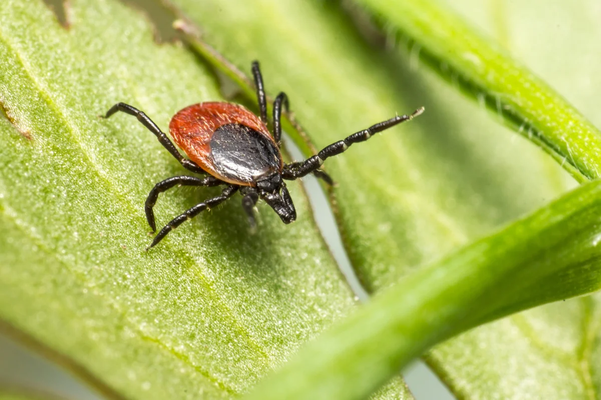
Tularemia, commonly known as rabbit fever, is a rare but potentially serious bacterial infection that affects thousands of people worldwide each year. This tick-borne bacterial disease caused by Francisella tularensis, can lead to several forms of infection, depending on how the bacteria enter the body. Understanding the causes and early symptoms of tularemia—and knowing when to seek treatment can make a significant difference in recovery outcomes. What Is Tularemia? Tularemia is an acute bacterial disease caused by Francisella tularensis, a highly infectious pathogen that naturally occurs in wild animals and their environment. This zoonotic infection spreads from animals to humans through multiple routes, posing a particular risk to those who spend time outdoors or work with animals. The bacterium is remarkably infectious, requiring only a small number of organisms to cause disease in humans. According to data from the Centers for Disease Control and Prevention (CDC), the average number of tularemia cases reported between 2011 and 2022 was 56% higher than during 2001 to 2010, indicating a marked rise in incidence over the past decade. Types of Tularemia Understanding the different tularemia types helps in recognising symptoms and seeking appropriate treatment. Each form presents unique characteristics: Ulceroglandular tularemia: The most common form, featuring a skin ulcer at the infection site with nearby swollen lymph nodes Glandular tularemia: Characterised by swollen lymph nodes without visible skin ulcers Oculoglandular tularemia: Affects the eyes, causing redness, pain, and swollen lymph nodes near the ears or jaw Oropharyngeal tularemia: Affects the mouth, throat, and gastrointestinal tract following ingestion of contaminated food or water Pneumonic tularemia: The most severe form affecting the lungs, often resulting from inhaling contaminated particles Typhoidal tularemia: A systemic form with high fever but no obvious localised symptoms, making diagnosis challenging Causes of Tularemia Tularemia causes stem from infection with Francisella tularensis, a bacterium that thrives in various environmental conditions. This pathogen naturally circulates among wild animals, particularly rabbits, hares, rodents, and various arthropods including ticks and deer flies. The bacteria can survive for weeks in water, soil, and decaying animal matter, creating multiple exposure opportunities. Two main subspecies exist — Type A strains, common in North America, tend to cause more severe illness, whereas Type B strains, found mainly in Europe and Asia, usually result in milder disease. Understanding these tularemia causes helps identify risk factors and implement appropriate prevention strategies. How Tularemia Spreads The transmission pathways for this tick-borne disease are diverse, reflecting the bacteria's adaptability: Tick bites from infected rabbits, dogs, or ticks such as the wood tick or lone star tick Bites from infected deer flies or other blood-feeding insects Direct contact with infected animal tissues through cuts or breaks in the skin Inhalation of contaminated dust particles during outdoor activities like mowing or farming Ingestion of contaminated water or undercooked meat from infected wild animals Laboratory exposure when handling infected cultures without appropriate biosafety precautions Is Tularemia Contagious Between People? Tularemia is not contagious between humans. Health authorities, including the World Health Organization (WHO), confirm that person-to-person transmission has never been documented. This means you cannot catch tularemia from someone who has the infection, even through close contact. The disease spreads exclusively through environmental or animal exposure, making it fundamentally different from many other infectious diseases that spread through respiratory droplets or direct contact. Tularemia Symptoms Recognising tularemia symptoms early is crucial for prompt treatment. The clinical presentation varies depending on the route of infection and the specific tularemia types involved: Sudden onset fever and chills: Often the first signs to appear Severe headache and malaise: Contributing to an overall feeling of illness Muscle and joint pain: Affecting daily activities and mobility Loss of appetite and weight loss: Particularly noticeable in prolonged cases Skin ulcers or sores: Typically appearing at the site of tick bites or animal contact Swollen, painful lymph nodes: Most commonly in the neck, armpit, or groin areas Eye symptoms: Including redness, pain, and discharge in oculoglandular forms Respiratory symptoms: Such as dry cough and chest pain in pneumonic cases Symptoms by Tularemia Type Tularemia Type Route of Infection Key Local Symptoms Systemic Symptoms Ulceroglandular Tick bite, animal contact Painful skin ulcer, swollen regional lymph nodes Fever, chills, headache, muscle aches Glandular Tick bite, animal contact Swollen lymph nodes without visible ulcer Fever, malaise, headache Oculoglandular Eye contamination Red, painful eye, swollen facial lymph nodes Fever, headache, general malaise Oropharyngeal Ingestion Sore throat, mouth ulcers, neck lymph nodes Fever, nausea, abdominal pain Pneumonic Inhalation Dry cough, chest pain, breathing difficulty High fever, severe fatigue Typhoidal Various routes Often no localising signs High fever, weakness, abdominal symptoms Risk Factors for Tularemia Certain activities and circumstances increase your likelihood of encountering this tick-borne disease: Outdoor occupations: Such as farming, landscaping, veterinary work, and wildlife management Recreational activities: Such as hunting, fishing, camping, and hiking in endemic areas Geographic location: Living in or visiting areas with high tick populations Seasonal factors: Higher risk during spring and summer months when ticks are most active Animal exposure: Handling wild rabbits, rodents, or their carcasses Laboratory work: Involving Francisella tularensis cultures or infected specimens Complications of Untreated Tularemia Pneumonia and respiratory failure: Particularly dangerous in pneumonic forms Meningitis: Infection spreading to the brain and spinal cord coverings Pericarditis: Inflammation of the heart's protective membrane Osteomyelitis: Bone infections requiring prolonged antibiotic therapy Sepsis: Life-threatening systemic infection requiring immediate medical intervention When to Seek Emergency Care High fever above 39°C (102°F) with severe chills Difficulty breathing or chest pain suggesting pneumonic involvement Signs of sepsis, including confusion, rapid heartbeat, or low blood pressure Severe eye symptoms with vision changes or intense pain Rapidly spreading skin infection or multiple ulcers How Tularemia Is Diagnosed Clinical assessment: Evaluating symptoms, exposure history, and physical examination findings Laboratory testing: Including blood cultures, serology, and molecular diagnostics Imaging studies: Chest X-rays for suspected pneumonic cases Tissue sampling: Collecting specimens from ulcers or lymph nodes when appropriate Differential diagnosis: Ruling out similar conditions like plague or Lyme disease Tests for Tularemia Blood cultures: Culturing bacteria from blood samples using specialised media Serology tests: Detecting antibodies through tube agglutination or ELISA methods PCR testing: Detects bacterial DNA from blood, tissue, or respiratory samples CBC Test: Assessing white blood cell counts and inflammatory markers ESR Automated Blood Test: Measuring inflammation levels in the body Liver Function Test (LFT): Evaluating potential liver involvement Electrolytes Test: Monitoring fluid balance and organ function Treatment Options for Tularemia Effective tularemia treatment relies primarily on appropriate antibiotic therapy. Streptomycin remains the gold-standard antibiotic for tularemia treatment, typically administered intramuscularly for 10-14 days. Alternative antibiotics include gentamicin, doxycycline, and ciprofloxacin, depending on the specific tularemia types and patient factors. Early treatment significantly improves outcomes, with most patients recovering completely when therapy begins promptly. Supportive care addresses symptoms like fever and pain, while monitoring ensures treatment effectiveness and identifies potential complications. Hospital Care for Severe Tularemia Severe cases of this tick-borne disease may require hospitalisation: Intravenous antibiotic administration: Ensuring optimal drug levels and absorption Respiratory support: Including oxygen therapy or mechanical ventilation for pneumonic cases Fluid management: Maintaining proper hydration and electrolyte balance Intensive monitoring: Tracking vital signs and organ function Isolation precautions: Standard precautions only, as person-to-person transmission doesn't occur Is There a Tularemia Vaccine? Currently, there is no licensed vaccine for tularemia available for public use. Research continues into developing effective vaccines, but prevention through risk reduction remains the primary strategy for avoiding this glandular infection. How to Prevent Tularemia Avoid direct contact with wild animals, especially rabbits and rodents Use protective equipment when handling potentially infected animals Implement tick prevention measures during outdoor activities Treat pets with appropriate tick and flea control products Maintain clean water sources and avoid drinking from potentially contaminated sources Practice food safety by thoroughly cooking wild game meat Use insect repellents containing DEET when outdoors in endemic areas Tick Bite Prevention Tips Wear appropriate clothing: Long sleeves, long trousers, and closed shoes when outdoors Use effective repellents: Apply EPA-registered insect repellents to skin and clothing Stay on cleared paths: Avoid walking through tall grass and dense vegetation Perform regular tick checks: Examine your body thoroughly after outdoor activities Remove ticks promptly: Use fine-tipped tweezers to extract attached ticks completely Tularemia and Pets: Can Animals Transmit It? While pets rarely transmit tularemia directly to humans, they can serve as transport hosts for infected ticks, making comprehensive tick control essential for household protection. Rabbits, dogs, and cats may carry infected ticks into home and surrounding areas, or develop tularemia themselves after exposure. Tularemia vs. Lyme Disease vs. Plague Condition Causative Agent Primary Vector Key Symptoms Geographic Distribution Tularemia Francisella tularensis Ticks, deer flies Skin ulcers, swollen lymph nodes Northern hemisphere, rural areas Lyme Disease Borrelia burgdorferi Ixodes ticks Erythema migrans rash, joint pain Temperate regions worldwide Plague Yersinia pestis Fleas Bubonic swellings, severe systemic illness Endemic foci globally Global Occurrence & High-Risk Regions Tularemia occurs across the Northern Hemisphere, with significant regional variations in incidence and tularemia types. The United States reports approximately 200 cases annually, primarily from Arkansas, Kansas, and Oklahoma. European countries, particularly Scandinavia and Eastern Europe, experience periodic outbreaks. High-risk regions typically feature abundant wildlife, suitable tick habitats, and frequent human outdoor activity increasing exposure risk. Climate change and ecological factors continue to influence the geographic distribution of this tick-borne disease. Tularemia in Children Children face similar risks as adults but may have different clinical presentations. Young children often develop more severe symptoms and require careful monitoring during tularemia treatment. Parents should be particularly vigilant about tick prevention when children play outdoors and seek medical attention promptly if children develop fever after potential exposure. This glandular infection can progress more rapidly in paediatric patients, making early recognition and treatment crucial for optimal outcomes. Tularemia in Pregnancy Pregnant women with tularemia require specialised medical care to protect both maternal and foetal health. Certain antibiotics used for tularemia treatment may not be suitable during pregnancy, necessitating careful drug selection. The infection itself can potentially affect pregnancy outcomes, making prompt diagnosis and appropriate therapy essential. Pregnant women should take extra precautions to avoid exposure, particularly during outdoor activities in endemic areas. When to Consult a Doctor You should seek medical attention if you develop fever, headache, or other concerning symptoms after potential exposure to ticks, infected animals, or contaminated environments. Early consultation is particularly important if you notice skin ulcers, swollen lymph nodes, or respiratory symptoms. Healthcare providers can assess risk factors, evaluate symptoms, and initiate appropriate diagnostic testing and treatment. Don't delay seeking care, as early tularemia treatment significantly improves outcomes and reduces complication risks. Conclusion Understanding tularemia empowers you to recognise risks and seek appropriate care when needed. At Metropolis Healthcare, we support your health journey with comprehensive diagnostic services spanning over 4,000 tests and profiles. Our extensive network of 220+ laboratories and 10,000+ touchpoints across India ensures accessible, accurate testing when you need it most. From routine blood work to specialised infection panels, our home sample collection service brings convenient, reliable diagnostics directly to you. FAQs What is tularemia, and how do you get it? Tularemia is a bacterial infection caused by Francisella tularensis, spread through tick or fly bites, contact with infected animals, inhalation, or contaminated food or water. What are the first signs of tularemia? Early symptoms include sudden fever, chills, headache, muscle aches, and sometimes a skin ulcer or swollen lymph nodes. How serious is tularemia? It can be serious if untreated, but prompt antibiotic treatment usually leads to full recovery. Can tularemia be cured? Yes, tularemia is curable with appropriate antibiotics, especially when treated early. Is tularemia contagious from person to person? No, it does not spread between people. How fast do tularemia symptoms appear? Symptoms usually develop within 3–5 days but can appear anytime between 1 and 14 days. What animals carry tularemia? Rabbits, rodents, ticks, deer flies, and occasionally domestic animals like cats can carry the bacteria. How can you prevent tularemia if you work outdoors? Use insect repellent, wear protective clothing, avoid handling wild animals, and check for ticks regularly. References https://pubmed.ncbi.nlm.nih.gov/32989563/ https://www.cdc.gov/mmwr/volumes/73/wr/mm735152a1.htm https://www.webmd.com/a-to-z-guides/what-is-tularemia
Babesiosis: A Complete Guide to This Tick-Borne Infection
Babesiosis is an emerging tick-borne infection that attacks your red blood cells, causing symptoms that range from mild, flu-like illness to severe haemolytic anaemia. This parasitic disease, transmitted primarily through infected blacklegged tick bites, remains largely unknown to many people despite its increasing prevalence in certain regions. Understanding the causes and early symptoms of babesiosis—and knowing when to seek medical attention—can significantly improve outcomes can make a significant difference in your health outcomes. What Is Babesiosis? Babesiosis is an infectious disease caused by protozoan parasites of the genus Babesia, which specifically target and destroy red blood cells. These microscopic organisms invade your erythrocytes, multiply inside them, and eventually cause the cells to burst, leading to haemolytic anaemia in severe cases. The most common species affecting humans is Babesia microti, primarily found in certain regions where blacklegged ticks thrive. Once these parasites enter your bloodstream through a tick bite, they begin their destructive cycle within your red blood cells. This red blood cell infection can range from completely asymptomatic to life-threatening, depending on various factors including your immune system strength and overall health status. Unlike many other infections, babesiosis can persist in your system for months or even years if left untreated, making early diagnosis of babesiosis crucial for effective management. How Babesiosis Spreads Understanding how this infection transmits helps you take appropriate preventive measures: Tick bites: The primary transmission occurs through bites from infected blacklegged (deer) ticks carrying Babesia parasites Blood transfusions: Contaminated blood products can transmit the infection because Babesia parasites can survive in stored blood Organ transplantation: Rarely, infected donor organs may transmit the infection to recipients Mother-to-child transmission: Pregnant women can pass the infection to their babies during pregnancy or delivery No casual contact spread: You cannot catch babesiosis through touching, coughing, or everyday contact with infected individuals How Long It Takes to Get Babesiosis After a Tick Bite After an infectious tick bite, babesiosis symptoms typically begin appearing 1-6 weeks later. The incubation period varies significantly between individuals, with some people developing symptoms within a week while others may not notice any signs for several months. This delayed onset often makes it challenging to connect the illness with the original tick exposure, especially if you don't remember being bitten. Where Is Babesiosis Found? Babesiosis occurs most commonly in temperate regions where blacklegged ticks establish populations. In the United States, human cases concentrate in the Northeast and upper Midwest states, particularly areas where Lyme disease is also endemic. The infection is considered emerging, with reported cases increasing due to expanding tick populations, climate changes, and improved diagnostic recognition. Globally, different Babesia species cause disease in Europe and Asia, often associated with cattle or wildlife reservoirs. As environmental conditions change and tick ranges expand, babesiosis may appear in previously unaffected areas. Babesiosis Symptoms Many people with babesiosis never develop noticeable symptoms, but when they occur, they often resemble flu-like illness: Fever (often high and persistent) Chills and sweats (particularly night sweats) Fatigue and weakness (sometimes severe) Headache (can be intense) Body aches and muscle pain Loss of appetite Nausea and sometimes vomiting General malaise (feeling unwell) Dark urine or jaundice (indicating haemolytic anaemia) Shortness of breath (in severe cases) Mild vs. Severe Symptoms of Babesiosis Mild Babesiosis Severe Babesiosis Low-grade fever, manageable fatigue High persistent fever, profound weakness Mild anaemia, stable condition Significant haemolytic anaemia, jaundice Normal breathing patterns Shortness of breath, respiratory distress Stable blood pressure Low blood pressure, possible shock Normal organ function Potential organ failure Usually outpatient treatment Often requires hospitalisation Causes & Pathophysiology Babesiosis is caused by protozoan parasites that invade and multiply within your red blood cells. After entering through tick bites, these organisms reproduce asexually inside erythrocytes, eventually causing cell destruction and haemolytic anaemia. This process releases cellular contents into your bloodstream, triggering inflammatory responses and potentially leading to organ dysfunction in severe cases. The parasites' ability to hide within red blood cells makes them particularly challenging for your immune system to eliminate, explaining why some infections persist for extended periods. Risk Factors for Severe Babesiosis Certain groups face higher risks for developing severe babesiosis symptoms: Adults over 50 years of age are more likely to experience complications People without spleens (asplenic individuals) cannot effectively clear infected red blood cells and are especially susceptible to severe infection Immunocompromised patients including those with HIV, cancer, or taking immunosuppressive medications Individuals with chronic illnesses such as heart disease or diabetes Those taking certain medications that affect immune function Complications of Babesiosis Severe babesiosis can lead to serious complications requiring immediate medical attention: Severe haemolytic anaemia requiring transfusions Kidney failure from damaged red blood cells Liver dysfunction and jaundice Acute respiratory distress syndrome (ARDS) Heart failure or cardiac complications Blood clotting disorders Multi-organ failure in extreme cases When to Seek Emergency Care Contact emergency services immediately if you experience: Difficulty breathing or shortness of breath at rest Chest pain or rapid heartbeat Severe weakness or inability to stand Dark brown or cola-coloured urine Yellow skin or eyes (jaundice) High fever above 39°C (102°F) with severe symptoms Confusion or altered mental state How Babesiosis Is Diagnosed Healthcare providers use several methods for accurate babesiosis diagnosis: Blood smear examination: Microscopic examination reveals parasites inside red blood cells Molecular testing (PCR): Detects parasite DNA with high accuracy Antibody testing: Measures immune response to Babesia infection Complete blood count: Shows signs of haemolytic anaemia and low platelet counts Clinical assessment: Evaluates symptoms, tick exposure history, and risk factors Your doctor will also inquire about any recent travel to tick-endemic areas, outdoor activities, and whether you've noticed any tick bites recently. Treatment for Babesiosis Babesiosis is treated when symptoms are present or when the parasite remains detectable on testing for an extended period. According to research summarised by the National Library of Medicine, most mild to moderate infections are managed with a combination of atovaquone and azithromycin, which is generally effective and better tolerated. Quinine with clindamycin may be used as an alternative but is more likely to cause side effects. Treatment typically lasts 7–14 days, with adjustments depending on clinical response. People with weakened immunity or recurring infection may need longer therapy until blood tests confirm parasite clearance. In cases that do not improve, different drug combinations may be used, though no single option is clearly superior. Treatment for Severe Babesiosis Severe cases require more intensive babesiosis treatment approaches: Hospitalisation for close monitoring and supportive care Intravenous clindamycin plus quinine (or quinidine with cardiac monitoring) Exchange transfusion may be required for parasitaemia ≥10%, severe anaemia, or evidence of organ dysfunction Blood transfusions to correct significant haemolytic anaemia Organ support, such as dialysis for kidney failure Intensive care monitoring, as respiratory distress and multi-organ complications are common in severe disease Can Babesiosis Go Away on Its Own? While some people with strong immune systems might clear mild babesiosis infections without treatment, this approach carries significant risks. Untreated infections can persist for months or years, potentially leading to chronic symptoms and serious complications. The parasites may persist at low levels and reactivate during periods of immune suppression, making proper medical treatment essential for complete recovery. Babesiosis Prevention Use insect repellents containing DEET, picaridin, or permethrin on skin and clothing Wear protective clothing including long sleeves, long trousers, and closed shoes when in tick habitats Perform thorough daily tick checks on yourself, your children, and pets after outdoor activities Shower within two hours of coming indoors to wash off unattached ticks Keep grass short and remove leaf litter around your home Create barriers using wood chips or gravel between wooded areas and your garden How to Remove a Tick Safely Use fine-tipped tweezers to grasp the tick as close to your skin as possible Pull steadily upward with even pressure; don't twist or jerk the tick Clean the bite area thoroughly with soap and water or an antiseptic solution Dispose of the tick by flushing it down the toilet or placing it in alcohol Monitor the bite site for signs of infection or unusual symptoms over the following weeks Babesiosis vs. Lyme Disease vs. Anaplasmosis Feature Babesiosis Lyme Disease Anaplasmosis Primary symptoms Flu-like, haemolytic anaemia Bull's-eye rash, joint pain Fever, headache, muscle aches Diagnostic tests Blood smear, PCR Antibody tests, clinical signs Blood tests, PCR Treatment Atovaquone + azithromycin Doxycycline Doxycycline Complications Severe anaemia, organ failure Arthritis, neurological issues Low white blood cell count Babesiosis and Co-Infections Approximately 20% of babesiosis patients also contract Lyme disease, as the same ticks can carry multiple pathogens simultaneously. Co-infections typically result in more severe symptoms and prolonged illness duration. If you're diagnosed with one tick-borne disease, your healthcare provider will likely test for others to ensure comprehensive treatment. Babesiosis in Children Children usually experience milder babesiosis symptoms than adults, often resembling common viral infections. However, very young children and those with compromised immune systems face higher risks for severe complications. Parents should watch for persistent fever, unusual fatigue, or breathing difficulties following potential tick exposure. Babesiosis in Pregnancy Pregnant women with babesiosis face risks of transmitting the infection to their unborn babies. The condition can also complicate pregnancy through severe anaemia or other complications. Prompt diagnosis and treatment during pregnancy are crucial to protect both maternal and foetal health. Global Distribution & Climate Change Impact Climate change is expanding tick habitats and extending their active seasons, potentially increasing babesiosis transmission in previously unaffected regions. Warmer temperatures and changing precipitation patterns create favourable conditions for tick populations to establish in new areas, making awareness and prevention even more important. When to Consult a Doctor Seek medical attention if you develop flu-like symptoms within six weeks of potential tick exposure, especially if you've been in areas where babesiosis is endemic. Early consultation allows for prompt babesiosis diagnosis and treatment, significantly improving your prognosis and reducing complication risks. Conclusion Babesiosis represents a serious but manageable tick-borne infection that requires awareness, prevention, and prompt medical attention. The key to managing this red blood cell infection lies in prevention through tick bite avoidance and seeking immediate medical care when symptoms develop. At Metropolis Healthcare, we support your health journey with comprehensive diagnostic services designed for accuracy and convenience. Our extensive portfolio of over 4,000 tests includes specialised panels for infectious diseases like babesiosis, ensuring precise diagnosis when you need it most. Through our network of 10,000+ touchpoints across India, we bring reliable testing directly to your doorstep with our home sample collection service. FAQs What causes babesiosis in humans? Babesiosis is caused by Babesia parasites transmitted primarily through infected blacklegged tick bites. These microscopic organisms invade red blood cells, multiply within them, and cause cell destruction leading to various symptoms ranging from mild flu-like illness to severe haemolytic anaemia. How serious is babesiosis? The severity of babesiosis varies significantly between individuals. Many people experience no symptoms at all, while others develop mild flu-like illness. However, vulnerable populations including older adults, people without spleens, and immunocompromised individuals can develop life-threatening complications. What are the first signs of babesiosis? Early babesiosis symptoms typically include fever, chills, sweats, fatigue, headache, and body aches—very similar to flu symptoms. These signs usually appear 1-6 weeks after an infected tick bite. Can babesiosis be cured? Yes, babesiosis can be effectively treated with appropriate antibiotic combinations, typically atovaquone plus azithromycin. Early treatment leads to better outcomes, while severe cases may require hospitalisation and additional supportive care. How long does babesiosis last? With proper treatment, most people recover from babesiosis within 1-2 weeks. However, untreated infections can persist for months or even years, with symptoms potentially lasting 6-8 weeks. Can you get babesiosis twice? Reinfection with babesiosis is possible but uncommon. Previous infection may provide some immunity, but this protection isn't complete or permanent. People can potentially contract different Babesia species or experience reactivation of persistent infections during periods of immune suppression. Will doxycycline treat babesiosis? Doxycycline is not the standard treatment for babesiosis. The preferred treatment combines atovaquone with azithromycin, or alternatively clindamycin with quinine for severe cases. How do you prevent babesiosis from tick bites? Prevention focuses on avoiding tick bites through protective clothing, insect repellents containing DEET or picaridin, daily tick checks after outdoor activities, prompt tick removal, and maintaining tick-free environments around your home by keeping grass short and removing leaf litter. References https://www.ncbi.nlm.nih.gov/books/NBK430715/#article-18082.s8 https://my.clevelandclinic.org/health/diseases/24809-babesiosis https://www.cdc.gov/dpdx/babesiosis/index.html https://www.publichealthontario.ca/en/Diseases-and-Conditions/Infectious-Diseases/Vector-Borne-Zoonotic-Diseases/Babesiosis

Cyclospora Infection: Symptoms, Causes & Prevention
Cyclospora infections are becoming increasingly recognised as a significant cause of foodborne diarrhoeal disease, particularly during warmer months when fresh produce consumption peaks. This microscopic parasite affects thousands of people annually, causing prolonged stomach cramps, diarrhoea, and other debilitating symptoms. Understanding the symptoms, causes, and prevention of cyclospora empowers you to protect yourself and your family from this persistent intestinal infection. What Is Cyclospora? Cyclospora cayetanensis is a single-celled intestinal parasite that specifically targets the human small intestine, causing an illness called cyclosporiasis. The World Health Organization (WHO) confirms that unlike many other intestinal parasites, cyclospora infects only humans and requires a maturation period outside the body before becoming infectious. This unique characteristic affects how the parasite spreads and makes direct person-to-person transmission unlikely. How Cyclospora Spreads The microscopic nature of cyclospora makes it invisible to the naked eye, yet its impact on digestive health can be substantial. Understanding transmission helps you take appropriate preventive measures: Contaminated fresh produce: Especially imported herbs, leafy greens, and berries consumed raw Contaminated water: Untreated or inadequately filtered drinking water in endemic regions Environmental contamination: Oocysts from human faeces reach soil or water, becoming infectious after days to weeks Poor sanitation practices: Inadequate sewage treatment leading to crop contamination during growing or processing Recreational water exposure: Swimming in contaminated lakes or rivers in affected areas Ice made from contaminated water: A common oversight when travelling Common Foods Linked to Cyclospora Outbreaks Fresh leafy greens: Lettuce, spinach, and salad mixes Fresh herbs: Particularly cilantro, basil, and parsley Imported berries: Raspberries, blackberries, blueberries, and mixed berry products Raw vegetable mixes: Especially those from tropical or subtropical regions Where Is Cyclospora Found? Cyclospora thrives in tropical and subtropical regions where sanitation infrastructure may be inadequate, allowing human waste to contaminate water sources and agricultural areas. Many infections in developed countries occur in travellers returning from Central and South America, the Caribbean, and parts of Asia where the parasite is endemic. However, seasonal outbreaks now occur regularly in North America and Europe, typically during late spring and summer months, linked to imported fresh produce contaminated before or during export. Cyclospora Symptoms Recognising cyclospora symptoms helps ensure prompt medical attention: Watery diarrhoea: Often frequent, voluminous, and occasionally explosive Severe stomach cramps: Persistent abdominal pain that may worsen after eating Bloating and excessive gas: Uncomfortable digestive distention Loss of appetite: Significant reduction in food interest Unintentional weight loss: Often substantial over the infection period Persistent nausea: May be accompanied by occasional vomiting Overwhelming fatigue: Persistent tiredness that can significantly interfere with daily activities Low-grade fever: Mild temperature elevation in some cases Flu-like symptoms: Body aches, headache, and general malaise Severe Symptoms of Cyclospora Infection While many experience mild to moderate illness, cyclospora can cause severe complications requiring immediate medical attention. Prolonged diarrhoea can cause severe dehydration, with some patients losing nearly 10 kilograms during their illness. Immunocompromised individuals are particularly at risk and may experience prolonged, relapsing symptoms that significantly impact quality of life. Warning signs of severe dehydration include dizziness, dry mouth, reduced urination, confusion, and inability to keep fluids down, all requiring urgent medical care. Causes & Pathophysiology Cyclospora infection occurs after ingestion of Cyclospora cayetanensis oocysts through contaminated food or water. Once swallowed, the oocysts release sporozoites in the small intestine, where they invade the cells lining the small intestine and multiply. This process disrupts normal intestinal absorption and fluid balance, leading to malabsorption, watery diarrhoea, and abdominal cramps. The oocysts passed in stool are initially non-infectious and must mature in the environment over several days to weeks before they can cause infection again. This delayed maturation period explains why direct person-to-person transmission is extremely uncommon. Incubation Period of Cyclospora The incubation period for cyclospora typically ranges from 2 to 14 days, with most people developing symptoms approximately one week after exposure. This delay reflects the time needed for the parasite to establish infection in your small intestine and for your body to mount an inflammatory response. Risk Factors for Cyclospora Infection Several factors increase your likelihood of contracting cyclospora: Travel to endemic regions: Higher exposure in areas with poor sanitation Consuming raw produce: Unwashed fruits and vegetables may carry the parasite Drinking untreated water: Contaminated water can transmit Cyclospora Weakened immune system: Reduced ability to fight infection Seasonal timing: Higher risk during warmer or rainy months Age factors: Children and older adults may be more vulnerable Complications of Cyclospora Severe dehydration: Requiring hospitalisation and intravenous fluid replacement Significant weight loss: Potentially exceeding 10% of body weight Nutritional deficiencies: Due to malabsorption and prolonged poor appetite Chronic fatigue: Lasting weeks to months after initial infection Prolonged illness: Symptoms persisting for several months without treatment Relapsing symptoms: Recurring episodes of diarrhoeal disease How Cyclospora Is Diagnosed Healthcare providers use specific diagnostic steps for accurate cyclospora diagnosis: Medical history review: Discussing symptoms, travel history, and recent food consumption Physical examination: Assessing hydration status and abdominal tenderness Stool sample collection: Obtaining fresh specimens for laboratory analysis Microscopic stool examination: Identifies Cyclospora oocysts using specialised staining methods Specialised staining techniques: Using modified acid-fast stains for better visualisation Molecular testing (PCR): Offers enhanced sensitivity and specificity for confirming diagnosis Treatment for Cyclospora Effective cyclospora treatment typically involves antibiotic therapy with trimethoprim-sulfamethoxazole (TMP-SMX), the first-line treatment for this diarrhoeal disease. Most patients notice improvement within 2-4 days of starting treatment, with complete resolution occurring within 7-10 days. Supportive care includes maintaining adequate hydration through oral rehydration solutions or intravenous fluids in severe cases. Your healthcare provider may recommend probiotics to restore normal gut bacteria after antibiotic treatment. What If You Are Allergic to Sulfa Drugs? If you cannot tolerate trimethoprim-sulfamethoxazole due to allergies, alternative cyclospora treatment options exist, though they may be less effective. Ciprofloxacin has shown limited success for cyclospora infections, primarily in immunocompromised patients. However, treatment duration may be longer, and symptom resolution might be slower compared to standard therapy. Can Cyclospora Go Away on Its Own? While some healthy individuals may eventually recover without treatment, this approach carries significant risks. Untreated infections often persist for weeks to months, causing substantial weight loss, dehydration, and nutritional deficiencies. The cyclical nature of symptoms means you might feel better temporarily, only to experience recurring episodes of severe stomach cramps and diarrhoea. How Long Does Cyclospora Last? With appropriate antibiotic therapy, most people experience significant improvement within 3-7 days and complete recovery within 2 weeks. However, some individuals, particularly those with compromised immune systems, may require longer treatment courses or experience prolonged recovery periods. How to Prevent Cyclospora Rinse all fruits and vegetables thoroughly under running water, even those you plan to peel Limit consumption of imported fresh herbs and berries during outbreak seasons Opt for thoroughly cooked meals when dining out Wash hands frequently with soap and water When travelling to endemic areas, drink only sealed bottled water Avoid ice unless made from safe water sources Travel Safety Tips Drink only bottled or boiled water Avoid raw vegetables and fruits Choose reputable and hygienic restaurants Carry hand sanitizer Pack oral rehydration salts for quick diarrhoeal treatment Cyclospora vs. Giardia vs. Cryptosporidium Feature Cyclospora Giardia Cryptosporidium Incubation Period 2-14 days 1-14 days 2-10 days Primary Symptoms Watery diarrhoea, stomach cramps Greasy stools, bloating Watery diarrhoea, fever Treatment TMP-SMX Metronidazole Nitazoxanide Duration (untreated) Weeks to months 2-6 weeks 1-3 weeks Cyclospora in Children Children may experience more severe symptoms because of their developing immune systems and smaller body size, making dehydration a particular concern. Young patients often present with more pronounced stomach cramps and may become dehydrated more quickly than adults. Paediatric cyclospora treatment requires careful dosing adjustments and close monitoring for treatment response and potential side effects. Cyclospora in Pregnancy Pregnant women with cyclospora infection face unique risks, as severe diarrhoea and dehydration can threaten both maternal and foetal health. While trimethoprim-sulfamethoxazole is generally avoided during the first trimester due to potential birth defects, healthcare providers must carefully weigh the risks and benefits of treatment options. Outbreak Tracking & Public Health Alerts Public health agencies actively monitor cyclospora outbreaks, particularly those linked to imported produce. The WHO and national health departments issue alerts when contaminated food products are identified, helping consumers make informed choices. These surveillance systems have identified recurring patterns, with most outbreaks occurring during summer months when fresh produce consumption peaks. When to Seek Medical Care Persistent diarrhoea lasting more than three days Signs of dehydration including dizziness, dry mouth, or reduced urination Severe stomach cramps interfering with daily activities High fever above 38.5°C (101.3°F) Blood in stool or severe abdominal pain Inability to keep fluids down due to vomiting Conclusion Understanding cyclospora infection empowers you to recognise symptoms early, seek appropriate treatment, and implement effective prevention strategies. This diarrhoeal disease, while challenging, responds well to prompt medical intervention with proper cyclospora diagnosis and treatment. At Metropolis Healthcare, we support your health journey with comprehensive diagnostic services designed to detect various infections accurately. Our extensive network of over 4,600 service centres, reaching 10,000+ touchpoints across India, ensures convenient access to advanced diagnostic testing, including stool examinations for intestinal parasites. FAQs What is Cyclospora and how do you get it? Cyclospora cayetanensis is a microscopic parasite causing cyclosporiasis, a diarrhoeal disease affecting the small intestine. You contract cyclospora by consuming contaminated food or water containing mature oocysts. What are the first signs of Cyclospora? Early cyclospora symptoms include watery diarrhoea, stomach cramps, and loss of appetite, typically appearing 2-14 days after exposure. You might also experience bloating, nausea, and fatigue as initial signs of this diarrhoeal disease, with symptoms often developing gradually rather than suddenly. How long does Cyclospora last? Without treatment, cyclospora infections can persist for weeks to months with recurring episodes of symptoms. With proper cyclospora treatment using antibiotics, most people recover within 7-10 days, though some may experience longer recovery periods depending on their immune status. What kills Cyclospora parasites? Trimethoprim-sulfamethoxazole is the most effective cyclospora treatment, eliminating the parasite from your intestinal tract. Proper cooking temperatures can kill cyclospora in food, while chlorination and boiling water destroy oocysts in contaminated water sources. What foods are commonly contaminated with Cyclospora? Fresh herbs like cilantro and basil, leafy greens including lettuce and spinach, and imported berries are most commonly associated with cyclospora outbreaks. Can Cyclospora be cured? Yes, cyclospora responds well to appropriate antibiotic treatment, with most patients experiencing complete recovery. Early cyclospora diagnosis and treatment prevent complications and reduce symptom duration, making prompt medical attention essential for suspected infections. Is Cyclospora contagious between people? Direct person-to-person transmission of cyclospora is rare because freshly shed oocysts require environmental maturation to become infectious. However, poor hygiene practices could potentially spread the infection. How is Cyclospora infection diagnosed? Cyclospora diagnosis requires specialised stool examination using modified acid-fast staining techniques to identify characteristic oocysts. Your healthcare provider may order multiple stool samples since oocyst shedding can be intermittent, and molecular testing methods offer enhanced diagnostic accuracy. Can Cyclospora return after treatment? While properly treated cyclospora infections typically resolve completely, reinfection can occur through new exposure to contaminated sources. References https://my.clevelandclinic.org/health/diseases/17957-cyclosporiasis https://www.cdc.gov/cyclosporiasis/index.html https://www.fda.gov/food/foodborne-pathogens/cyclospora

Campylobacter Infection: Causes, Symptoms & Treatment
Campylobacter infection ranks among the leading causes of foodborne illness worldwide, affecting millions of people annually. According to the World Health Organization (WHO), diarrhoeal diseases are the most common illnesses resulting from unsafe food, with 550 million people falling ill yearly. This includes 220 million children under five years. This bacterial gastroenteritis can cause significant discomfort, but understanding Campylobacter causes, symptoms, and treatment options empowers you to protect yourself and your family. What Is Campylobacter Infection? Campylobacter infection, medically known as campylobacteriosis, is a bacterial intestinal infection primarily caused by Campylobacter jejuni. This foodborne illness affects your intestinal tract, leading to inflammation that triggers diarrhoea, abdominal cramps, and fever. The infection typically develops after consuming contaminated food or water, particularly undercooked or improperly handled poultry products. Most Campylobacter infections resolve within a week with proper care. However, certain individuals, especially those with weakened immune systems, may experience severe complications. Understanding how this bacterial gastroenteritis develops helps you recognise symptoms early and seek appropriate Campylobacter treatment when needed. How Campylobacter Infection Happens Several transmission routes can lead to Campylobacter infection. The most common transmission routes include: Consuming undercooked poultry contaminated with Campylobacter bacteria Cross-contamination when raw poultry juices touch ready-to-eat foods, utensils, or kitchen surfaces Drinking unpasteurised milk from infected animals during milking or handling Drinking contaminated well or surface water exposed to animal faeces Eating contaminated produce that contacted dirty water or infected animal products Direct contact with infected animals, including farm animals, puppies, and kittens How Campylobacter Spreads Understanding transmission patterns helps prevent Campylobacter infection effectively: Foodborne transmission through undercooked meat and poultry Kitchen cross-contamination via cutting boards, knives, and preparation surfaces Contaminated drinking water or recreational water sources Raw dairy products from infected livestock Direct contact with infected pets or farm animals Person-to-person transmission through poor hand hygiene after using the toilet or handling diapers Who Is at Highest Risk? Certain groups face increased vulnerability to severe Campylobacter infection: Children under five years due to developing immune systems Adults over 65 years with naturally declining immunity Immunocompromised individuals including those with HIV/AIDS or cancer Pregnant women, who may experience more severe illness and potential complications People with chronic conditions such as diabetes or liver disease Agricultural workers frequently exposed to livestock International travellers visiting areas with poor sanitation Campylobacter Infection Symptoms Recognising Campylobacter symptoms enables prompt treatment and prevents complications. Common signs include: Diarrhoea that may range from watery to bloody Severe abdominal cramps and persistent pain Fever typically ranging from mild to moderate Nausea and vomiting causing additional discomfort Fatigue and general malaise affecting daily activities Headache and body aches in some cases Loss of appetite and reduced food intake Early vs. Severe Symptoms Early/Mild Symptoms Severe/Complicated Symptoms Gradual abdominal cramps High fever (≥38.9°C) or persistent fever Watery diarrhoea without blood Severe bloody diarrhoea or frequent stools Mild nausea and occasional vomiting Signs of dehydration (dark urine, dry mouth) Low-grade fever and fatigue Inability to keep fluids down Mild headache or body aches Severe abdominal pain or tenderness Symptoms gradually improving over several days Symptoms lasting more than a week How Long After Exposure Do Symptoms Start? Campylobacter symptoms typically appear 2-5 days after exposure, though the incubation period can range from 1-10 days. Most people initially experience general malaise, fever, and cramping, followed by diarrhoea that may become bloody within 24-48 hours. Some individuals with strong immune systems may experience minimal symptoms or mistake the infection for a brief stomach upset. Complications of Campylobacter Infection While most cases resolve without complications, severe outcomes can occur: Dehydration from persistent diarrhoea and vomiting Bacteraemia where Campylobacter spreads into the bloodstream Reactive arthritis causing painful joint inflammation that may last weeks to months Guillain–Barré syndrome, a rare autoimmune neurological disorder that can cause weakness and paralysis Pregnancy complications, including possible miscarriage or preterm labour Inflammation of the liver (hepatitis) or pancreas (pancreatitis) in severe cases When to See a Doctor Seek immediate medical attention if you experience: Bloody stools or persistent blood in diarrhoea High fever above 38.9°C or lasting more than 24 hours Signs of dehydration including dizziness, dry mouth, or decreased urination Severe abdominal pain that worsens or doesn't improve Symptoms persisting beyond one week Inability to keep fluids down due to persistent vomiting How Campylobacter Is Diagnosed Healthcare providers use a structured approach to accurately diagnose Campylobacter infection and rule out other causes of gastrointestinal illness. The diagnostic process typically includes the following steps: Medical history review: Your doctor asks about recent food consumption, particularly undercooked poultry, unpasteurised milk, or contaminated water, as well as any recent travel or known exposure risks. Physical examination: This helps assess hydration status, fever, and abdominal tenderness, which can indicate the severity of infection. Stool sample collection: A fresh stool sample is collected for detailed laboratory analysis to detect bacterial pathogens. Laboratory testing: Specific tests are performed to identify Campylobacter bacteria and confirm the diagnosis. Additional tests: Further investigations may be ordered if complications or severe infection are suspected. Tests for Campylobacter Infection Laboratory diagnostics play a vital role in confirming Campylobacter infection and guiding appropriate treatment. Based on symptoms, duration of illness, and risk factors, your doctor may recommend one or more of the following tests: Stool culture: Considered the gold standard for Campylobacter detection, this test allows direct identification of the bacteria and helps assess antibiotic sensitivity when required Campylobacter antigen detection: Identifies specific bacterial antigens in stool samples, offering faster results than traditional cultures PCR testing: A highly sensitive molecular method that detects Campylobacter DNA, especially valuable for early or severe infections Blood tests: Performed if bacteraemia is suspected, particularly in immunocompromised individuals Complete blood count (CBC): Helps evaluate infection severity by assessing markers of inflammation. Treatment for Campylobacter Infection Most Campylobacter infections resolve without antibiotics. Supportive care focuses on preventing dehydration through adequate fluid and electrolyte replacement. Oral rehydration solutions containing electrolytes help maintain proper hydration. Severe cases may require hospitalisation for intravenous fluid therapy. Antibiotics are typically reserved for severe infections, immunocompromised patients, or those with complications. Azithromycin is the preferred antibiotic for severe or prolonged cases. Fluoroquinolones may be used in specific situations, though resistance is increasingly common. Can Campylobacter Go Away Without Treatment? Yes, most healthy individuals recover from Campylobacter infection without specific medical treatment. The immune system typically clears the bacteria within 5-7 days. However, supportive care including rest, hydration, and symptom management remains essential. Avoiding anti-diarrhoeal medications initially allows your body to eliminate the bacteria naturally. Monitor symptoms closely and seek medical care if complications develop. How Long Does Campylobacter Infection Last? Typical Campylobacter infections last 5-7 days in healthy individuals. Symptoms usually peak within the first 2-3 days before gradually improving. Complete recovery, including normal bowel function, may take 1-2 weeks. Factors affecting duration include your immune status, bacterial load, and promptness of supportive care. Some people may experience lingering fatigue or digestive sensitivity for several weeks following acute illness. How to Prevent Campylobacter Infection Effective prevention strategies include: Cook poultry thoroughly to an internal temperature of at least 74°C (165°F) Avoid cross-contamination by using separate cutting boards for raw meat Wash hands thoroughly with soap and water, especially after handling raw poultry or pet animals Drink pasteurised milk and avoid raw dairy products Use safe water sources and avoid untreated well water Practice good hygiene when caring for pets or farm animals Store and refrigerate foods promptly at safe temperatures (below 5°C/41°F) When to Consult a Doctor or Gastroenterologist Consult healthcare providers immediately if you develop severe Campylobacter symptoms including bloody diarrhoea, high fever, or dehydration signs. Seek gastroenterology consultation if symptoms persist beyond two weeks, recurrent infections, or suspected complications like reactive arthritis. Immunocompromised individuals should seek prompt medical evaluation for any suspected Campylobacter infection due to increased complication risks. Conclusion Understanding Campylobacter infection empowers you to protect your family's health through proper food safety practices and early symptom recognition. Most infections resolve completely with appropriate care, but knowing when to seek medical attention ensures optimal outcomes. At Metropolis Healthcare, we support your health journey with comprehensive diagnostic services designed around your needs. Our extensive portfolio of over 4,000 tests includes specialised stool cultures and antigen detection for accurate Campylobacter diagnosis. With our convenient at-home sample collection service spanning 10,000+ touchpoints across India, you can access reliable testing without leaving your home. Take charge of your digestive health today. Book your home sample collection today and experience healthcare designed around your needs. FAQs What is the most common cause of Campylobacter infection? Consuming undercooked or contaminated poultry represents the leading cause of Campylobacter infection. Cross-contamination during food preparation and consuming unpasteurised dairy products are other significant sources. What are the first signs of Campylobacter? Initial Campylobacter symptoms typically include fever, general malaise, and abdominal cramping, followed by diarrhoea within 24-48 hours. Some people may experience nausea and headache early in the infection. Is Campylobacter infection contagious? Yes, Campylobacter can spread person-to-person through the faecal-oral route, particularly with poor hand hygiene. However, foodborne transmission remains far more common than direct human transmission. How long does Campylobacter last? Most Campylobacter infections resolve within 5-7 days with supportive care. Complete recovery, including normal digestive function, typically occurs within 1-2 weeks in healthy individuals. What foods commonly cause Campylobacter? Campylobacter causes can be attributed to undercooked poultry, unpasteurised milk, contaminated water, and cross-contaminated foods. Raw or undercooked chicken poses the highest risk for Campylobacter infection. Can Campylobacter be cured? While there's no specific cure, most infections resolve naturally with supportive care. Antibiotics may be prescribed for severe cases or high-risk patients to prevent complications. Do antibiotics help Campylobacter? Antibiotics can reduce symptom duration and prevent complications in severe cases, but they're not routinely prescribed for uncomplicated infections. The decision depends on symptom severity and patient risk factors. How to prevent Campylobacter infection from poultry? Cook chicken to 74°C internal temperature, avoid cross-contamination, wash hands after handling raw poultry, and store chicken properly. These measures significantly reduce Campylobacter transmission risk. References https://www.cdc.gov/campylobacter/index.html https://www.nhs.uk/conditions/food-poisoning/ https://my.clevelandclinic.org/health/diseases/15251-campylobacter-infection

Fifth Disease in Children: Symptoms & Care Tips
Fifth disease is one of the most common childhood infectious diseases, affecting thousands of children across India and millions worldwide each year. Also known as erythema infectiosum, this viral condition causes the distinctive "slapped cheek" appearance that often worries parents. Understanding the symptoms, causes, and care tips for fifth disease helps you manage this usually mild but sometimes concerning illness with confidence. What Is Fifth Disease? Fifth disease, medically known as erythema infectiosum, is a viral childhood illness caused by human parvovirus B19. This This parvovirus infection primarily affects school-aged children between 5 and 15 years of age, though adults can contract it too. The condition gets its name from being historically classified as the fifth of six classic childhood rash diseases. The hallmark of fifth disease is the bright red rash on the cheeks that gives the appearance of having been "slapped". This distinctive appearance, combined with a subsequent lacy body rash, makes fifth disease relatively easy to identify once the rash stage begins. What Causes Fifth Disease? Fifth disease occurs due to infection with human parvovirus B19, a DNA virus that specifically targets humans. Unlike the parvovirus that affects dogs and cats, parvovirus B19 cannot be transmitted between humans and animals. After entering the body through respiratory droplets, the virus travels to the bone marrow, where it infects red blood cell precursors. This brief interruption in red blood cell production is harmless in most healthy children but can cause complications in those with existing blood disorders or weakened immune systems. How Fifth Disease Spreads Respiratory droplets: The virus spreads when infected individuals cough, sneeze, or talk, releasing contaminated droplets into the air Direct contact: Sharing drinks, utensils, or toys with someone carrying the virus Surface contamination: Touching objects contaminated with fresh respiratory secretions Close proximity: Spending time in confined spaces with infected individuals, particularly during the early symptomatic phase Blood transmission: Rarely through blood transfusions or needle stick injuries Contagious Period of Fifth Disease Children are most contagious during the early phase—before the rash appears. During this period, which typically lasts 4-7 days, your child may seem to have a mild cold with low fever, headache, and general tiredness. Interestingly, once the distinctive "slapped cheek" rash develops, children are generally no longer contagious. This means by the time you recognise fifth disease symptoms, your child can typically return to school or day care, provided they feel well enough to participate in normal activities. Symptoms of Fifth Disease Fifth disease symptoms typically develop in two distinct phases, making recognition easier once you know what to look for: Early symptoms (before rash appears): Low-grade fever (usually under 38.5°C) Mild headache and body aches Runny or stuffy nose Sore throat General fatigue and irritability Possible mild nausea Rash phase symptoms: Bright red "slapped cheek" facial rash Lacy, pink rash spreading over the trunk, arms, and legs Mild itching, particularly with the body rash Rash that fades and reappears with heat, sunlight, or physical activity Possible joint pain in older children and teenagers Stages of Fifth Disease Rash The rash of fifth disease follows a predictable pattern. This helps distinguish fifth disease from other childhood infectious diseases with similar early symptoms. Incubation period (4-21 days): Your child is infected but shows no symptoms Prodromal phase (4-7 days): Mild cold-like symptoms appear; child is most contagious Facial rash stage (1-4 days): Distinctive bright red cheek rash develops Body rash stage (1-3 weeks): Lacy rash appears on trunk and limbs, may fade and reappear Fifth Disease in Adults While primarily affecting children, adults can contract this parvovirus infection, especially if they haven't been previously exposed. Adult fifth disease symptoms often differ from those in children, with less prominent facial rash but more significant joint pain and swelling. In adults, joint symptoms may persist for several weeks or even months, particularly affecting hands, wrists, knees, and ankles. Pregnant women should seek medical advice promptly, as maternal parvovirus infection can, in rare cases, affect the developing baby. Fifth Disease in Children Children typically handle fifth disease better than adults, with most experiencing only mild discomfort. The condition is most common among primary school-aged children due to close contact in classroom settings and developing immune systems. Healthy children typically recover completely without specific treatment, developing lasting immunity that prevents future infection. The main concerns for parents are typically the dramatic appearance of the rash and ensuring their child remains comfortable during recovery. Risk Factors Several factors increase susceptibility to this childhood infectious disease: Age: Children aged 5-15 years face highest risk School attendance: Close contact environments facilitate transmission Compromised immunity: Children with weakened immune systems Blood disorders: Those with sickle cell disease or other anaemias face increased complications Pregnancy exposure: Pregnant women working with children Healthcare settings: Exposure in medical environments Seasonal patterns: Higher transmission rates during late winter and spring Complications of Fifth Disease Most children experience uncomplicated recovery, but certain groups face increased risks: Severe anaemia: In children with blood disorders like sickle cell disease Arthritis-like symptoms: Joint pain and swelling, more common in teenagers Pregnancy complications: Studies in the National Library of Medicine indicate a risk of intrauterine foetal demise, miscarriage, or hydrops fetalis Immune system complications: Prolonged infection in immunocompromised children Aplastic crisis: A temporary cessation of red blood cell production, usually seen in children with chronic anaemia When to See a Doctor High fever above 39°C or persistent fever Severe headache or neck stiffness Difficulty breathing or chest pain Extreme fatigue or weakness Signs of severe anaemia (pale skin, rapid heartbeat) Joint swelling or severe pain Rash accompanied by other concerning symptoms How Fifth Disease Is Diagnosed Clinical examination: Assessing the characteristic rash pattern and symptoms Medical history: Reviewing recent symptoms and potential exposures Blood tests: Checking for parvovirus B19 antibodies if diagnosis is unclear Differential diagnosis: Ruling out other conditions with similar rashes In most cases, doctors can diagnose fifth disease based on its distinctive rash and typical symptom pattern. Treatment for Fifth Disease No specific antiviral treatment exists for fifth disease, as it usually resolves naturally. Management focuses on symptom relief and comfort measures. Most children recover with simple home care including adequate rest, increased fluid intake, and age-appropriate pain relief for headaches or joint discomfort. For children with underlying health conditions, closer medical monitoring may be necessary. Those with blood disorders might require specialist care to manage potential complications. The vast majority of healthy children need only supportive care while their immune system clears the parvovirus infection. Is Fifth Disease Dangerous? For most healthy children, fifth disease poses minimal danger and resolves without lasting effects. The dramatic appearance of the rash often causes more concern than the actual illness warrants. However, certain groups including pregnant women, individuals with compromised immune systems, and those with blood disorders should take additional precautions. How to Prevent Fifth Disease Hand hygiene: Regular thorough handwashing with soap and water Respiratory etiquette: Teaching children to cover coughs and sneezes Avoiding sharing: Don't share drinks, utensils, or personal items Surface cleaning: Regular disinfection of frequently touched surfaces Isolation when symptomatic: Keeping children home during the contagious early phase Vaccination awareness: There is currently no vaccine available for parvovirus B19 Is Reinfection Possible? The immune system typically clears parvovirus infection within 7–10 days, after which most people develop lifelong immunity. This means your child is unlikely to contract fifth disease again after recovering from their first infection. When to Consult a Paediatrician Contact your paediatrician if you're unsure about your child's symptoms or if they have underlying health conditions that might complicate parvovirus infection. Children with blood disorders, immune system problems, or chronic illnesses require professional medical assessment. Conclusion The key to managing fifth disease lies in recognising fifth disease symptoms early, providing comfort care during recovery, and knowing when to seek professional medical advice. Most children bounce back quickly from this parvovirus infection, and develops lifelong immunity thereafter. When health concerns arise, having access to reliable diagnostic services provides invaluable peace of mind. With a comprehensive portfolio of over 4,000 tests and profiles—including specialised panels for infectious diseases—Metropolis Healthcare is equipped to support your family’s health journey. Our home sample collection service—spanning over 10,000 touchpoints across India—ensures accessible, accurate testing from the comfort of your home, ensures you can access quality diagnostics from the comfort of your home. FAQs What is fifth disease, and how serious is it? Fifth disease, or erythema infectiosum, is a mild viral childhood infectious disease caused by parvovirus B19. For healthy children, it's typically not serious, causing a distinctive rash and mild symptoms that resolve within 1-3 weeks without specific treatment. What are the first signs of fifth disease? Early fifth disease symptoms resemble a mild cold, including low fever, headache, runny nose, and general tiredness. These symptoms appear 4-21 days after infection and last about a week before the characteristic rash develops. Is fifth disease contagious? Yes, fifth disease is contagious, but primarily during the early cold-like phase before the rash appears. Once the distinctive facial rash develops, children are typically no longer contagious and can return to normal activities. How long does the fifth disease rash last? The facial rash usually lasts 1-4 days, while the body rash can persist for 1-3 weeks. The rash may fade and reappear with heat, sunlight, exercise, or emotional stress for several weeks after initial appearance. Can adults get fifth disease? Yes, adults can contract this parvovirus infection, though it's less common than in children. Adult symptoms often include more prominent joint pain and less obvious facial rash compared to children's presentations. How is fifth disease treated? No specific fifth disease treatment exists. Management focuses on comfort measures including rest, fluids, and appropriate pain relief for symptoms. Can you get fifth disease twice? Reinfection is extremely rare. After recovering from erythema infectiosum, most people develop lifelong immunity against parvovirus B19. Is fifth disease dangerous during pregnancy? While most pregnant women who contract parvovirus infection have healthy babies, there is a risk of foetal complications including anaemia or even miscarriage. Consult your healthcare provider if you have been exposed to the disease. References https://www.cdc.gov/parvovirus-b19/about/index.html https://www.ncbi.nlm.nih.gov/books/NBK513309/ https://www.nhs.uk/conditions/slapped-cheek-syndrome/ https://www.mayoclinic.org/diseases-conditions/parvovirus-infection/symptoms-causes/syc-20376085

Republic Day: Why Preventive Health Checkups Matter for a Healthy India
Why Republic Day is More Than a Celebration Republic Day on January 26th commemorates India's transformation into a sovereign democratic republic in 1950, when our Constitution came into effect. Beyond the magnificent parades, flag-hoisting ceremonies, and vibrant cultural parade showcasing our unity, Republic Day symbolizes our ongoing journey toward justice, liberty, and progress. The Constitution enshrined fundamental principles that continue guiding our nation today. These include ensuring dignity for every citizen, promoting welfare, and building a society where everyone can thrive. Republic Day reminds us that true national strength emerges when every individual can contribute their best to society. The Link Between National Progress and Public Health A healthy population forms the foundation of national progress—driving productivity, innovation, and economic growth. When citizens enjoy good health, they contribute more effectively to society, reducing healthcare burdens and strengthening our economy. Public health underpins the democratic ideals enshrined in our Constitution. Healthy citizens can participate more fully in democratic processes, pursue education and employment opportunities, and support their families and communities. This aligns perfectly with initiatives like Aatmanirbhar Bharat, which begin with individual well-being. Republic Day 2026 Theme & Relevance According to a press release by the Ministry of Defence, the Republic Day 2026 will celebrate two core themes through competitions: Painting Contest – Samriddhi Ka Mantra: Aatmanirbhar Bharat’, Essay Competition – ‘Swatantrata Ka Mantra - Vande Mataram’, and Singing Competition – ‘Vande Mataram in Various Renditions’. These themes emphasise indigenous innovation, economic strength, and national progress through collective effort. What Are Preventive Health Checkups? Preventive health checkups are comprehensive medical screenings designed to detect potential health issues before symptoms appear. These systematic evaluations include various diagnostic tests that India's healthcare system recommends for maintaining optimal wellness across different age groups. Unlike reactive medical care that addresses existing problems, preventive checkups focus on early identification and risk assessment. They typically include blood pressure monitoring, cholesterol testing, blood sugar screening, and targeted examinations based on individual risk factors. The goal is to catch diseases in their earliest, most treatable stages. Why Preventive Healthcare is Crucial for India Today India faces a complex healthcare landscape marked by rising chronic and lifestyle-related diseases alongside demographic change. Preventive health care provides a strategic response aligned with India’s national health awareness goals. Non-communicable diseases such as diabetes, hypertension, and heart disease often progress silently, making regular checkups vital for early detection and control. Preventive care slows disease progression, reduces patient suffering, and eases system pressure. Economically, early prevention lowers long-term costs by avoiding expensive advanced treatments and prolonged hospitalization for families and health systems. Common Health Challenges Faced by Indians Cardiovascular diseases: Heart conditions and hypertension affect millions of Indians, often remaining undetected until serious complications develop. High-sodium diets, sedentary lifestyles, and stress contribute significantly to these conditions. Diabetes and metabolic disorders: India has one of the world's largest diabetic populations, with many cases remaining undiagnosed. Genetic predisposition combined with lifestyle factors creates a substantial risk across age groups. Cancer: Common forms, including breast, cervical, and colorectal cancers, often go undetected in early stages when treatment outcomes are most favorable. Obesity and related complications: Changing dietary patterns and reduced physical activity contribute to rising obesity rates, increasing risks for multiple health conditions. Mental health concerns: Stress, anxiety, and depression affect increasing numbers of Indians, yet often remain unaddressed due to stigma and limited awareness. How Preventive Health Checkups Help Build a Healthy India Preventive checkups create a foundation for national health awareness by enabling early intervention and promoting health consciousness across communities. When individuals prioritise regular screening, they contribute to broader public health goals while protecting their personal well-being. These checkups help identify high-risk individuals who can benefit from targeted interventions, preventing progression to advanced disease stages. This approach reduces healthcare system overload, allowing medical resources to be utilised more efficiently and effectively. Key Preventive Health Checkups Everyone Should Consider Essential preventive checkups include blood pressure measurement, Cholesterol Test, Fasting Blood Sugar (FBS) Test, body mass index assessment, and Complete Blood Count (CBC) Test. These basic tests provide valuable insights into your overall health status and risk factors. Age-specific screenings become crucial as you mature. Women should consider mammograms and Pap Smear Test by Conventional Method, whilst men might need the PSA-Total Prostate Specific Antigen Serum Test. Both sexes benefit from colonoscopies, Bone Profile, and eye examinations as they age. The specific tests you need depend on your family history, lifestyle factors, and existing health conditions. Preventive Health Checkups for Young Adults Young adults aged 18-39 should focus on establishing baseline health measurements and identifying early risk factors. Essential screenings include blood pressure monitoring, Cholesterol Test, body mass index assessment, and STD (Sexually Transmitted Diseases) Profile–1/2/3 for sexually active individuals. Vaccinations remain important during young adulthood, including annual flu shots, HPV vaccines, and travel-related immunizations as needed. Mental health screening also becomes increasingly important during this life stage. Preventive Health Checkups for Working Professionals Working professionals aged 40–64 face unique health challenges related to occupational stress, sedentary work environments, and demanding schedules. Annual checkups should include diabetes screening through HbA1c Test and Lipid Profile–3 (Screening: Fasting/Non-Fasting), Cardiac Test (Heart Screen), and vision examinations. Stress-related health issues become more common during these years, making blood pressure monitoring and mental health assessments particularly important. Cancer screenings also become age-appropriate, including mammograms and the Cervical Screening Profile - 1/2 for women and the Cancer Profile Test for men. Preventive Health Checkups for Senior Citizens Senior citizens aged 65 years and older require more frequent and comprehensive screenings to maintain health and independence. Essential tests include Kidney Function Test (KFT), Cardiac Injury Profile-Maxi/Mini, Osteoporosis Profile–1/2/3, Cancer Marker Profile, Pneumonia Panel by FilmArray (Biofire), Sputum, BAL, or Endotracheal Secretions Panel, and cognitive function assessments, such as Alzheimer's Disease Screening Profile. Routine CBC (Complete Blood Count) tests become increasingly important for monitoring chronic conditions common in this age group. Fall risk assessments, hearing and vision tests, and medication reviews help maintain quality of life and safety. How Often Should Preventive Health Checkups Be Done? Screening frequency depends on age, risk factors, family history, and existing health conditions. Generally, healthy adults should undergo comprehensive checkups every 1-3 years, with high-risk individuals requiring annual assessments. Specific screenings follow different schedules: blood pressure checks annually and cholesterol testing every 3–5 years for low-risk individuals; mammograms annually after age 40; and colonoscopies every 10 years starting at age 45. Your healthcare provider can create personalised screening schedules based on your individual risk profile. Preventive Healthcare: A Responsibility Toward Family & Nation Prioritizing preventive care extends beyond personal benefit—it represents a commitment to family welfare and national progress. When you maintain good health, you reduce the emotional and financial burden on loved ones while contributing productively to society. This responsibility aligns perfectly with Republic Day values of collective welfare and social progress. Healthy citizens create stronger communities, more robust economies, and more resilient nations. Your personal health choices ripple outward, influencing family members, colleagues, and community members to prioritise their own wellness. This Republic Day, Commit to a Healthier India As we celebrate Republic Day 2026 under the themes of Vande Mataram and Aatmanirbhar Bharat, let's pledge to prioritise our health as an act of patriotism. Scheduling a preventive health checkup represents a meaningful step toward building the strong, self-reliant India our Constitution envisions. This Republic Day health awareness call to action isn't just about individual wellness; it's about collective responsibility. When millions of Indians commit to preventive care, we create a healthier, more productive nation capable of achieving ambitious development goals. At Metropolis Healthcare, we support your health journey with comprehensive diagnostic services designed around your needs. Our network of over 220 laboratories and more than 4,600 service centers ensures that quality health care remains accessible across India. With our convenient at-home sample collection service reaching over 10,000 touchpoints nationwide, you can prioritise your health without disrupting your routine. FAQs Why are preventive health checkups important? They detect diseases early, prevent complications, reduce treatment costs, and improve health outcomes, enabling healthier lives with less burden on the healthcare system. How often should preventive health checkups be done? Every 1-3 years for most healthy adults, annually for high-risk individuals; frequency depends on age, gender, and family history. Which tests are included in a basic health checkup? Blood pressure measurement Cholesterol test/ Lipid Profile 1/2/3 Fasting blood glucose (FBS) test/HbA1c test Complete blood count (CBC) BMI assessment and physical examination Can preventive health checkups detect diseases early? Yes, screenings like mammograms and colonoscopies detect cancers and chronic conditions before symptoms appear, thereby significantly improving treatment outcomes. Are preventive health checkups necessary without symptoms? Yes, many serious conditions like hypertension and high cholesterol often remain asymptomatic while causing progressive damage throughout the body. How does preventive healthcare benefit the nation? It reduces disease prevalence, boosts workforce productivity, decreases health care spending, and builds a healthier population for sustained national progress.