


Web Stories


Latest Blogs
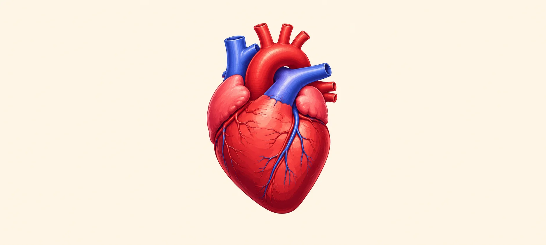
Artificial Heart: What It Is And How It Works To Save Lives
An artificial heart is a mechanical device that helps replace the pumping work of a severely damaged heart. It is usually used when both lower chambers of the heart, called ventricles, are no longer able to pump enough blood to the body. For many people with end-stage heart failure, an artificial heart can be a life-saving bridge while they wait for a donor heart transplant. It helps maintain blood circulation, supports vital organs and gives the body time to become stronger before transplant surgery. Although the term artificial heart may sound futuristic, this technology has been used for decades in selected patients with advanced heart failure disease. Research continues to make these devices smaller, safer and more suitable for long-term use. Understanding Heart Failure And The Need For Artificial Hearts Your heart works like a pump. It sends oxygen-poor blood to the lungs and oxygen-rich blood to the rest of your body. This steady pumping action keeps your brain, kidneys, liver, muscles and other organs functioning. Heart failure occurs when the heart cannot pump blood as well as it should. It does not mean the heart has stopped. It means the heart function has become too weak or stiff to meet the body’s needs. In mild to moderate heart failure, medicines, lifestyle changes, devices and surgery may help. However, in severe biventricular heart failure, both the right and left ventricles may fail. When medicines and other support devices are not enough, a total artificial heart may be considered. What Causes Heart Failure? Common causes of heart failure include: Coronary artery disease Heart attack Long-standing high blood pressure Cardiomyopathy, which means disease of the heart muscle Heart valve disease Congenital heart defects Severe abnormal heart rhythms Myocarditis, which means inflammation of the heart muscle Diabetes related heart disease Certain infections Damage from some cancer treatments Failed previous heart transplant Failed ventricular assist device in selected cases Symptoms Of Heart Failure Symptoms may develop slowly or appear suddenly. They may include: Breathlessness during activity or while lying down Fatigue and weakness Swelling in the feet, ankles, legs or abdomen Fast or irregular heartbeat Reduced ability to exercise Persistent cough or wheezing Sudden weight gain due to fluid retention Loss of appetite or nausea Dizziness or fainting Difficulty sleeping flat Confusion or reduced alertness in severe cases If you have chest pain, severe breathlessness, fainting or sudden weakness, seek emergency medical help. The History Of Artificial Hearts The idea of replacing the failing human heart with a mechanical pump developed over many decades. Scientists, surgeons and engineers worked together to design a device that could keep blood moving safely through the body. Early artificial hearts were large, complex and difficult to manage. Over time, improvements in materials, surgery, power systems and blood clot prevention made these devices more practical for selected patients. Today, a total artificial heart is mainly used as a bridge to transplant in people with severe heart failure who are waiting for a donor heart. Early Pioneers Of Artificial Heart Technology Early artificial heart research involved animal studies, experimental pumps and the development of mechanical circulatory support systems. These efforts helped doctors understand how an artificial pump could take over heart function. Several pioneers contributed to this field by developing early heart-lung machines, artificial ventricles and mechanical blood pumps. Their work created the foundation for modern total artificial heart systems. First Successful Artificial Heart Transplant The first human total artificial heart implant was performed in 1969 as a temporary bridge to transplant. Later, in 1982, a permanent artificial heart implant received global attention. These early cases were medically and ethically complex, but they helped advance research. Over the following years, artificial hearts became more focused on helping patients survive until a suitable donor heart became available. Current State Of Artificial Heart Research Artificial heart research is moving in three main directions: better survival, better quality of life and longer device support. Current research is looking at: Smaller artificial hearts for people with smaller chest sizes Devices that may be suitable for adolescents and selected children More durable pumps Safer blood flow patterns Lower risk of blood clots and bleeding Quieter and more portable external drivers Fully implantable systems in the future Artificial hearts that may one day work as long-term destination therapy Newer experimental devices are also being studied in early feasibility trials. Some designs use magnetic levitation and continuous-flow technology to reduce wear and improve durability. These are promising developments, but they are still under specialist evaluation. Challenges In Artificial Heart Development Creating an artificial heart is difficult because the human heart performs complex work every second. A device must pump enough blood, respond to body needs and avoid damaging blood cells. Major challenges include: Preventing blood clots Reducing bleeding risk from blood-thinning medicines Preventing infection where tubes exit the body Avoiding stroke Reducing device size Making power systems more convenient Improving long-term durability Supporting patients with small chest size Managing emotional stress during transplant waiting Making the technology accessible and affordable These challenges explain why artificial hearts are used only in carefully selected patients at advanced cardiac centres. How Long Can An Artificial Heart Function? A total artificial heart can support a person for several months while they wait for a donor heart. Some people have lived with artificial heart support for more than a year in special situations. How long it can function depends on the device, the person’s condition, complications, transplant availability and ongoing care. It is important to understand that most currently used total artificial hearts are not meant to be routine permanent replacements. They are usually used as a bridge to transplant. Your heart specialist can explain the expected timeline based on the specific device and your medical condition. How Artificial Hearts Impact The Cardiovascular System A total artificial heart replaces the damaged ventricles and takes over the main pumping function. It connects to the heart’s upper chambers, called atria, and to major blood vessels such as the aorta and pulmonary artery. It helps by: Sending blood to the lungs for oxygenation Sending oxygen-rich blood to the body Improving blood flow to organs Reducing symptoms caused by poor circulation Helping the body recover strength before transplant Supporting kidney, liver and brain function by improving circulation Because the device changes how blood moves through the body, patients need close monitoring. Blood-thinning medicines are often needed to reduce clot risk. Regular checkups help doctors monitor device function, infection risk and organ health. Artificial Heart Vs. Mechanical Circulatory Support Devices Artificial hearts and mechanical circulatory support devices are related, but they are not the same. A total artificial heart replaces both lower chambers of the heart. It is used when both ventricles are severely damaged or when other devices are not suitable. A ventricular assist device, such as an LVAD, supports one ventricle, usually the left ventricle. It works with the patient’s own heart rather than replacing both ventricles. In simple terms: An artificial heart replaces both ventricles. An LVAD helps one weakened ventricle pump blood. A BiVAD supports both ventricles but does not replace the heart in the same way as a total artificial heart. ECMO may provide short-term heart and lung support in critical care. Your cardiac team decides the right option based on your heart function, right heart strength, valve health, body size, transplant eligibility and overall condition. Post-Surgery Lifestyle With An Artificial Heart Life after artificial heart surgery requires careful daily care and close medical follow-up. You may need to adapt to the device, medicines and activity limits. Important lifestyle steps may include: Attending regular checkups with your cardiac team Taking blood-thinning medicines exactly as prescribed Watching for signs of infection Keeping driveline areas clean and dry Learning how to manage the external driver and batteries Following cardiac rehabilitation if advised Eating a heart-healthy diet Getting enough sleep and rest Avoiding smoking and alcohol misuse Staying physically active within safe limits Monitoring weight, temperature and symptoms Seeking help for anxiety, low mood or stress Keeping emergency contact details available Family members and caregivers are usually trained to help with device care and emergency steps. Life Expectancy After Receiving An Artificial Heart Life expectancy after receiving an artificial heart varies widely. Many people receive the device because they are already critically ill with advanced heart failure. Their outcome depends on age, overall health, organ function, infection risk, transplant availability and complications. For many patients, the main goal is to survive long enough to receive a donor heart. Once a successful heart transplant is completed, long-term outlook depends on transplant health, medicines, infection prevention and regular monitoring. Some patients may not survive the surgery or may develop complications. This is why artificial heart implantation is considered only after detailed evaluation by a specialist heart team. Artificial Heart And Organ Donation Artificial hearts are closely linked with organ donation. Most total artificial hearts are used as a bridge to transplant. This means the device helps a person stay alive while waiting for a donated human heart. The shortage of donor hearts is one reason artificial heart technology is so important. Many people with end-stage heart failure wait for a suitable donor organ. A total artificial heart can help maintain circulation during this waiting period. Organ donation remains essential. Artificial hearts can support patients, but for many people, a human heart transplant is still the final treatment goal. Future Of Artificial Hearts: What’s Next? The future of artificial hearts is focused on safer, smaller and longer-lasting devices. Researchers are working on systems that may reduce complications and improve day-to-day comfort. Future developments may include: Fully implantable artificial hearts Wireless energy transfer systems Smaller devices for more body sizes Better materials that reduce clotting Smarter pumps that adjust to activity levels Longer battery life Lower infection risk Better use in children and adolescents More options for patients who cannot receive donor hearts These advances may one day make artificial hearts a more practical long-term option. For now, their main role remains highly specialised care for advanced heart failure. Artificial Heart In Children And Adolescents Artificial hearts in children and adolescents are more complex because of body size, growth and congenital heart disease. Smaller chest size can make device placement difficult. In selected children or adolescents with severe heart failure, mechanical circulatory support may be used while waiting for transplant. Some newer smaller artificial heart systems are being studied for patients who cannot be supported by standard adult-sized devices. Paediatric use requires highly specialised teams, careful patient selection and close family counselling. Are Artificial Hearts A Long-Term Solution? At present, most total artificial hearts are not routine long-term solutions. They are mainly used as temporary support until heart transplant. In some cases, long-term or destination therapy is being studied for people who are not transplant candidates. This remains an evolving field. Long-term artificial heart use must address infection, clotting, bleeding, durability, power supply and quality of life. So, while artificial hearts can save lives, they are usually part of a larger treatment pathway rather than a permanent cure. Cultural And Ethical Considerations Of Artificial Hearts Artificial hearts raise important cultural, emotional and ethical questions. Some people may feel hopeful about advanced technology. Others may feel anxious about living with a mechanical device inside the body. Important considerations include: Informed consent Quality of life Cost and access Emotional readiness Family support Religious or cultural beliefs about body integrity Fair allocation of donor hearts End-of-life planning in advanced disease Device use in children Long-term dependence on technology Doctors, counsellors, family members and spiritual support teams can help patients make decisions that match their values and medical needs. How Artificial Hearts Are Changing The Face Of Heart Disease Treatment Artificial hearts have changed the way doctors manage severe heart failure. They give selected patients a chance to survive when medicines, standard surgery or other devices are no longer enough. They also help bridge the gap between heart failure and transplant. For patients who are too weak to wait safely, an artificial heart can restore circulation, improve organ function and create time. This progress also reminds us why heart health matters. Preventing and detecting heart disease early can reduce the risk of advanced heart failure. Regular heart tests, blood pressure checks, cholesterol monitoring, diabetes screening and timely care for symptoms can support better outcomes. Conclusion An artificial heart is a major medical advancement for people with severe heart failure. It can replace the pumping work of both ventricles and help maintain blood circulation while a patient waits for a heart transplant. It is not a routine treatment for all heart problems. It is used only in selected cases of end-stage heart failure when other options are not enough. The surgery is complex, and patients need close monitoring, medicines, device care and emotional support. For everyday heart health, early detection is still vital. If you have risk factors such as high blood pressure, diabetes, high cholesterol, obesity, smoking history or a family history of heart disease, regular screening can help you stay informed. Metropolis Healthcare offers heart tests, full body checkups, accurate reports, home sample collection, quick turnaround time and easy booking through the website, app, call and WhatsApp. With 4,000 plus tests, expert pathologists, NABL and CAP accredited labs, and a strong home collection network, Metropolis Healthcare supports preventive healthcare and ongoing wellness monitoring. FAQ How Long Can A Person Live With An Artificial Heart? A person may live with an artificial heart for several months while waiting for a donor heart transplant. Some people may be supported for longer in special cases. Survival depends on the device, the patient’s health, complications and transplant availability. What Are The Side Effects Of An Artificial Heart? Possible risks include bleeding, infection, blood clots, stroke, anaemia, kidney problems, liver problems, device-related issues and complications from major surgery. Patients also need blood-thinning medicines and careful device care. Can An Artificial Heart Be Used As A Permanent Solution? Most total artificial hearts are currently used as a temporary bridge to transplant. Permanent or long-term use is being studied, but it is not yet the standard option for most patients. Are Artificial Hearts Safe For Children? Artificial hearts may be used in selected children or adolescents with severe heart failure, but this is highly specialised. Safety depends on body size, diagnosis, device availability and the child’s overall condition. A paediatric heart failure and transplant team must assess each case carefully. References National Heart, Lung, and Blood Institute. Total Artificial Heart. Updated 2023. National Heart, Lung, and Blood Institute. Heart Failure. Updated 2022. Henn MC, Mokadam NA. Total artificial heart as a bridge to transplantation. Curr Opin Organ Transplant. 2022;27(3):222-228. PMID: 35649113. Villa CR, Morales DLS. The Total Artificial Heart in End-Stage Congenital Heart Disease. Front Physiol. 2017;8:131. PMID: 28536530. Cook JA, Shah KB, Quader MA, Cooke RH, Kasirajan V, Rao KK, Smallfield MC, Tchoukina I, Tang DG. The total artificial heart. J Thorac Dis. 2015;7(12):2172-2180. PMID: 26793338. Copeland JG, Smith RG, Arabia FA, Nolan PE, McClellan D, Tsau PH, Sethi GK, Bose RK, Banchy ME, Covington D, Slepian MJ, McCarthy MS. Cardiac replacement with a total artificial heart as a bridge to transplantation. N Engl J Med. 2004;351(9):859-867. PMID: 15329425. Tang DG, Shah KB, Hess ML, Kasirajan V. Implantation of the SynCardia total artificial heart. J Vis Exp. 2014;(89):e51377. PMID: 25046690. Cleveland Clinic. Artificial Heart. Medically reviewed. Updated 2024.

Semaglutide vs Liraglutide: Which Is Better For Weight Loss?
Semaglutide and liraglutide are both GLP-1 receptor agonists used in weight management and type 2 diabetes care. They help reduce appetite, improve fullness, and support better blood sugar control. The main difference is that semaglutide is usually taken once weekly as an injection for weight loss, while liraglutide is taken once daily as an injection. Clinical studies suggest that semaglutide generally leads to greater average weight loss than liraglutide when both are used with diet and exercise. However, the better choice depends on your health condition, weight loss goal, side effect tolerance, cost, availability, and doctor’s recommendation. What Is Semaglutide vs Liraglutide? Semaglutide and liraglutide are medicines from the GLP-1 agonist group. GLP-1 is a natural hormone that helps control appetite, digestion, and blood sugar. Semaglutide is used for weight management and type 2 diabetes, depending on the brand and dose prescribed. For weight loss, it is commonly used as a once weekly injection. Liraglutide is also used for weight management and type 2 diabetes, depending on the product and dose. For weight loss, liraglutide is usually prescribed as a once daily injection. Both medicines should be used only under medical supervision. They are not casual slimming injections. How Do Semaglutide And Liraglutide Work For Weight Loss? Semaglutide and liraglutide mimic the action of GLP-1 in your body. They help reduce appetite, slow stomach emptying, and increase the feeling of fullness after meals. This can help you eat smaller portions and reduce overall calorie intake. They may also help improve blood sugar control by increasing insulin release when blood sugar is high and reducing excess glucose production by the liver. For weight loss, these medicines work best when combined with a reduced calorie diet, regular physical activity, better sleep, and routine health monitoring. Semaglutide vs Liraglutide Weight Loss: Key Differences Feature Semaglutide Liraglutide Medicine class GLP-1 receptor agonist GLP-1 receptor agonist Common weight loss dosing pattern Once weekly injection Once daily injection Usual weight management maintenance dose 2.4 mg once weekly, depending on product and tolerance 3.0 mg once daily, depending on product and tolerance Appetite effect Strong appetite reduction in many people Appetite reduction in many people Average weight loss in head to head trial Higher average weight loss Lower average weight loss compared with semaglutide Convenience More convenient for many people due to weekly dosing Requires daily consistency Dose escalation Gradual increase over several weeks Gradual increase over several weeks Common side effects Nausea, vomiting, diarrhoea, constipation, abdominal discomfort Nausea, vomiting, diarrhoea, constipation, abdominal discomfort Prescription requirement Required Required Effectiveness: Which Leads To More Weight Loss? Semaglutide generally leads to more weight loss than liraglutide in clinical studies. In the STEP 8 trial, adults with overweight or obesity without diabetes lost more weight with once weekly semaglutide 2.4 mg than with once daily liraglutide 3.0 mg. Average body weight reduction was about 15.8% with semaglutide compared with about 6.4% with liraglutide over 68 weeks. Semaglutide may be more suitable when greater weight loss is needed and the person can tolerate it. Liraglutide may still be useful for people who respond well to it, prefer daily routine, or cannot use semaglutide. Results vary from person to person. Neither medicine works well without lifestyle changes. Dosage And Frequency: Weekly vs Daily Injections Semaglutide for weight loss is commonly taken once weekly as an injection. Liraglutide for weight loss is commonly taken once daily as an injection. Semaglutide may be easier for people who find daily injections difficult. Liraglutide may suit people who prefer a daily routine or need closer dose adjustment. Both medicines are started at a low dose and increased gradually. Do not increase the dose faster than prescribed. Do not use semaglutide and liraglutide together unless a specialist specifically advises it. Speed Of Results: How Fast Do They Work? Some people notice reduced appetite within the first few weeks. Visible weight loss usually takes longer. Since both medicines start at low doses and increase gradually, early weight loss may be modest. Semaglutide often takes several months to show its full weight loss effect because dose escalation is gradual. Liraglutide may reach its maintenance dose faster, but the average total weight loss may be lower than semaglutide. Your doctor may review progress after a defined period to decide whether the medicine is working well enough to continue. Side Effects Comparison: Semaglutide vs Liraglutide Side Effect Or Risk Semaglutide Liraglutide Nausea Common Common Vomiting Common Common Diarrhoea Common Common Constipation Common Common Abdominal pain Can occur Can occur Indigestion or acidity Can occur Can occur Reduced appetite Common Common Injection site reaction Can occur Can occur Gallbladder disease Possible risk Possible risk Acute pancreatitis Rare but serious risk Rare but serious risk Kidney problems from dehydration Possible, especially with vomiting or diarrhoea Possible, especially with vomiting or diarrhoea Low blood sugar More likely if combined with insulin or sulfonylureas More likely if combined with insulin or sulfonylureas Thyroid tumour warning Warning applies Warning applies Diabetic retinopathy concern May need caution in people with diabetic eye disease Less commonly highlighted, but diabetes monitoring is still important Common Side Effects Common side effects of semaglutide and liraglutide may include: Nausea Vomiting Diarrhoea Constipation Stomach pain Bloating Indigestion Acidity Gas Reduced appetite Headache Tiredness Dizziness Injection site redness or discomfort These side effects are often stronger when treatment starts or when the dose is increased. Eating smaller meals, avoiding greasy foods, drinking enough water, and stopping when full may help. Serious Risks And Warnings Seek urgent medical help if you develop: Severe stomach pain that may spread to the back Persistent vomiting Symptoms of acute pancreatitis Signs of gallbladder disease, such as severe upper abdominal pain, fever, nausea, or yellowing of the skin Severe dehydration after vomiting or diarrhoea Reduced urination or swelling of the feet or hands Severe allergic reaction A lump in the neck, hoarseness, trouble swallowing, or breathing difficulty New or worsening mood changes Suicidal thoughts Very low blood sugar symptoms, especially if you also take insulin or sulfonylureas Do not ignore severe or persistent symptoms. Which Is Safer For Long-Term Use? Both semaglutide and liraglutide can be used long term in suitable people under medical supervision. Safety depends on your medical history, dose, side effects, other medicines, kidney function, gallbladder health, blood sugar, and response to treatment. Semaglutide may offer greater weight loss, but some people may experience stronger digestive side effects. Liraglutide may be better tolerated by some people, but it requires daily injections and may produce less average weight loss. Long term use needs regular follow up. Your doctor may monitor weight, waist size, blood pressure, blood sugar, HbA1c, cholesterol, kidney function, liver function, digestive symptoms, and overall tolerance. Cost Comparison: Semaglutide vs Liraglutide Cost Factor Semaglutide Liraglutide General cost pattern Often costly, especially branded weight loss products Can also be costly, but some markets may have more pricing variation Dosing cost consideration Weekly dosing, but higher product cost may apply Daily dosing, so monthly usage can be substantial Insurance or coverage Depends on indication, plan, and region Depends on indication, plan, and region Availability May vary by city, pharmacy, and prescription use May vary by city, pharmacy, and prescription use Affordability decision Depends on dose, duration, product, and coverage Depends on dose, duration, product, and coverage Do not choose between these medicines based only on cost. The safer choice is the one that suits your medical profile and can be monitored properly. Availability And Prescription Requirements Both semaglutide and liraglutide require a prescription. They should be used only after a doctor checks your BMI, medical history, current medicines, blood sugar status, kidney function, gallbladder history, digestive health, and pregnancy plans. Availability may differ by country, city, pharmacy, and approved indication. Some products are approved for diabetes, some for weight management, and some for both depending on the brand and dose. Do not use a diabetes medicine for weight loss unless your doctor has prescribed it for that purpose. Who Should Choose Semaglutide? Semaglutide may be considered if: You need greater average weight loss You prefer a once weekly injection You have difficulty following daily injections Your doctor feels you are suitable for semaglutide You can tolerate gradual dose escalation You can manage the cost and availability You are willing to follow diet, exercise, and monitoring advice You do not have contraindications such as a personal or family history of medullary thyroid cancer or MEN 2 Who Should Choose Liraglutide? Liraglutide may be considered if: You prefer or can manage a daily injection routine Your doctor feels liraglutide is safer or more suitable for you You have tolerated liraglutide well in the past You need a medicine that can be adjusted through daily dosing Semaglutide is not suitable, available, or affordable You are using it for a prescribed diabetes or weight management indication You can follow regular medical review You do not have contraindications such as a personal or family history of medullary thyroid cancer or MEN 2 Semaglutide vs Liraglutide: Pros And Cons Medicine Pros Cons Semaglutide Greater average weight loss, once weekly injection, strong appetite reduction, useful for suitable people with obesity or overweight related risks Can cause digestive side effects, may be costly, may not suit people with certain medical conditions, needs gradual dose escalation Liraglutide Useful for weight management and diabetes depending on dose and product, daily dosing may allow routine consistency, may be suitable when semaglutide is not preferred Requires daily injection, average weight loss may be lower than semaglutide, can cause digestive side effects, may still be costly Can You Switch Between Semaglutide And Liraglutide? Yes, switching may be possible, but only under medical guidance. You should not switch on your own. Your doctor may consider switching if you do not lose enough weight, have side effects, face availability issues, or need a different dosing schedule. The timing of the switch, starting dose, and monitoring plan must be decided carefully to reduce the risk of nausea, vomiting, low blood sugar, dehydration, or other complications. Do not take both medicines together. Lifestyle Factors That Improve Results Medicines can support weight loss, but your routine determines how sustainable the results are. Follow a balanced GLP-1 Diet if you are using GLP-1 medicines Eat protein with each main meal Include vegetables, pulses, fruits, whole grains, nuts, curd, eggs, fish, lean meat, paneer, or tofu based on your diet preference Avoid crash diets Reduce sugary drinks, sweets, fried foods, and ultra processed snacks Eat slowly and stop when comfortably full Drink enough water Walk regularly if medically suitable Add strength training two to three times a week Sleep well Manage stress Track weight weekly Monitor waist size Keep follow up appointments Check blood sugar, cholesterol, kidney, liver, and other health markers as advised Doctor’s Recommendation: Which Is Better For You? For weight loss, semaglutide is generally more effective than liraglutide based on clinical evidence. It also offers the convenience of once weekly dosing. However, it is not automatically better for every person. Liraglutide may be more suitable for some people depending on tolerance, medical history, cost, availability, and doctor preference. Both medicines can cause side effects and both need regular monitoring. Metropolis Healthcare supports proactive health management with full body checkups, 4,000 tests, speciality testing, home sample collection, accurate results, quick turnaround time, and easy booking through website, call, app, and WhatsApp. With a strong home collection network and 10,000 touchpoints, Metropolis can help you monitor important markers such as blood sugar, HbA1c, cholesterol, kidney function, liver function, thyroid health, and overall wellness. If you are starting or already using GLP-1 Agonists, options such as the GLP 1 Test Package and GLP-1 Monitor Package can help you stay informed about your health markers during your weight management journey. References Rubino DM, Greenway FL, Khalid U, O’Neil PM, Rosenstock J, Sørrig R, et al. Effect of weekly subcutaneous semaglutide vs daily liraglutide on body weight in adults with overweight or obesity without diabetes: The STEP 8 randomized clinical trial. JAMA. 2022;327(2):138-150. PMID: 35015037. DailyMed. Wegovy, semaglutide injection, prescribing information. National Library of Medicine. DailyMed. Saxenda, liraglutide injection, prescribing information. National Library of Medicine. Wilding JPH, Batterham RL, Calanna S, Davies M, Van Gaal LF, Lingvay I, et al. Once weekly semaglutide in adults with overweight or obesity. N Engl J Med. 2021;384(11):989-1002. PMID: 33567185. Pi-Sunyer X, Astrup A, Fujioka K, Greenway F, Halpern A, Krempf M, et al. A randomized, controlled trial of 3.0 mg of liraglutide in weight management. N Engl J Med. 2015;373(1):11-22. PMID: 26132939.

How GLP-1 Injections Work In The Body?
What Is GLP-1 And Why Is It Important In The Body? GLP-1 stands for glucagon-like peptide-1. It is a natural hormone released by your gut after you eat. This hormone helps your body manage blood sugar, digestion, appetite, and fullness. It tells the pancreas to release insulin when blood sugar rises. It also helps reduce glucagon, a hormone that raises blood sugar. At the same time, GLP-1 slows how quickly food leaves your stomach and sends fullness signals to your brain. Because of these actions, GLP-1 plays an important role in diabetes management, appetite control, and weight regulation. How Does GLP-1 Work In The Body? GLP-1 works by helping your pancreas release insulin, reducing excess sugar release from the liver, slowing digestion, and signalling fullness to the brain. In simple words, GLP-1 helps your body handle food more steadily after a meal. It supports blood sugar control, reduces hunger, helps you feel full for longer, and may reduce overall calorie intake. GLP-1 injections work by mimicking this natural hormone and activating GLP-1 receptors in different parts of the body. How Do GLP-1 Medications Work For Weight Loss? GLP-1 medications can support weight loss by acting on appetite, digestion, and food intake. They may help by: Reducing hunger signals Increasing the feeling of fullness Slowing stomach emptying Reducing cravings in some people Helping you eat smaller portions Supporting better blood sugar control Reducing calorie intake over time Supporting long term weight management when combined with diet, exercise, and medical monitoring GLP-1 medicines are not a quick fix. They work best when combined with a structured eating plan, regular physical activity, strength training, sleep management, and follow up with your doctor. How GLP-1 Affects The Brain, Gut, And Pancreas GLP-1 injections act on several organs at the same time. They affect: The Brain: They act on appetite centres and help reduce hunger. The Gut: They slow gastric emptying, so food stays in the stomach longer. The Pancreas: They help release insulin when blood sugar rises. The Liver: They reduce excess glucose release by lowering glucagon. The Digestive System: They may cause nausea, fullness, constipation, or diarrhoea in some people because digestion slows down. Role Of GLP-1 In Insulin Secretion And Blood Sugar Control After you eat, your blood sugar rises. GLP-1 helps your pancreas release insulin in response to this rise. Insulin helps move sugar from your blood into your cells, where it can be used for energy. GLP-1 also reduces glucagon. Glucagon normally tells the liver to release stored sugar. By lowering glucagon, GLP-1 helps prevent excess sugar from entering the blood. This is why GLP-1 medications are useful in type 2 diabetes. They help improve blood sugar control in a glucose-dependent way, which means they work more when blood sugar is high. How GLP-1 Slows Digestion And Controls Appetite GLP-1 slows gastric emptying. This means food leaves your stomach more slowly and enters the small intestine at a slower pace. This slower movement helps you feel full for longer after meals. It may also reduce sudden hunger and help prevent overeating. This effect is one reason GLP-1 medicines can support weight loss. However, slower digestion can also cause side effects such as nausea, bloating, burping, constipation, or vomiting, especially when treatment starts or when the dose increases. How GLP-1 Injections Signal Fullness In The Brain GLP-1 injections act on brain areas that help control appetite and satiety. Satiety means the feeling of being satisfied after eating. When GLP-1 receptors are activated in the brain, you may feel less hungry and may feel full after eating less food. Some people also notice fewer cravings for high calorie foods. This brain effect is important because weight management is not only about willpower. Hunger, fullness, hormones, sleep, stress, and metabolism all influence eating behaviour. How Fast Do GLP-1 Injections Start Working In The Body? GLP-1 injections start acting in the body after the medicine is absorbed, but the visible effects take time. Some people notice reduced hunger or early fullness within the first few days or weeks. Blood sugar improvements may also begin early in people with type 2 diabetes. Weight changes usually happen gradually over several weeks to months. Doctors often start with a low dose and increase it slowly. This gradual approach helps reduce side effects and gives your body time to adjust. What Happens In Your Body After A GLP-1 Injection? A GLP-1 injection usually follows a step-by-step process in the body. The Medicine Is Injected Under The Skin Most GLP-1 injections are given under the skin of the abdomen, thigh, or upper arm. Your doctor or healthcare provider will guide you on the correct method. The Medicine Is Absorbed Slowly The medicine enters your bloodstream over time. Some GLP-1 injections are taken once daily, while others are taken once weekly. It Activates GLP-1 Receptors The medicine binds to GLP-1 receptors in organs such as the pancreas, brain, stomach, and gut. It Supports Insulin Release When blood sugar rises after a meal, it helps the pancreas release insulin. It Reduces Glucagon It helps lower excess glucagon, which reduces extra sugar release from the liver. It Slows Stomach Emptying Food stays in the stomach longer, which can help you feel full. It Reduces Hunger Signals It acts on appetite pathways in the brain, which may reduce cravings and overeating. It Supports Gradual Weight Loss Over time, reduced calorie intake and better appetite control may support weight loss. Step-By-Step: How GLP-1 Travels And Acts In The Body Injection Site: The medicine is injected into fatty tissue under the skin. Absorption: It slowly moves into the bloodstream. Circulation: It travels through the body. Pancreas Action: It helps insulin release when blood sugar is high. Liver Action: It reduces excess sugar release by lowering glucagon. Gut Action: It slows gastric emptying. Brain Action: It increases fullness and reduces hunger. Metabolic Effect: It supports blood sugar control and weight management over time. Benefits Of GLP-1 Medications Beyond Weight Loss GLP-1 medications may offer benefits beyond weight loss in selected people. Possible benefits include: Better blood sugar control in type 2 diabetes Reduced appetite Support for long term weight management Lower HbA1c in people with diabetes Better control of post-meal blood sugar spikes Improvement in some heart health markers Possible benefit for blood pressure in some people Possible improvement in cholesterol markers in some people Support for metabolic health when combined with lifestyle changes These benefits depend on the medicine used, your health status, dose, response, and medical supervision. Common Side Effects And Why They Occur GLP-1 side effects are usually linked to slower digestion and changes in appetite. Common side effects include: Nausea Vomiting Diarrhoea Constipation Bloating Burping Acidity Stomach pain Reduced appetite Early fullness Fatigue Dizziness Headache Injection site irritation These effects are more common during the first few weeks or after dose increases. They may improve as your body adjusts. Less common but serious risks may include pancreatitis, gallbladder disease, severe dehydration, kidney stress, allergic reactions, and worsening digestive symptoms. People with diabetes may also need monitoring for low blood sugar if GLP-1 medicines are used with insulin or sulfonylureas. Who Should And Should Not Use GLP-1 Injections? GLP-1 injections should be used only under medical supervision. They may be considered for: People with type 2 diabetes who need better blood sugar control People with obesity who meet clinical criteria People who are overweight with weight related health risks, if advised by a doctor People who need structured metabolic health monitoring People who can follow diet, exercise, and follow up plans They may not be suitable for: People with a personal or family history of medullary thyroid carcinoma People with Multiple Endocrine Neoplasia syndrome type 2 People with previous severe allergic reaction to the medicine People with active pancreatitis People with severe gastroparesis or serious digestive motility problems Pregnant women, unless specifically advised by a specialist People planning pregnancy People using the medicine only for cosmetic or rapid weight loss without medical need People who cannot follow medical monitoring Your doctor will decide suitability based on your medical history, BMI, blood sugar status, medicines, family history, and lab reports. Are GLP-1 Injections Safe For Long-Term Use? GLP-1 injections have been studied for diabetes and weight management, and many people use them safely under medical supervision. However, long term safety depends on the specific medicine, your health condition, dose, response, and monitoring. Regular follow up is important. Your doctor may monitor blood sugar, HbA1c, kidney function, liver function, lipid profile, weight, nutrition status, digestive symptoms, and other markers based on your condition. You should also tell your doctor if you are planning surgery, endoscopy, or any procedure under anaesthesia, because GLP-1 medicines can slow stomach emptying. Global Availability And Prescription Guidelines GLP-1 medicines are available in many countries, but their approved use, brand names, doses, and eligibility criteria may differ. Some are approved for type 2 diabetes, some for chronic weight management, and some may have both uses depending on local regulations. In most places, GLP-1 injections require a prescription. They should be started only after medical evaluation. Buying or using them without supervision can be unsafe, especially if you have diabetes, kidney disease, digestive problems, gallbladder disease, thyroid cancer risk, or are taking other medicines. If you are considering GLP-1 treatment, speak with a qualified doctor. Testing and follow up can help make treatment safer and more personalised. Conclusion GLP-1 injections work by copying the action of a natural gut hormone. They help regulate blood sugar, reduce hunger, slow digestion, and increase fullness. This is why they can support type 2 diabetes care and medically supervised weight management. However, GLP-1 medicines are not suitable for everyone. They need the right prescription, gradual dosing, healthy eating, exercise, and regular monitoring. Metropolis Healthcare can support your wellness journey with preventive health checkups, speciality testing, accurate reports, expert pathologists, NABL and CAP accredited labs, and convenient home sample collection. With 4,000+ tests and easy booking through the website, app, call, and WhatsApp, Metropolis makes health monitoring simple and accessible. If you are exploring GLP-1 Agonists, following a GLP-1 Diet, or considering a GLP 1 Test Package or GLP-1 Monitor Package, regular testing can help you and your doctor track important health markers such as blood sugar, HbA1c, kidney function, liver function, lipid profile, and nutritional status. FAQ Can GLP-1 Injections Cause Dumping Syndrome? GLP-1 injections do not usually cause dumping syndrome. Dumping syndrome happens when food moves too quickly from the stomach into the small intestine. GLP-1 medicines usually do the opposite by slowing stomach emptying. However, some side effects such as nausea, diarrhoea, dizziness, sweating, or weakness may feel similar. If you have these symptoms after meals, speak to your doctor. Why Do GLP-1 Medications Cause Nausea Similar To Dumping Syndrome? GLP-1 medicines can cause nausea because they slow stomach emptying and affect appetite signals in the brain. Your stomach may stay full for longer, so large meals, oily foods, or eating too fast can trigger discomfort. This can feel similar to some digestive symptoms of dumping syndrome, but the underlying process is different. How Can You Reduce Digestive Side Effects Of GLP-1 Drugs? You can reduce digestive side effects by: Eating smaller meals Eating slowly Stopping when full Avoiding fried and oily foods Limiting very sweet foods Drinking water through the day Avoiding large meals late at night Reducing alcohol Following the dose schedule given by your doctor Reporting severe or persistent symptoms early What Foods Should Be Avoided While Taking GLP-1 Injections? You may need to limit foods that worsen nausea, bloating, or reflux. These may include: Fried foods Greasy foods Very spicy foods Very sweet foods Sugary drinks Large heavy meals Alcohol Carbonated drinks if they cause bloating High fat desserts Fast food You do not need to avoid all these foods forever. The goal is to understand what triggers your symptoms and choose balanced meals that your body tolerates well. Do GLP-1 Medications Affect Gastric Emptying Permanently? GLP-1 medications slow gastric emptying while they are active in the body. This effect may be stronger when treatment begins and may reduce over time in some people. It is not usually considered permanent. If you have persistent vomiting, severe fullness, inability to eat, or symptoms of gastroparesis, contact your doctor. References Drucker DJ. Mechanisms of action and therapeutic application of glucagon-like peptide-1. Cell Metab. 2018;27(4):740-756. PMID: 29617641. Ard J, Fitch A, Fruh S, Herman L. Weight loss and maintenance related to the mechanism of action of glucagon-like peptide 1 receptor agonists. Adv Ther. 2021;38(6):2821-2839. PMID: 33977495. Collins L, Costello RA. Glucagon-like peptide-1 receptor agonists. StatPearls. Updated 2024. U.S. Food and Drug Administration. Wegovy prescribing information. Revised 2026. Kim JA, Yoo HJ. Exploring the side effects of GLP-1 receptor agonist: to ensure its optimal positioning. Diabetes Metab J. 2025;49(4):525-541. Kindel TL, Wang AY, Wadhwa A, et al. Multisociety clinical practice guidance for the safe use of glucagon-like peptide-1 receptor agonists in the perioperative period. Clin Gastroenterol Hepatol. 2025;23(12):2083-2085. PMID: 39480373.

Mounjaro For Weight Loss: Complete Guide
Mounjaro is a prescription injection that contains tirzepatide. It is mainly used for type 2 diabetes management in many countries, but tirzepatide is also known for its strong effect on weight loss. Mounjaro for weight loss should only be used under medical supervision. It is not a cosmetic slimming injection or a short cut for quick fat loss. It works best when combined with a balanced diet, regular physical activity, proper sleep, and routine health monitoring. What Is Mounjaro Weight Loss And How Does It Work? Mounjaro contains tirzepatide, a medicine that acts on two natural hormone pathways in the body: GIP and GLP-1. These hormones help regulate appetite, digestion, insulin release, and blood sugar control. Because Mounjaro affects hunger and fullness, many people eat less while taking it. This can lead to gradual weight loss when the medicine is used with healthy lifestyle changes. Mounjaro injection for weight loss should be prescribed only after a doctor checks your BMI, medical history, current medicines, diabetes status, digestive health, kidney function, gallbladder history, and weight related health risks. How Mounjaro Weight Loss Targets Appetite And Fat Loss Mounjaro may support weight loss by: Reducing appetite Helping you feel full sooner Helping you stay full for longer after meals Slowing stomach emptying Reducing overeating and cravings in some people Supporting better blood sugar control Improving insulin sensitivity Helping reduce calorie intake when paired with a structured diet Supporting fat loss over time when used with exercise and lifestyle changes It does not melt fat directly. It helps create conditions that make weight loss easier and more sustainable. Mounjaro Injection For Weight Loss: How It Helps Reduce Body Weight Mounjaro injection for weight loss works by changing appetite signals and slowing how quickly food leaves the stomach. You may feel less hungry, feel satisfied with smaller meals, and find it easier to reduce calorie intake. This can help people with obesity or excess weight lose body weight gradually. However, the response is different for each person. Your results depend on dose, diet, activity, sleep, stress, medical conditions, other medicines, and how consistently you follow the treatment plan. How To Use Mounjaro Injection For Weight Loss Use It Once Weekly Mounjaro is usually injected once a week on the same day each week. Choose The Injection Site It is injected under the skin of the abdomen, thigh, or upper arm. Rotate Injection Sites Do not inject into the exact same spot every time. Rotation helps reduce skin irritation. Use It With Or Without Food Mounjaro can usually be taken with or without meals. Follow The Prescribed Dose Do not increase, reduce, or skip doses without medical advice. Use A New Needle Or Device As Directed Follow the instructions given for your specific pen or device. Store It Correctly Follow the storage instructions on the product label and your pharmacist’s advice. Track Symptoms Note nausea, vomiting, diarrhoea, constipation, abdominal pain, appetite changes, dizziness, or signs of dehydration. Recommended Dosage For Mounjaro Weight Loss The dose is increased slowly to help reduce digestive side effects. Your doctor will decide the right dose for you. Treatment Stage Usual Dose Pattern Starting dose 2.5 mg once weekly for 4 weeks First increase 5 mg once weekly Further increases Dose may rise by 2.5 mg steps every 4 weeks if needed and tolerated Common maintenance doses 5 mg, 10 mg, or 15 mg once weekly, based on response and tolerance Maximum dose 15 mg once weekly The 2.5 mg dose is usually used for treatment initiation. It is not usually considered the main long term maintenance dose. How Effective Is Mounjaro For Weight Loss? Tirzepatide has shown significant weight loss results in clinical studies when used with diet and exercise. In major trials, people with obesity or excess weight lost a meaningful percentage of body weight over several months. Higher doses generally produced greater weight loss, but they may also increase the chance of side effects. The goal is not to reach the highest dose quickly. The goal is to find a dose that is effective and safe for you. Average Weight Loss Results With Mounjaro Clinical study results suggest that: Weight loss is gradual rather than immediate Appetite reduction may begin earlier than visible fat loss Many people lose more weight as the dose is increased Significant results often take several months Some people may lose around 15% to 20% or more of their starting body weight in clinical trial settings Results are usually better when medicine is combined with nutrition, exercise, and follow up care Weight regain can happen after stopping treatment if lifestyle and medical support are not continued Your personal result may be higher or lower than trial averages. Mounjaro Weight Loss Results Timeline: What To Expect Mounjaro weight loss usually happens in stages. Do not expect dramatic changes in the first few days. Weeks 1 To 4 You usually start with the lowest dose. Appetite may reduce, but weight loss may be mild. Nausea, fullness, or digestive changes may appear. Weeks 5 To 8 Your doctor may increase the dose if tolerated. You may notice better portion control and early weight changes. Weeks 9 To 16 Weight loss may become more visible. Clothes may feel looser, and waist measurement may reduce. Months 4 To 6 Many people see clearer changes in weight, appetite, and eating patterns. Regular monitoring becomes important. Months 6 To 12 Weight loss may continue if you remain consistent with treatment and lifestyle changes. Beyond 12 Months Your doctor may review whether long term use is suitable, whether the dose should continue, and how your health markers are responding. Week-by-Week Mounjaro Weight Loss Progress Week 1: You may notice reduced hunger or mild nausea Week 2: Your body continues adjusting to the medicine Week 3: Portion sizes may reduce naturally Week 4: Your doctor may review tolerance before increasing the dose Weeks 5 To 8: Weight loss may become more noticeable Weeks 9 To 12: Appetite control may improve further Weeks 13 To 16: Waist size and weight may show clearer changes After Week 16: Continued progress depends on dose, diet, activity, sleep, and follow up care Do not compare your progress with another person’s. Weight loss response is individual. Who Is Eligible For Mounjaro Weight Loss Treatment? Doctors may consider Mounjaro or tirzepatide based treatment for: Adults with obesity Adults with excess weight and weight related health risks People with type 2 diabetes who may benefit from better blood sugar control and weight reduction People who have not achieved enough weight loss with lifestyle changes alone People who can follow regular medical monitoring People who can manage possible side effects People who are not pregnant or planning pregnancy People who do not have contraindications such as certain thyroid cancer risks Eligibility depends on local approvals, clinical judgement, and your health profile. Who Should Avoid Mounjaro For Weight Loss? Mounjaro may not be suitable if you: Are pregnant Are planning pregnancy Are breastfeeding, unless your doctor advises otherwise Have a personal or family history of medullary thyroid cancer Have Multiple Endocrine Neoplasia syndrome type 2 Have had a serious allergic reaction to tirzepatide Have a history of pancreatitis Have severe stomach emptying problems or gastroparesis Have active gallbladder disease Have repeated vomiting or dehydration Have significant kidney impairment Take insulin or sulfonylureas without close monitoring Are already using another GLP-1 or GIP based medicine Are considering it only for rapid cosmetic weight loss Your doctor may ask for blood tests before and during treatment. Mounjaro Weight Loss Side Effects You Should Know Most Mounjaro side effects are related to the digestive system. They are often more common when starting treatment or increasing the dose. Possible side effects include: Nausea Vomiting Diarrhoea Constipation Stomach pain Indigestion Heartburn Bloating Gas Reduced appetite Tiredness Dizziness Injection site reaction Low blood sugar, especially if used with insulin or certain diabetes medicines Common Side Effects During Mounjaro Weight Loss Common side effects may include: Nausea after meals Early fullness Reduced appetite Loose stools Constipation Mild stomach cramps Acid reflux Burping Headache Fatigue These symptoms may improve as your body adjusts. Speak to your doctor if they continue, worsen, or interfere with eating and hydration. Serious Risks Of Mounjaro Weight Loss Treatment Serious side effects are less common, but they need urgent medical attention. These include severe abdominal pain, persistent vomiting, symptoms of acute pancreatitis, gallbladder disease, severe dehydration, kidney problems, serious allergic reaction, and very low blood sugar if used with diabetes medicines. Mounjaro carries a warning about thyroid C-cell tumours based on animal studies. It should not be used by people with a personal or family history of medullary thyroid cancer or Multiple Endocrine Neoplasia syndrome type 2. Can Mounjaro Weight Loss Cause Dumping Syndrome? Mounjaro is not usually described as a direct cause of dumping syndrome. Dumping syndrome happens when food moves from the stomach to the small intestine too quickly, often after stomach or weight loss surgery. Mounjaro usually slows stomach emptying, so it works differently from dumping syndrome. However, some digestive side effects, such as nausea, cramps, diarrhoea, sweating, weakness, or discomfort after meals, may feel similar. If you have had bariatric surgery or you develop repeated post meal symptoms, speak to your doctor. Symptoms Similar To Dumping Syndrome During Mounjaro Weight Loss Symptoms that may feel similar include: Nausea after eating Stomach cramps Diarrhoea Sweating Dizziness Weakness Bloating Fast heartbeat Feeling shaky after meals Sudden fullness after small meals These symptoms can have many causes. They should be medically reviewed if persistent. Tips To Avoid Digestive Issues During Mounjaro Weight Loss To reduce digestive discomfort: Eat smaller meals Eat slowly Stop eating when comfortably full Avoid greasy foods Reduce spicy and very heavy meals Limit sugary foods and sweet drinks Avoid large late night meals Drink enough water Include protein in each meal Avoid lying down immediately after eating Do not increase the dose faster than prescribed Report persistent vomiting or diarrhoea early Mounjaro Weight Loss vs Other Weight Loss Medications Medicine Active Ingredient Main Hormone Target Usual Weight Loss Dosing Pattern Key Point Mounjaro Tirzepatide GIP and GLP-1 Once weekly injection Used for type 2 diabetes in many countries, often discussed for weight loss because of tirzepatide’s strong effect Zepbound Tirzepatide GIP and GLP-1 Once weekly injection Tirzepatide product specifically approved for chronic weight management in some countries Semaglutide Semaglutide GLP-1 Usually once weekly injection for weight management Strong weight loss evidence and widely used in obesity care Liraglutide Liraglutide GLP-1 Once daily injection Daily GLP-1 option for weight management in suitable people Orlistat Orlistat Fat absorption in gut Usually taken with meals Works differently by reducing fat absorption The best medicine depends on your BMI, health risks, side effect tolerance, cost, availability, medical history, and doctor’s judgement. Diet Plan To Support Mounjaro Weight Loss A healthy diet can improve results and reduce side effects. Eat protein with each main meal Include vegetables daily Choose whole grains instead of refined grains Add pulses, beans, curd, paneer, tofu, eggs, fish, or lean meat based on your food preferences Include fruits in moderate portions Eat fibre rich foods to support digestion Drink enough water Keep meals small and balanced Avoid skipping meals if it leads to overeating later Follow a GLP-1 Diet plan if advised by your doctor or dietitian Track your food intake if it helps you stay consistent Avoid crash dieting The goal is steady fat loss while preserving nutrition and muscle health. Foods To Avoid While On Mounjaro Weight Loss Treatment Try to limit: Fried foods Greasy foods Sugary drinks Sweets and desserts Ultra processed snacks Large portions of refined carbs Alcohol Very spicy meals if they worsen acidity Large meals late at night Foods that trigger nausea or bloating Carbonated drinks if they worsen gas You do not need an extreme diet. You need a sustainable eating pattern. Mounjaro Weight Loss Cost, Availability And Prescription Mounjaro cost and availability can vary based on country, city, pharmacy, dose, supply, and prescription indication. Insurance or reimbursement may also differ depending on whether it is prescribed for type 2 diabetes or weight management. Because Mounjaro is a prescription medicine, it should not be bought or used without a doctor’s supervision. Avoid unverified online sellers, compounded products, or medicines without proper packaging and prescription support. Your doctor or pharmacist can guide you on legitimate availability, correct storage, and safe use. Is Mounjaro Weight Loss Safe For Long-Term Use? Mounjaro may be used long term in suitable people under medical supervision. Obesity is a chronic condition, so weight management may need long term support. Long term treatment needs regular review because your doctor must check whether the medicine is helping, whether side effects are manageable, and whether your health markers are safe. Monitoring may include blood sugar, HbA1c, cholesterol, liver function, kidney function, thyroid related history, gallbladder symptoms, weight, waist size, and blood pressure. Stopping treatment suddenly may lead to increased appetite and weight regain in some people. Your doctor can help plan the safest long term approach. Final Verdict: Is Mounjaro Weight Loss Right For You? Mounjaro can support significant weight loss in suitable people because tirzepatide acts on both GIP and GLP-1 pathways. It may reduce appetite, improve fullness, support better blood sugar control, and help with gradual weight reduction when combined with lifestyle changes. However, it is not right for everyone. It can cause digestive side effects and may carry serious risks in some people. It should only be used with a valid prescription and regular follow up. Metropolis Healthcare supports proactive health management with full body checkups, 4,000 tests, speciality testing, home sample collection, accurate results, quick turnaround time, and easy booking through website, call, app, and WhatsApp. With a strong home collection network and 10,000 touchpoints, Metropolis can help you monitor important health markers such as blood sugar, HbA1c, cholesterol, kidney function, liver function, thyroid health, and overall wellness. If you are starting or already using GLP-1 Agonists or similar weight management medicines, options such as the GLP 1 Test Package and GLP-1 Monitor Package can help you stay informed about your health markers during your weight loss journey. References DailyMed. Mounjaro, tirzepatide injection, prescribing information. National Library of Medicine. Jastreboff AM, Aronne LJ, Ahmad NN, Wharton S, Connery L, Alves B, et al. Tirzepatide once weekly for the treatment of obesity. N Engl J Med. 2022;387(3):205-216. PMID: 35658024. Aronne LJ, Sattar N, Horn DB, Bays HE, Wharton S, Lin WY, et al. Continued treatment with tirzepatide for maintenance of weight reduction in adults with obesity: The SURMOUNT-4 randomized clinical trial. JAMA. 2024;331(1):38-48. PMID: 38078870. Aronne LJ, Horn DB, le Roux CW, Sattar N, Batterham RL, Wharton S, et al. Tirzepatide as compared with semaglutide for the treatment of obesity. N Engl J Med. 2025. PMID: 40353578.

What Is OCD And How Does It Affect Your Daily Life?
What Is OCD? (Obsessive Compulsive Disorder Explained) Obsessive Compulsive Disorder, or OCD, is a mental health condition in which you experience repeated unwanted thoughts, fears, urges, or images. These are called obsessions. To reduce the anxiety caused by these thoughts, you may feel driven to repeat certain actions or mental rituals. These are called compulsions. OCD is not the same as being very neat, careful, or organised. It can be distressing, time consuming, and difficult to control. It may affect your work, studies, relationships, sleep, routine, and emotional well-being. The good news is that OCD can be treated and managed with the right support. OCD Full Form In Medical Terms OCD full form in medical terms is Obsessive Compulsive Disorder. Obsessive refers to repeated unwanted thoughts or urges. Compulsive refers to repeated behaviours or mental acts that you feel forced to do. OCD Disease: Is It A Mental Health Disorder? Yes, OCD is a mental health disorder. It is sometimes casually called an OCD disease, but the more accurate term is Obsessive Compulsive Disorder. It affects how your brain processes fear, uncertainty, discomfort, and the need for relief. People with OCD often know that their thoughts or rituals may not be logical, but still find it very hard to stop them. What Are The Common OCD Symptoms? OCD symptoms can vary from person to person. Some people mostly have intrusive thoughts, while others have visible rituals. Many people have both. Common OCD symptoms include: Repeated unwanted thoughts that cause anxiety Fear of germs, dirt, or contamination Repeated handwashing or cleaning Repeated checking of doors, gas knobs, switches, locks, or appliances Fear of harming yourself or others, even when you do not want to Strong need for symmetry, order, or exactness Repeating words, prayers, numbers, or phrases silently Asking for reassurance again and again Avoiding people, places, or objects that trigger fear Spending a lot of time on rituals Feeling temporary relief after doing a compulsion Feeling guilty, ashamed, or exhausted because of symptoms Obsessions Vs Compulsions: What’s The Difference? Feature Obsessions Compulsions Meaning Unwanted thoughts, urges, fears, or images Repeated actions or mental rituals How They Feel Disturbing, intrusive, and anxiety provoking Driven, repetitive, and difficult to resist Purpose They trigger fear or discomfort They are done to reduce fear or prevent something bad Examples Fear of germs, fear of harm, doubt, unwanted taboo thoughts Washing, checking, counting, repeating, arranging, reassurance seeking Early Warning Signs Of OCD Early signs of OCD may be easy to miss. You may think you are simply being careful, but the pattern becomes concerning when it starts taking too much time or causing distress. Early warning signs include: Taking much longer than usual to complete daily tasks Repeating the same action until it feels right Avoiding normal activities due to fear or doubt Feeling unable to stop checking or cleaning Asking the same question repeatedly for reassurance Becoming very upset if things are not arranged in a certain way Feeling trapped in a cycle of fear and relief Hiding rituals due to shame or embarrassment Losing sleep because of intrusive thoughts 7 Types Of OCD You Should Know OCD can appear in different forms. These are not always separate diagnoses, but common symptom patterns. Contamination OCD This involves fear of germs, dirt, bodily fluids, chemicals, or illness. It may lead to excessive washing, cleaning, or avoidance. Checking OCD This involves repeated checking to prevent harm or danger. You may check locks, switches, appliances, messages, or mistakes again and again. Symmetry And Ordering OCD This involves a strong need for things to feel even, balanced, arranged, or perfect. You may keep adjusting items until they feel right. Harm OCD This involves unwanted thoughts or images about harming yourself or others. These thoughts can be very distressing because they go against your values. Religious Or Moral OCD This involves intense fear of doing something morally wrong, sinful, or offensive. It may include repeated praying, confessing, or seeking reassurance. Relationship OCD This involves repeated doubts about your relationships, feelings, attraction, or whether you are making the right choice. Pure Obsessional OCD This often involves intrusive thoughts with mental rituals rather than visible compulsions. Mental checking, reviewing, repeating, or reassurance seeking may still be present. Some OCD related conditions may include Body Dysmorphic Disorder, Hoarding Disorder, Excoriation, and Olfactory Reference Disorder. These conditions can overlap with OCD patterns, but they need proper evaluation by a mental health professional. What Causes OCD? The exact cause of OCD is not fully known. It usually develops due to a combination of biological, genetic, psychological, and environmental factors. Possible factors include changes in brain circuits linked with fear, habit, and decision making. Family history may increase risk. Stressful life events, trauma, childhood experiences, and learned behaviours may also contribute. OCD is not caused by weakness, lack of willpower, or poor personality. It is a recognised mental health condition, and seeking help is an important step towards recovery. How OCD Affects Daily Life OCD can affect daily life in many ways. The impact depends on the severity of symptoms, the type of obsessions, and the time spent on compulsions. For example, if you have contamination fears, you may avoid public transport, handshakes, shared meals, or public toilets. If you have checking OCD, you may spend a long time checking locks or appliances before leaving home. If you have intrusive harm thoughts, you may avoid being around loved ones even though you care deeply about them. OCD may affect: Work or school attendance Focus and productivity Family relationships Social life Sleep Personal hygiene routines Eating patterns Travel Self-confidence Emotional health OCD can also occur along with anxiety, depression, panic disorder, Eating Disorders, tic disorders, or body image concerns. If symptoms are affecting your routine, early help can make recovery easier. When Should You Seek Help For OCD? You should seek help if unwanted thoughts or repetitive behaviours are taking up time, causing distress, affecting relationships, or stopping you from doing everyday activities. You should also speak to a mental health professional if you feel ashamed, exhausted, trapped, or unable to control rituals even when you know they are not helpful. Seek urgent help if you have thoughts of self-harm, feel unsafe, or feel unable to cope. Immediate support can protect you and help you get the right care. OCD Diagnosis: How Is OCD Identified? OCD is diagnosed by a qualified mental health professional through a detailed assessment. The process may include: Discussion Of Symptoms The doctor or therapist may ask about intrusive thoughts, fears, rituals, avoidance, and distress. Time And Impact Assessment They may check how much time symptoms take and how they affect your work, relationships, studies, or daily routine. Mental Health Evaluation They may look for anxiety, depression, panic disorder, Eating Disorders, tic disorders, trauma, or other related conditions. Medical History Review Your doctor may ask about medicines, sleep, substance use, family history, and physical health. Use Of Screening Tools Some professionals use structured questionnaires to understand severity. Rule Out Other Conditions Symptoms may be compared with conditions such as Body Dysmorphic Disorder, Hoarding Disorder, Excoriation, Olfactory Reference Disorder, or general anxiety. There is no single blood test or scan that confirms OCD. Diagnosis is mainly based on symptoms and clinical assessment. OCD Treatment Options: What Works Best? OCD treatment usually includes psychotherapy, medicines, or a combination of both. The right plan depends on your symptoms, severity, age, medical history, and personal needs. Common treatment options include: Cognitive Behavioral Therapy Exposure and Response Prevention Medicines such as SSRIs Family education and support Stress management Sleep improvement Regular follow up Support groups Treatment for related conditions such as anxiety, depression, panic disorder, or Eating Disorders Treatment takes time, but many people improve with the right care. Cognitive Behavioral Therapy (CBT) For OCD Cognitive Behavioral Therapy, or CBT, is one of the most effective therapies for OCD. A specific type of CBT called Exposure and Response Prevention, or ERP, is especially helpful. In ERP, you gradually face feared thoughts, objects, or situations while learning not to perform the compulsion. This helps your brain learn that anxiety can reduce on its own without rituals. ERP should be done with a trained therapist, especially if symptoms are severe. Medications For OCD Treatment Medicines can help reduce OCD symptoms for many people. Common medication options include: Selective serotonin reuptake inhibitors, also called SSRIs Clomipramine in selected cases Additional medicines if OCD occurs with anxiety, depression, or tic symptoms Medicine adjustments if symptoms do not improve enough Medication should always be prescribed and monitored by a qualified doctor. Do not start, stop, or change psychiatric medicines on your own. Lifestyle Changes To Manage OCD Lifestyle changes cannot replace medical treatment, but they can support recovery. Helpful habits include: Following your therapy plan regularly Avoiding reassurance seeking as much as possible Practising stress reduction Sleeping at a regular time Reducing caffeine if it worsens anxiety Exercising regularly Eating balanced meals Building a predictable daily routine Talking to trusted family members Joining a support group Avoiding alcohol or substance use as coping methods If physical symptoms such as fatigue, sleep issues, appetite changes, or sudden weight changes are present, your doctor may also advise health checkups to rule out related medical concerns. Can OCD Be Cured Or Managed Long-Term? OCD is often a long term condition, but it can be managed well. Some people have mild symptoms that improve with therapy. Others may need ongoing treatment and follow up. The goal is to reduce distress, reduce rituals, improve daily functioning, and help you live with more freedom. Early treatment, family support, therapy, medicines when needed, and consistent practice can make a major difference. OCD does not define you. With the right care, you can regain control over your routine and quality of life. Conclusion OCD is a mental health disorder that causes unwanted intrusive thoughts and repetitive behaviours or mental rituals. It is more than being neat or careful. It can affect daily life, relationships, sleep, work, studies, and emotional health. If you or someone you know has OCD symptoms, it is important to seek help from a mental health professional. Effective treatment options such as CBT, ERP, and medicines can help manage symptoms and improve quality of life. Metropolis Healthcare supports preventive healthcare and overall wellness with reliable diagnostic testing, full body checkups, accurate reports, expert pathologists, NABL and CAP accredited labs, and convenient home sample collection. While OCD diagnosis needs a mental health assessment, routine health checks can help your doctor understand your overall health and rule out physical factors that may affect mood, energy, sleep, and well-being. FAQ Can You Overcome OCD? Yes, many people can manage OCD well with proper treatment. CBT, especially ERP, and medicines can reduce symptoms and improve daily life. Some people may not become completely symptom free, but they can learn to control symptoms instead of letting symptoms control them. Is OCD A Brain Disorder? OCD is a mental health disorder linked with brain circuits involved in fear, habits, decision making, and uncertainty. It is not a weakness or personality flaw. Brain function, genetics, stress, and learning patterns may all play a role. What Is The Hardest OCD To Treat? There is no single hardest type of OCD for everyone. OCD can be harder to treat when symptoms are severe, hidden, linked with poor insight, or occur with depression, anxiety, tic disorders, trauma, or substance use. Early diagnosis and specialised ERP therapy can improve outcomes. What Are The 4 Stages Of OCD? A common way to understand the OCD cycle is through four stages: obsession, anxiety, compulsion, and temporary relief. First, an intrusive thought appears. Then anxiety rises. Next, a compulsion is performed to reduce distress. Finally, relief comes for a short time, but the cycle often returns. What Is The Root Cause Of OCD? There is no single root cause of OCD. It may develop due to a mix of brain chemistry, genetics, family history, stress, trauma, personality traits, and learned behaviours. A mental health professional can help identify your triggers and create a treatment plan.

Blood Tests For Cancer: How They Detect Different Types Of Cancer
A blood test for cancer can give important clues about your health. It can show abnormal blood cell levels, tumour markers, organ function changes, proteins, or cancer related genetic material in the blood. However, blood tests do not diagnose most cancers on their own. They are usually one part of a larger evaluation that may include physical examination, imaging, biopsy, bone marrow tests, genetic tests, and specialist review. Blood tests are especially useful for detecting some blood cancers, monitoring treatment response, checking recurrence, and understanding how cancer or treatment is affecting the body. What Is A Blood Test For Cancer And How Does It Work? A blood test for cancer is a laboratory test that checks your blood sample for signs that may be linked with cancer. These signs may include unusual blood cell counts, abnormal proteins, tumour markers, circulating tumour cells, or fragments of cancer DNA. Some blood tests look for direct changes in blood cells. Others look for substances released by cancer cells or by the body in response to cancer. A blood test for cancer markers may help doctors decide whether further tests are needed. It may also help monitor treatment progress or check if cancer has returned after treatment. Why Blood Tests Are Used In Cancer Detection Blood tests may be used to: Check for abnormal blood cell counts Detect possible signs of blood cancer Measure tumour markers Assess liver and kidney function before treatment Understand how the body is responding to cancer treatment Track whether cancer is reducing, stable, or progressing Monitor for recurrence after treatment Support diagnosis along with imaging and biopsy Help guide treatment decisions in selected cancers Check overall health before surgery, chemotherapy, radiation, or targeted therapy Blood tests are helpful, but they are not a replacement for a full medical evaluation. Which Blood Test For Cancer Is Most Commonly Used? The most commonly used blood test in cancer evaluation is the Complete Blood Count, also called CBC Test. A CBC Test measures red blood cells, white blood cells, haemoglobin, haematocrit, and platelets. It can help detect possible blood cancers such as leukaemia, lymphoma, and multiple myeloma. It can also show anaemia, infection, bleeding risk, or treatment related changes. For solid cancers, such as prostate cancer, ovarian cancer, breast cancer, lung cancer, or colon cancer, a CBC alone is usually not enough. Doctors may use tumour marker tests, imaging, biopsy, or other specialised tests based on symptoms and risk factors. Types Of Blood Tests For Cancer Detection Common blood tests used in cancer care include: Complete Blood Count: Measures blood cells and can help detect blood cancers or treatment related changes. Tumour Marker Tests: Measure substances that may be higher in some cancers, such as PSA, CA-125, CEA, AFP, or CA 19-9. Blood Protein Tests: Check abnormal protein patterns that may be seen in multiple myeloma and some other blood disorders. Peripheral Blood Smear: Allows specialists to look at blood cells under a microscope for abnormal shapes, sizes, or immature cells. Flow Cytometry: Helps identify certain types of leukaemia and lymphoma by studying markers on blood or bone marrow cells. Liquid Biopsy: Looks for circulating tumour DNA or cancer related genetic changes in the blood. Circulating Tumour Cell Tests: Detect cancer cells that have broken away from a tumour and entered the bloodstream. Liver And Kidney Function Tests: Do not diagnose cancer directly, but help assess organ function before and during treatment. What Types Of Cancer Can Be Detected Through Blood Tests? Blood tests may help in the evaluation of several cancers, including: Leukaemia: A CBC may show very high or very low white blood cells, anaemia, low platelets, or abnormal immature cells. Lymphoma: Blood tests may show abnormal blood counts, high LDH, or other changes, but biopsy is usually needed for confirmation. Multiple Myeloma: Blood protein tests, serum electrophoresis, calcium levels, kidney function tests, and blood counts may provide important clues. Prostate Cancer: PSA testing may show raised prostate specific antigen levels. However, high PSA can also occur due to non cancer causes. Ovarian Cancer: CA-125 Test may be used in selected cases, especially for monitoring treatment or recurrence. It is not a standalone screening test for everyone. Liver Cancer: AFP may be used along with imaging and clinical assessment. Colorectal Cancer: CEA may help monitor treatment response or recurrence, but it is not used alone to diagnose colon cancer. Testicular Cancer: AFP, beta hCG, and LDH may help in diagnosis, staging, and monitoring. Some newer blood tests aim to detect signals from multiple cancers, but many are still used carefully and need follow up testing. How Accurate Is A Blood Test For Cancer Detection? The accuracy of a blood test for cancer depends on the type of test, the cancer type, the stage of disease, and the reason for testing. Some tests are useful for certain cancers but not for others. For example, a CBC can help detect blood cancers, but it cannot reliably detect most solid tumours. Tumour markers may rise in cancer, but they can also rise due to non cancer conditions such as inflammation, infection, benign growths, liver disease, prostate enlargement, or endometriosis. A normal blood test also does not always rule out cancer. This is why doctors interpret blood test results with symptoms, examination findings, imaging, biopsy, and medical history. Can Blood Tests Alone Diagnose Cancer? In most cases, no. Blood tests alone cannot confirm cancer. A confirmed cancer diagnosis usually needs tissue or cell examination. This may be done through a biopsy, bone marrow test, surgical sample, or cytology, depending on the suspected cancer. Blood tests can raise suspicion, support diagnosis, guide further testing, help monitor treatment, and detect recurrence. They are an important part of cancer care, but they are rarely the only test needed. When Should You Consider A Blood Test For Cancer? You may need a blood test if you have: Unexplained weight loss Persistent fatigue Fever that does not settle Night sweats Unusual bleeding or bruising Long lasting swelling of lymph nodes Repeated infections Persistent abdominal bloating Blood in urine or stool Change in bowel habits Persistent pain without clear reason Loss of appetite Family history of certain cancers Previous cancer treatment and need for monitoring Abnormal imaging result Doctor advised screening or follow up These symptoms do not always mean cancer. Many common conditions can cause similar symptoms. A doctor can help decide which blood test for cancer or other test is suitable. How To Prepare For A Blood Test For Cancer Follow Your Doctor’s Instructions Some tests need fasting, while others do not. Confirm this before your appointment. Share Your Medical History Tell your doctor about current symptoms, past illnesses, surgeries, cancer history, and family history. List All Medicines And Supplements Some medicines may affect test results. Do not stop any medicine unless your doctor advises it. Stay Hydrated Drink water unless fasting instructions say otherwise. Avoid Heavy Exercise Before Testing Intense exercise may affect some blood values. Carry Previous Reports Older blood reports, imaging scans, biopsy reports, or treatment records help doctors compare results. Ask About Follow Up Understand when results are expected and what the next step may be. What Happens After An Abnormal Cancer Blood Test Result? An abnormal result does not always mean cancer. Your doctor will review the result along with your symptoms, age, sex, risk factors, examination findings, and previous reports. You may be advised to repeat the blood test, do imaging such as ultrasound, CT, MRI, or PET scan, undergo biopsy, meet a specialist, or take additional tests such as tumour markers, bone marrow examination, or genetic testing. The next step depends on which value is abnormal and what cancer or non cancer condition is suspected. Benefits And Limitations Of Blood Tests For Cancer Benefits Limitations Simple and quick sample collection Cannot diagnose most cancers alone Helps detect blood cell abnormalities Normal results do not always rule out cancer Useful for blood cancers such as leukaemia and lymphoma Tumour markers can rise due to non cancer causes Helps monitor treatment response Some cancers do not produce measurable markers Helps check recurrence in selected cancers Results need expert interpretation Can assess kidney, liver, and overall health before treatment Additional tests such as imaging or biopsy may be needed Useful in follow up care Not every person needs every cancer blood test Are Blood Tests Used For Cancer Monitoring And Recurrence? Yes. Blood tests are often used after a cancer diagnosis to monitor treatment response, side effects, and recurrence. For example, tumour markers may be tracked over time in selected cancers. A falling marker may suggest response to treatment, while a rising marker may suggest recurrence or progression. However, one result is usually not enough. Doctors look at trends, symptoms, scans, and clinical findings. Blood tests may also help monitor anaemia, infection risk, platelet count, liver function, kidney function, and nutritional status during cancer treatment. Preventive Health Checkups And Early Cancer Detection Preventive health checkups can help identify health changes early. They may include a CBC Test, liver function, kidney function, blood sugar, and other routine markers. These tests may not screen for all cancers, but they can reveal abnormalities that need further evaluation. Cancer screening should be based on age, sex, family history, symptoms, lifestyle risk, and doctor advice. Some cancers need specific screening tests, such as Pap smear, HPV testing, mammography, colonoscopy, PSA testing, or targeted imaging. Metropolis Healthcare supports preventive health management with 4,000 tests, full body checkups, speciality testing, home sample collection, accurate results, quick turnaround time, and easy booking through website, call, app, and WhatsApp. With a strong home collection network and 10,000 touchpoints, Metropolis can help you monitor important health markers through tests such as CBC Test, Tumor Marker Test, and CA-125 Test when advised by your doctor. FAQ Can Cancer Be Detected During A Blood Test? Yes, some cancers can show signs in blood tests. Blood cancers such as leukaemia, lymphoma, and multiple myeloma may cause abnormal blood counts or abnormal proteins. Some solid cancers may show raised tumour markers. However, most cancers need imaging, biopsy, or other tests for confirmation. What Is The Biggest Indicator Of Cancer? There is no single biggest indicator of cancer. Warning signs may include unexplained weight loss, persistent fatigue, unusual bleeding, a lump, long lasting fever, night sweats, change in bowel habits, persistent pain, or abnormal test results. The most important step is to see a doctor if symptoms continue or worsen. What Are The Top 3 Causes Of Cancer? Cancer usually develops due to a mix of genetic, lifestyle, environmental, and biological factors. Major preventable risk factors include tobacco use, infections linked with cancer, unhealthy diet, excess body weight, alcohol use, physical inactivity, and UV radiation. Family history and ageing can also increase risk. What Food Kills Cancer Cells In The Body? No food can reliably kill cancer cells in the body. A healthy diet can support overall health, immunity, treatment recovery, and weight management, but it cannot replace medical cancer treatment. A balanced diet with vegetables, fruits, whole grains, pulses, nuts, lean proteins, and adequate hydration is helpful for general wellness. What Is The New Blood Test For Cancer? Newer blood tests include liquid biopsy and multi cancer early detection tests. These tests look for cancer related DNA, proteins, or other signals in the blood. Some are used in selected patients to guide treatment or monitor cancer. Others are still being studied for early detection. Any abnormal result needs careful follow up with a doctor. References National Cancer Institute. Tests and procedures used to diagnose cancer. Updated 2023. National Cancer Institute. Tumor markers in cancer diagnosis and treatment. Updated 2023. Mayo Clinic Staff. Cancer blood tests: Lab tests used in cancer diagnosis. Updated 2024. Cleveland Clinic. Blood tests for cancer: Diagnosis and screening. Updated 2024. National Cancer Institute. Tumor marker tests in common use. Updated 2023.

Best Fat Loss Exercises To Burn Belly Fat Fast
Belly fat is one of the most common fitness concerns. It can affect how you feel, how your clothes fit, and sometimes, how confident you feel in your body. But it is important to know one thing clearly: you cannot reduce belly fat only by doing stomach exercises. The best fat loss exercise plan is one that helps you burn overall body fat. This includes cardio, strength training, HIIT, full body movements, a balanced fat loss diet, good sleep, stress control, and regular consistency. You do not need extreme workouts or expensive equipment to start. Many effective fat loss exercises at home can help you build strength, burn calories, and improve your overall fitness when done correctly. What Are The Best Fat Loss Exercises To Burn Belly Fat Fast? The best exercises for fat loss are full body movements that increase your heart rate, use large muscle groups, and help you burn calories. These include brisk walking, jumping jacks, squats, lunges, mountain climbers, burpees, push ups, planks, cycling, swimming, and strength training exercises. HIIT workouts and compound strength movements are especially useful because they work many muscles at once. However, no exercise can directly melt fat from your belly alone. Belly fat reduces when your overall body fat reduces. For the best results, combine exercise with healthy eating, proper hydration, enough protein, and regular sleep. How Does Fat Loss Exercise Help Reduce Belly Fat? Fat loss exercise helps your body use more energy. When you burn more calories than you consume over time, your body starts using stored fat for fuel. This can include fat around your belly, hips, thighs, arms, and other areas. Exercise also helps improve insulin sensitivity, heart health, muscle strength, stamina, and mood. Strength training is especially helpful because muscle tissue uses more energy than fat tissue, even at rest. Core exercises like planks and bicycle crunches are useful too. They strengthen your abdominal muscles, improve posture, and support your back. But they should be part of a full body fat loss workout, not the only exercise you do. Types Of Fat Loss Exercises You Should Include A balanced fat loss workout should include different types of movement. This helps you burn calories, build muscle, protect your joints, and avoid boredom. Cardio-Based Fat Loss Exercises Cardio exercises increase your heart rate and help your body burn calories. These are often considered the best exercise for fat loss when combined with diet and strength training. Good cardio exercises include: Brisk walking Jogging Cycling Swimming Dancing Stair climbing Skipping Low impact aerobics Marching in place Fast walking on an incline Start with a pace that feels manageable. You should feel slightly breathless, but still able to speak in short sentences. Strength Training For Full Body Fat Loss Exercise Strength training helps you build lean muscle and improve body shape. It also supports long term fat loss by improving your resting energy use. Useful strength exercises include: Squats Lunges Push ups Glute bridges Dumbbell rows Shoulder presses Deadlifts Step ups Planks Resistance band exercises You can use body weight, dumbbells, resistance bands, or gym machines. If you are a beginner, focus on correct form before increasing intensity. HIIT Workouts For Faster Fat Burn HIIT stands for High-Intensity Interval Training. It includes short bursts of hard exercise followed by short recovery periods. Examples include: 30 seconds jumping jacks, 30 seconds rest 30 seconds mountain climbers, 30 seconds rest 20 seconds burpees, 40 seconds rest 30 seconds squat jumps, 30 seconds rest 30 seconds high knees, 30 seconds rest HIIT can be effective, but it is intense. If you have heart disease, joint pain, uncontrolled diabetes, high blood pressure, or are new to exercise, speak to your doctor before starting high intensity workouts. 10 Best Fat Loss Exercise At Home You can do these fat loss exercises at home without equipment. Start slowly and increase the number of rounds as your stamina improves. Jumping Jacks Stand tall, jump your feet out, raise your arms, and return to the starting position. This improves heart rate and warms up your full body. High Knees Run in place while lifting your knees towards your chest. Keep your core tight and move at a steady pace. Bodyweight Squats Stand with feet shoulder width apart. Bend your knees and push your hips back as if sitting on a chair. Squats work your thighs, hips, glutes, and core. Reverse Lunges Step one leg back and lower your body gently. Return to standing and repeat on the other side. Lunges help strengthen your lower body. Mountain Climbers Start in a plank position and bring one knee at a time towards your chest. This works your core, shoulders, and legs while raising your heart rate. Modified Burpees Stand tall, place your hands on the floor, step your feet back into a plank, step forward again, and stand up. Beginners can avoid the jump. Push Ups Push ups work your chest, shoulders, arms, and core. You can start with knee push ups if regular push ups feel difficult. Plank Hold your body in a straight line on your forearms or hands. Keep your stomach firm and avoid dropping your hips. Bicycle Crunches Lie on your back and move opposite elbow towards opposite knee. This strengthens the abdominal and side waist muscles. Glute Bridges Lie on your back with knees bent. Lift your hips, squeeze your glutes, and lower slowly. This supports your hips, lower back, and core. Full Body Fat Loss Workout Plan Before every workout, warm up for 5 to 10 minutes. You can march in place, do arm circles, walk slowly, or perform light mobility movements. After the workout, cool down and stretch gently. Beginner Fat Loss Workout Routine Do this 3 to 4 days a week. March in place: 1 minute Bodyweight squats: 10 reps Wall push ups: 10 reps Glute bridges: 12 reps Step ups or stair walking: 1 minute Plank: 15 to 20 seconds Rest: 60 seconds Repeat: 2 to 3 rounds Intermediate Fat Loss Workout Split Do this 4 to 5 days a week. Jumping jacks: 45 seconds Squats: 15 reps Reverse lunges: 10 reps each side Push ups: 10 to 12 reps Mountain climbers: 30 seconds Plank: 30 to 45 seconds Rest: 45 seconds Repeat: 3 to 4 rounds Advanced Fat Loss Workout Plan Do this 4 to 5 days a week, only if your fitness level allows it. Burpees: 10 reps Jump squats: 12 reps Push ups: 15 reps Mountain climbers: 45 seconds Walking lunges: 12 reps each side Plank shoulder taps: 30 seconds Bicycle crunches: 20 reps Rest: 30 to 45 seconds Repeat: 4 to 5 rounds Weekly Fat Loss Workout Schedule Day Workout Focus Monday Cardio and core Tuesday Strength training Wednesday Brisk walking or low intensity cardio Thursday HIIT or full body circuit Friday Strength training Saturday Long walk, cycling, swimming, or dance Sunday Rest, stretching, or yoga Common Mistakes That Slow Down Belly Fat Loss Small mistakes can slow your progress even when you exercise regularly. Ignoring Diet And Nutrition Exercise works best when your diet supports your goal. If you eat more calories than your body needs, belly fat loss becomes slower. Avoid these common diet mistakes: Skipping protein Drinking too many sugary drinks Eating large portions of fried snacks Depending only on a fat loss drink Using fat loss tablets without medical advice Expecting a fat loss belt to burn belly fat Eating very little and then overeating later A fat loss diet should include balanced meals with protein, vegetables, whole grains, fruits, healthy fats, and enough water. Overtraining Without Recovery More exercise is not always better. Your body needs rest to repair muscles, balance hormones, and maintain energy. Overtraining can lead to: Tiredness Poor sleep Body pain Low motivation Increased hunger Higher injury risk Take at least 1 to 2 lighter days each week. Sleep well and stretch after workouts. Only Doing Crunches For Belly Fat Crunches can strengthen your abdominal muscles, but they do not directly burn belly fat. If your routine includes only sit ups, crunches, or planks, your calorie burn may be limited. For better results, combine: Cardio Strength training HIIT Core work Healthy eating Daily movement How To Combine Diet And Fat Loss Exercise For Faster Results Exercise and diet work together. Exercise helps you burn calories and build strength. Food gives your body the nutrients it needs to recover and function well. To support fat loss: Eat protein with every main meal Add vegetables to lunch and dinner Choose whole grains over refined grains Limit sugary drinks and sweets Use healthy fats in small portions Avoid extreme crash diets Keep a regular meal pattern Track your progress through waist size, energy, strength, and clothes fit Protein Intake And Fat Loss Protein helps you feel full and supports muscle repair after exercise. Good protein choices include: Dal Lentils Beans Curd Paneer Eggs Fish Chicken Soy Tofu Nuts and seeds in small portions If you exercise regularly, your protein needs may be higher. A nutrition expert can help you choose the right amount for your body weight and health goals. Importance Of Sleep And Stress Management Poor sleep and high stress can make fat loss harder. They may increase hunger, cravings, tiredness, and emotional eating. Helpful habits include: Sleeping at a regular time Reducing screen use before bed Practising deep breathing Walking outdoors Doing yoga or stretching Taking rest days Avoiding late night heavy meals Stress control is not just good for your mind. It also supports better food choices and regular exercise. Hydration And Metabolism Water helps your body function well during exercise. Dehydration can make you feel tired, dizzy, or less focused. Simple hydration tips include: Drink water through the day Carry a bottle when you exercise Drink more in hot weather Limit sugary drinks Choose water instead of high calorie beverages You do not need a special fat loss drink to reduce belly fat. Plain water, balanced meals, and consistent movement are more useful. How Long Does It Take To Lose Belly Fat With Exercise? The time needed to lose belly fat depends on your starting weight, diet, age, hormones, sleep, stress, activity level, and medical history. Some people notice better stamina within 2 to 3 weeks. Visible waist changes often take longer. A steady approach is safer and more sustainable than rapid weight loss. Focus on progress signs such as: Better energy Improved sleep Looser clothes Better strength Lower waist measurement Improved blood test markers Better consistency with meals and workouts If you have sudden weight gain, unexplained weight loss, tiredness, irregular periods, diabetes, thyroid concerns, or other symptoms, speak to your doctor. Best Time To Do Fat Loss Exercise For Maximum Results The best time to exercise is the time you can follow consistently. Morning workouts may help some people stay disciplined. Evening workouts may suit others better because the body feels warmer and more active. Choose a time based on: Your work schedule Your energy level Meal timing Sleep routine Family responsibilities Safety and comfort Avoid intense exercise immediately after a heavy meal. Keep a small gap and listen to your body. Conclusion The best way to burn belly fat is to focus on overall fat loss, not only abdominal exercises. A complete plan should include cardio, strength training, HIIT when suitable, a balanced fat loss diet, good sleep, stress control, and regular health monitoring. Metropolis Healthcare can support your wellness journey with preventive health checkups, full body checkups, speciality testing, and reliable reports. With 4,000+ tests, expert pathologists, NABL and CAP accredited labs, easy booking through the website, call, app, and WhatsApp, and home sample collection through a strong network of 10,000 touchpoints, Metropolis makes proactive health monitoring simple and accessible. If you are working on weight management, routine blood tests can help you understand important health markers such as blood sugar, lipid profile, liver function, thyroid function, and vitamin levels. People exploring GLP-1 Agonists, a GLP-1 Diet, the GLP 1 Test Package, or the GLP-1 Monitor Package can also use structured health tracking to stay informed and make safer decisions with medical guidance. FAQ What Is The Most Effective Fat Loss Exercise? The most effective fat loss exercise is usually a combination of cardio, strength training, and full body movements. Brisk walking, squats, lunges, burpees, mountain climbers, push ups, and cycling are useful options. The best exercise is one you can do safely and regularly. Can I Do Fat Loss Exercise At Home Without Equipment? Yes, you can do fat loss exercise at home without equipment. Bodyweight exercises such as squats, lunges, push ups, planks, jumping jacks, high knees, mountain climbers, and glute bridges can help you burn calories and build strength. How Many Days A Week Should I Exercise For Fat Loss? Most adults can aim for at least 150 minutes of moderate intensity aerobic activity per week, along with muscle strengthening exercises on 2 or more days a week. If you are new to exercise, start with 3 days a week and build gradually. Do Ab Workouts Burn Belly Fat? Ab workouts strengthen your core, but they do not directly burn belly fat. Belly fat reduces when your total body fat reduces through calorie control, cardio, strength training, and consistent healthy habits. What Is GLP-1 (Glucagon-Like Peptide-1) And How Does It Support Fat Loss? GLP-1 is a natural hormone that helps regulate blood sugar, appetite, and fullness. GLP-1 Agonists are medicines that act like this hormone and may support weight management in selected people under medical supervision. They are not a replacement for healthy eating, exercise, or regular health monitoring. Can Rapid Weight Loss Cause Dumping Syndrome? Rapid weight loss itself does not usually cause dumping syndrome. Dumping syndrome is more commonly linked with rapid emptying of food from the stomach into the small intestine, often after stomach or weight loss surgery. Symptoms may include nausea, diarrhoea, bloating, cramps, dizziness, or tiredness after meals. If you have these symptoms, speak to your doctor. References World Health Organization. WHO guidelines on physical activity and sedentary behaviour. Geneva: World Health Organization; 2020. Parker T. What’s the best exercise to lose fat around your belly? British Heart Foundation. Updated 7 January 2026. Donnelly JE, Blair SN, Jakicic JM, Manore MM, Rankin JW, Smith BK. Appropriate physical activity intervention strategies for weight loss and prevention of weight regain for adults. Med Sci Sports Exerc. 2009;41(2):459-471. PMID: 19127177. Vispute SS, Smith JD, LeCheminant JD, Hurley KS. The effect of abdominal exercise on abdominal fat. J Strength Cond Res. 2011;25(9):2559-2564. PMID: 21804427. Ard J, Fitch A, Fruh S, Herman L. Weight loss and maintenance related to the mechanism of action of glucagon-like peptide 1 receptor agonists. Adv Ther. 2021;38(6):2821-2839. PMID: 33977495. National Institute of Diabetes and Digestive and Kidney Diseases. Definition and facts for dumping syndrome. Last reviewed January 2019.