

Web Stories


Latest Blogs

Pink Himalayan Salt Benefits: Is It Healthier Than Table Salt?
What Is Pink Himalayan Salt? Pink Himalayan salt is a natural mineral salt sourced from the ancient Khewra Salt Mine near the foothills of the Himalayas in Pakistan. Its distinctive pink hue comes from the presence of trace minerals, including iron, potassium, magnesium, and calcium. This salt was formed millions of years ago from prehistoric seas, making it one of the oldest and purest forms of salt in the world. Unlike heavily processed table salt, pink salt undergoes minimal refinement and is often marketed for its unique flavour profile, mineral content, and visually appealing colour. Himalayan pink salt benefits have gained popularity in culinary applications, wellness products, and decorative items like salt lamps. Pink Salt vs. Sea Salt – Which one is better? Both Himalayan salt and sea salt are less processed than table salt and contain some trace minerals. The main differences lie in their sources and appearance. Feature Pink Himalayan Salt Sea Salt Source Ancient sea beds in the Himalayan mountains Evaporated modern seawater Colour Pink White, grey, or pink, depending on minerals Texture Coarse Coarse or fine Processing Minimal, unrefined Minimal, sometimes refined Trace Minerals Iron, potassium, magnesium, calcium Varies based on source and pollution levels In terms of health impacts, the Himalayan pink salt benefits and sea salt benefits are nutritionally similar. Both consist mostly of sodium chloride. The trace minerals in Himalayan pink salt are not present in sufficient quantities to make a significant difference. Sea salt's mineral content depends on the water source and may have marine pollutants. Ultimately, both salts affect the body in the same way as regular salt. Himalayan Salt vs Table Salt – Key differences you should know The major distinction between Himalayan salt and table salt lies in their degree of processing. Table salt is typically mined from underground salt deposits and heavily refined to remove impurities and create fine crystals. Additives like iodine and anti-caking agents are then added. In contrast, pink salt is minimally processed to yield coarser crystals that retain the natural pink colour from trace minerals. However, the mineral content is not enough to provide substantial pink salt benefits over table salt. Feature Himalayan Pink Salt Table Salt Source The Himalayan mountains in Pakistan Underground salt mines Processing Minimal, unrefined Heavily refined, fortified with iodine Texture Coarse Fine Colour Pink White Minerals Trace amounts of iron, magnesium, potassium, and calcium Iodine added, trace minerals removed Uses Cooking, seasoning, decoration Cooking, seasoning, and food preservation While pink Himalayan salt is less artificial than table salt, both have the same basic nutritional value, as sodium chloride is the main component. The trace minerals in Himalayan salt, though often promoted as beneficial, are not present in high enough amounts to have a substantial health impact. Essential Minerals Present in Pink Himalayan Salt The main pink salt benefits are due to its mineral content, which gives the salt its pink colour. While Himalayan salt does contain more trace minerals than table salt, the amounts are very small. Here is the mineral content per gram in pink Himalayan salt versus regular table salt: Mineral Pink Himalayan Salt (mg) Table Salt (mg) Calcium 1.6 0.4 Potassium 2.8 0.9 Magnesium 1.06 0.0139 Iron 0.037 0.0101 Sodium 368 381 While these trace minerals are indeed present in pink Himalayan salt, they exist in very small quantities. As a result, they are unlikely to provide significant health benefits when the salt is consumed in typical dietary amounts. Top 15 Health Benefits of Pink Himalayan Salt Here are some of the Himalayan pink salt benefits: 1. Rich in minerals: Pink Himalayan salt contains small amounts of minerals such as calcium, magnesium, and potassium, which give it its pink colour. However, these minerals are present in such tiny amounts that they are unlikely to provide significant health benefits 2. Balances electrolytes: Its natural mineral content helps regulate fluid levels in the body, like all salts, himalayan pink salt contributes sodium, which is essential for hydration and nerve and muscle function. Its effect is the same as regular salt. It's especially beneficial for athletes or those who sweat heavily. 3. Improves hydration: Like all salts, pink salt provides sodium, which helps maintain fluid balance in the body. However, there is no evidence that pink salt hydrates better than plain water or other salts 4. Supports respiratory health: Inhaling salt air, through salt therapy or salt caves, can help clear mucus, reduce inflammation, and ease breathing difficulties associated with asthma, bronchitis, or allergies. 5. Aids digestion: Pink Himalayan salt stimulates digestive enzymes and stomach acid, aiding in food breakdown and nutrient absorption. It can also help reduce bloating and gas when consumed in small amounts. 6. Promotes pH balance: Some holistic practices claims that pink salt balances body pH are not supported by scientific evidence. The body regulates pH tightly through kidneys and lungs, not diet salt. 7. Detoxifies the body: Pink salt baths can help with relaxation and muscle soothing, but they do not detoxify the body. Detoxification is carried out by organs such as the liver and kidneys. This detox process can support better circulation and skin clarity over time. 8. Improves sleep quality: There is no reliable evidence that consuming pink Himalayan salt improves sleep. Adequate magnesium intake from food sources is more relevant for sleep regulation. 9.Supports healthy blood pressure: Excess sodium from any salt, including pink salt, can raise blood pressure. There is no evidence pink salt provides any benefit over table salt. Taken in moderation, it may help maintain more stable blood pressure levels without additives or chemicals. 10. Reduces muscle cramps: Its magnesium and potassium content help prevent muscle cramps by supporting proper muscle contraction and nerve function. Especially useful for those who experience night cramps or cramps during exercise. 11. Improves skin health: When used in scrubs or baths, pink salt can exfoliate and soothe skin. While it may improve dryness and texture, there is no strong evidence that it treats acne, psoriasis, or eczema. 12. Boosts respiratory function (halotherapy): Breathing in pink salt particles during halotherapy may reduce sinus congestion, inflammation, and lung irritation. This is often used in wellness spas for treating chronic respiratory issues naturally. 13. Natural air purifier (via salt lamps): Pink salt lamps are popular for ambience, but scientific evidence for air purification or health benefits is lacking, purifying the air by attracting dust and allergens. 14. May support hormonal balance: Pink salt does not contain enough iodine to support thyroid health. For thyroid support, iodized salt is recommended. This may assist in maintaining balanced energy, mood, and metabolism. 15. Promotes bone health: Calcium, magnesium, and other trace minerals in pink salt may contribute to maintaining strong, healthy bones. These minerals support bone density and joint function when part of a balanced diet. Possible Side Effects of Himalayan Pink Salt Excessive consumption of Himalayan salt can lead to health issues similar to those caused by other salts, due to its sodium content. Potential side effects include: High blood pressure Increased risk of heart disease and stroke Fluid retention and bloating Kidney strain in individuals with impaired kidney function Worsening of certain medical conditions, such as congestive heart failure It is crucial to use pink salt in moderation and consult with a doctor if you have concerns about your salt intake, especially if you have pre-existing health conditions. Why Does Your Body Need Salt? Importance & Functions Salt plays a vital role in various bodily functions, making it an essential nutrient when consumed in appropriate amounts. Some of the key functions of salt include: Maintaining fluid balance and hydration Regulating blood pressure and volume Supporting nerve and muscle function Aiding in digestion and nutrient absorption Balancing electrolytes like sodium, potassium, and chloride However, it is important to note that excessive salt intake can have detrimental effects on health, so moderation is key. The World Health Organisation recommends limiting daily sodium intake to less than 2 grams (equivalent to 5 grams of salt) for adults. How to Use Pink Himalayan Salt for Cooking & Wellness Pink Himalayan salt can be used in various ways for culinary and wellness purposes: As a cooking and finishing salt, it adds a visual appeal to dishes. In salt grinders for a coarser texture and adjustable grind size As an ingredient in homemade seasoning blends and rubs In salt blocks for grilling, searing, and serving food As a component in bath salts and body scrubs for relaxation and skin care In salt lamps and decorative items for ambient lighting and purported air purification When using pink salt for cooking or wellness, it is essential to remember that it is still primarily composed of sodium chloride and should be used in moderation as part of a balanced lifestyle. As a leading chain of diagnostic labs across India, Metropolis Healthcare is committed to providing accurate pathology testing and health check-up services to empower patients in prioritising their health. With a team of qualified blood collection technicians who make at-home visits and advanced diagnostic labs that process samples, Metropolis ensures reliable results and personalised care. Test reports are conveniently shared online via email and the user-friendly Metropolis TruHealth app, making it easy for you to take charge of your well-being. FAQs What makes Himalayan salt special? The main difference between Himalayan salt and regular table salt is that pink salt is less processed and contains trace amounts of additional minerals, which give it a pink colour. However, these minerals are not present in high enough amounts to provide significant health benefits. Who should avoid Himalayan salt? People with high blood pressure, heart disease, kidney disease, or other conditions that require sodium restriction should limit their intake of all types of salt, including pink Himalayan salt. Always follow your doctor's recommendations for salt intake. Is it safe to eat Himalayan salt daily? Eating pink Himalayan salt daily is safe as long as you keep your total sodium intake within the recommended limit of 2,300 mg per day. However, there is no added health benefit to choosing Himalayan salt over other types of salt. Use it in moderation as you would regular salt. Is Himalayan salt healthier than sea salt? Pink Himalayan salt and sea salt have similar nutritional profiles and health effects. While Himalayan salt may contain slightly higher trace mineral amounts, the quantities are insignificant compared to the high sodium content. Sea salt's mineral content varies depending on the water source. Which Is Healthier – Pink Salt or White Salt? Both the pink and white salt are similar in sodium content and nutrition. Although the pink salt contains trace minerals, they are minimal. However, moderation matters more than type for maintaining heart and overall health. References • https://nutritionsource.hsph.harvard.edu/salt-and-sodium/ • https://health.clevelandclinic.org/do-sea-salt-kosher-salt-and-pink-salt-beat-table-salt • https://newsnetwork.mayoclinic.org/discussion/mayo-clinic-minute-is-himalayan-sea-salt-a-healthy-alternative-video/
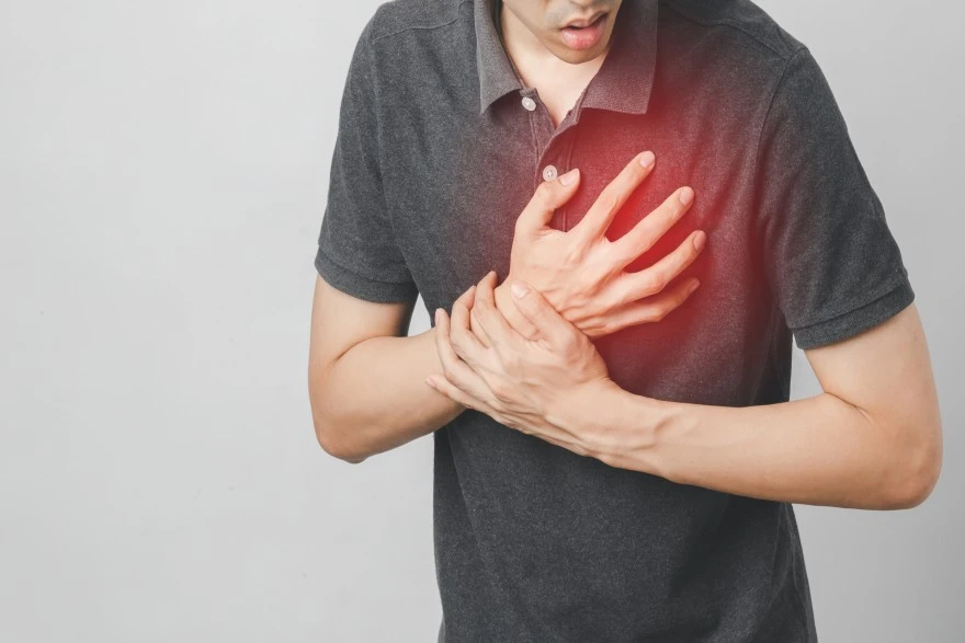
Chest Pain Due To Gas: Causes And Remedies
How Does Gas Buildup Cause Chest Pain? Gas can accumulate in the digestive system for various reasons. Some common causes of chest pain due to gas include: Swallowing excess air: Gas can build up in the digestive system when we swallow too much air, a condition known as aerophagia. This commonly happens when eating or drinking too quickly, chewing gum, drinking carbonated beverages, or smoking. The excess air may not escape easily and can become trapped in the digestive tract, leading to discomfort and bloating. Gas-producing foods: Certain foods are harder to digest and are more likely to cause gas as they break down. These include beans, lentils, and cruciferous vegetables such as broccoli, cabbage, and cauliflower. Onions and dairy products can also be problematic, especially for those with lactose intolerance. As these foods are fermented by gut bacteria, gas is released as a byproduct. Food intolerances and digestive disorders: Food intolerances, like lactose intolerance or coeliac disease, can interfere with proper digestion and lead to excessive gas production. Digestive conditions such as irritable bowel syndrome (IBS), gastro-oesophageal reflux disease (GERD), Crohn’s disease, and ulcerative colitis also increase the likelihood of gas buildup. These conditions often disrupt normal bowel function and cause gas to become trapped. When too much gas accumulates, it can cause the stomach or intestines to stretch and distend. This stretching creates pressure that may radiate into the chest area. Gas can also become trapped in certain bends of the colon, particularly near the upper abdomen, which can lead to sharp, localised pain. This left-sided chest pain due to gas can sometimes be mistaken for heart pain because the symptoms may overlap, but it is not due to shared nerve pathways Though chest pain due to gas is usually harmless, any unexplained chest pain should be taken seriously. Seek medical attention if the pain is severe, persistent, or accompanied by symptoms like shortness of breath or dizziness. What Does Gas-Induced Chest Pain Feel Like? Chest pain due to gas can feel surprisingly intense, making many people worry about their heart. However, some characteristic signs can help you identify gas pain: Location: The pain is often felt in the lower chest or upper abdomen and may shift to different spots. Pain character: It frequently manifests as a sharp, stabbing, or jabbing pain rather than the crushing or heavy sensation associated with heart-related pain. Some people describe it as a deep, dull ache or a feeling of pressure or tightness. Associated digestive symptoms: Gas, chest pain usually comes with other symptoms like abdominal bloating and distension, excessive burping or belching, and passing a lot of gas or flatulence. Triggers: You may notice the pain occurs after eating a large meal, especially one including hard-to-digest or gas-producing foods. It can also happen if you've been under a lot of stress, which affects digestion. Relieving factors: Unlike cardiac pain, gas pain often improves when you belch, pass gas, or have a bowel movement. Changing position, like going from lying down to sitting up, can also help shift the gas and relieve discomfort. Gas Pain vs. Heart Pain: Key Differences It's crucial to differentiate gas pain from heart-related pain. Here's a quick comparison: Feature Gas Pain in the Chest Heart Attack Chest Pain Character Sharp, stabbing, burning, jabbing Crushing, squeezing, heavy pressure Location The lower chest/upper abdomen may shift Centre or left side of chest Digestive symptoms Bloating, belching, flatulence, nausea Nausea, cold sweats, shortness of breath Relieving factors Belching, passing gas, changing position Not relieved by gas-related actions Radiation May spread to the abdomen May radiate to the arm, jaw, back, and neck Triggers Eating, drinking, swallowing air Physical exertion, emotional stress Duration Tends to pass once gas moves through More persistent, may worsen with time If you have chest pain with symptoms like crushing pressure, shortness of breath, cold sweats, or pain radiating into your arm, jaw, or back, treat it as a medical emergency and seek help immediately. Common Symptoms of Chest Pain from Gas If you're experiencing chest pain due to gas, watch out for these common symptoms: Tightness, pressure, or a dull ache in the chest Sharp, stabbing, or jabbing pains in the chest and upper abdomen A burning sensation in the chest, similar to indigestion or heartburn Pain that seems to move between the chest and abdomen Excessive burping or belching, sometimes bringing up bitter liquid or partially digested food. Bloating and abdominal distension, with your belly feeling full and tight Increased flatulence or passing a lot of gas Nausea and reduced appetite Symptoms can vary from person to person and may overlap with other conditions. If you have persistent or intense chest pain, it's best to get it checked by a doctor to rule out any serious issues. How Doctors Diagnose Chest Pain Due to Gas To diagnose the cause of chest pain and distinguish between gas pain and more serious conditions, doctors follow a thorough and step-by-step approach: Medical history: Your doctor will begin by asking about your eating habits, recent meals, speed of eating, and whether you consume gas-producing foods like beans or carbonated drinks. They’ll also enquire about smoking, chewing gum, and any history of food intolerances or digestive conditions like IBS or GERD. Symptom assessment: You'll be asked to describe your pain in detail—its location, intensity, timing, and whether it's sharp, dull, or cramping. Associated symptoms such as bloating, belching, flatulence, heartburn, nausea, or changes in bowel habits help narrow down the cause. Physical examination: The doctor will gently press on areas of your abdomen to check for tenderness, bloating, or discomfort. They may also listen to your abdomen with a stethoscope to detect bowel sounds that indicate movement of gas. Rule out serious conditions: Because chest pain can signal a heart attack or other serious issues, doctors usually rule out cardiac causes first. This may involve an electrocardiogram (ECG), chest X-ray, and blood tests to check for signs of heart damage, especially if the pain radiates or is accompanied by shortness of breath or dizziness. Digestive investigations: If gas pain is suspected, the doctor may order: Stool tests to detect infections, blood, or inflammation. Imaging tests like X-rays, ultrasound, or CT scans are used to identify excess gas or intestinal issues. Tests for food intolerances like lactose intolerance or coeliac disease Evaluation for conditions like GERD, IBS, or inflammatory bowel diseases Your doctor may also recommend an elimination diet or other lifestyle. Changes to identify triggers for gas buildup. With the right diagnosis and treatment plan, most people can effectively manage their chest pain due to gas symptoms. Effective Medical Treatments for Gas-Induced Chest Pain When chest pain due to gas becomes frequent or severe, medical intervention may be necessary. Treatment focuses on relieving gas buildup and addressing any underlying digestive issues. Over-the-Counter Medications Several OTC medications can effectively alleviate chest pain due to gas: Simethicone: This medication works by breaking up gas bubbles in the gut, making them easier to expel. Antacids: If gas pain is related to indigestion or acid reflux, antacids can neutralise stomach acid and provide relief. Activated charcoal: Some people use activated charcoal for gas relief, but clinical evidence is weak and it is not routinely recommended. Lactase supplements: For those with lactose intolerance, these supplements aid in digesting dairy products, reducing gas symptoms. Alpha-galactosidase: This enzyme assists in digesting certain carbohydrates, which can minimise gas production after eating beans or cruciferous vegetables. Probiotics: These beneficial bacteria may support gut health and alleviate symptoms in some individuals with recurring gas issues. Always check with your doctor before starting any new medication, and be sure to follow the package instructions carefully. Dietary Modifications Making strategic changes to your diet can significantly reduce the formation of gas and related chest discomfort: Identify and avoid trigger foods: Common culprits include beans, lentils, cruciferous vegetables (like broccoli and cabbage), onions, carbonated drinks, and artificial sweeteners such as sorbitol. Reduce lactose intake: If you're lactose intolerant, opt for lactose-free alternatives or take lactase supplements when consuming dairy. Monitor gluten intake: For those with coeliac disease or gluten sensitivities, avoiding gluten-containing products can prevent gas and other digestive symptoms. Eat smaller, more frequent meals: Large meals can overburden digestion and increase gas production. Increase dietary fibre gradually: While fibre is beneficial, suddenly increasing your intake can cause excessive gas. Introduce fibre-rich foods like whole grains, fruits, and vegetables slowly. Stay hydrated: Drinking enough water supports healthy digestion and prevents constipation, which can worsen gas buildup. Lifestyle Modifications In addition to dietary changes, certain lifestyle habits can help minimise the frequency and severity of chest pain due to gas: Eat slowly and chew thoroughly: Eating too quickly and not chewing food properly can cause you to swallow excess air, a condition called aerophagia. This extra air increases gas buildup in the digestive system, leading to discomfort. Avoid chewing gum: These habits make you swallow more air than usual, which contributes to gas accumulation and bloating. Quit smoking: Smoking not only introduces air into the digestive tract but can also irritate the digestive system, worsening symptoms like gas and chest pain. Exercise regularly: Physical activity helps stimulate digestion and encourages the movement of gas through the intestines, which can relieve pressure and pain. Manage stress: High stress and anxiety levels can disrupt gut motility, causing slower digestion and increased gas production, which may intensify chest discomfort. Maintain a healthy weight: Excess weight, especially around the abdomen, can increase pressure on the stomach and oesophagus, worsening acid reflux and other digestive problems that contribute to gas pain. By combining these lifestyle habits with a proper diet, many people experience significant relief from gas-related chest pain. Best Home Remedies to Relieve Gas Pain in the Chest In addition to medical treatments and lifestyle modifications, here are several home remedies for chest pain due to gas: Warm compress: Applying a warm compress or heating pad to your upper abdomen or chest area can help relax muscles and ease the pressure caused by trapped gas. The heat improves blood flow, which may also reduce pain. Herbal teas: Drinking herbal teas such as peppermint, ginger, chamomile, or fennel can soothe the digestive tract, reduce bloating, and promote the movement of gas through the intestines. These teas have natural anti-inflammatory and carminative properties. Gentle exercise: Light physical activity like walking, yoga, or stretching encourages movement of gas through the intestines and can relieve pressure and pain. Avoid vigorous exercise immediately after eating. Deep breathing exercises: Practising slow, deep breathing helps relax your diaphragm and abdominal muscles, which can reduce tension and discomfort linked to gas buildup in the chest area. Avoid carbonated beverages: Cutting out fizzy drinks prevents the ingestion of extra gas and helps reduce bloating and chest pain. Stay hydrated: Drinking plenty of water supports digestion and helps prevent constipation, which can cause gas to build up and worsen chest discomfort. Eat smaller meals: Eating smaller, more frequent meals instead of large portions prevents overwhelming the digestive system and lowers the risk of excess gas formation. Avoid gas-triggering foods: Temporarily reducing intake of foods known to cause gas, like beans, onions, broccoli, and dairy (especially if lactose intolerant), can help minimise symptoms. Over-the-counter remedies: Antacids and anti-gas medications containing simethicone can break up gas bubbles and provide quick relief from chest pain. Combining these home remedies for chest pain due to gas with healthy lifestyle habits often provides effective relief from symptoms. However, persistent or severe symptoms warrant medical evaluation. Possible Complications of Chest Pain Caused by Gas While chest pain due to gas is usually harmless and self-limiting, complications can arise if the underlying cause is not addressed: Misdiagnosis: Gas pain can mimic more serious conditions like a heart attack or angina. Delayed recognition of cardiac issues can be life-threatening. Chronic digestive disorders: Persistent gas pain may indicate conditions such as gastro-oesophageal reflux disease (GERD), irritable bowel syndrome (IBS), inflammatory bowel disease (IBD), or food intolerances. Left untreated, these can lead to ongoing discomfort, malnutrition, and reduced quality of life. Secondary complications: Severe bloating and distension can cause pain or difficulty breathing. Nausea and vomiting may lead to dehydration. Constipation or diarrhoea may occur depending on the underlying cause. Recurring symptoms can also cause emotional distress and anxiety. Rare complications: In severe cases, chronic gas and bloating may signal intestinal obstruction or infection, requiring urgent medical care. If chest pain due to gas is frequent, persistent, or associated with weight loss, blood in the stool, fever, or severe abdominal pain, consult a healthcare provider for further evaluation. Duration: How Long Does Gas Pain in the Chest Last? Chest pain due to gas typically lasts from a few minutes up to several hours, depending on the amount of trapped gas and how quickly it moves through the digestive system. For most people, the pain is temporary and improves once the gas is released through belching or passing gas. The duration of chest pain due to gas symptoms can vary based on factors such as diet, digestion speed, and individual sensitivity. Eating large meals or gas-producing foods can prolong discomfort, while physical activity and certain home remedies often help shorten the duration. In some cases, if gas becomes trapped in the bends of the intestines or if there is an underlying digestive disorder like irritable bowel syndrome (IBS) or gastro-oesophageal reflux disease (GERD), gas pain may last longer or occur more frequently. This can lead to recurring episodes of chest discomfort. It’s important to recognise that gas-related chest pain usually improves with time and simple remedies, but if chest pain is severe, persistent, or accompanied by symptoms such as shortness of breath, dizziness, sweating, or pain radiating to the arm or jaw, immediate medical attention is necessary to rule out heart problems or other serious conditions. Overall, while gas pain in the chest can be uncomfortable and sometimes alarming, it generally resolves within a few hours with appropriate management. How to Know if It's Gas Pain or a Heart Attack? One of the most concerning aspects of left side chest pain due to gas is that it can sometimes mimic the symptoms of a heart attack. Here are some key factors to consider: Nature of the pain: Gas pain is often described as sharp, stabbing, or cramping, while heart attack pain typically feels like a squeezing, pressure, or heaviness in the chest. Location of the pain: Gas pain is usually localised to a specific area in the chest or upper abdomen, whereas heart attack pain may radiate to the arms, jaw, neck, or back. Triggering factors: Gas pain often occurs after eating certain foods or in relation to other digestive symptoms like bloating or belching. Heart attack pain is typically not related to eating and may be triggered by physical exertion or stress. Associated symptoms: Heart attack pain is often accompanied by shortness of breath, cold sweats, nausea, or lightheadedness. Gas pain is more likely to be associated with digestive symptoms like flatulence or abdominal discomfort. If you're unsure whether your chest pain is due to gas or a heart issue, it's always best to err on the side of caution and seek immediate medical attention. Conclusion At Metropolis Healthcare, we understand how concerning any type of chest pain can be. Our team of healthcare professionals can recommend appropriate diagnostic tests if needed, such as stool studies, breath tests, or blood work, to investigate persistent digestive issues. With just a simple blood sample collected right in your own home by our skilled technicians, you'll be on your way to getting the answers and care you need for better digestive health. Remember, you don't have to just live with chronic gas pain. References • https://www.niddk.nih.gov/health-information/digestive-diseases/gas-digestive-tract/symptoms-causes • https://www.mayoclinic.org/diseases-conditions/gas-and-gas-pains/symptoms-causes/syc-20372709 • https://my.clevelandclinic.org/health/diseases/7314-gas-and-gas-pain • https://openheart.bmj.com/content/5/2/e000859 • https://www.heart.org/en/health-topics/heart-attack/warning-signs-of-a-heart-attack • https://www.mayoclinic.org/diseases-conditions/gas-and-gas-pains/in-depth/gas-and-gas-pains/art-20044739
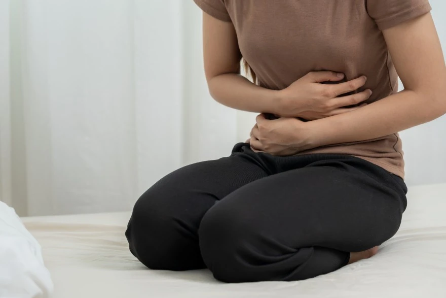
7 Common Types Of Stomach Pain In Women & When To Get Tested
As a woman, experiencing stomach pain can be worrying and uncomfortable. Abdominal pain in women can stem from various causes, ranging from harmless menstrual cramps to potentially serious conditions like endometriosis or ovarian cysts. Understanding the different types of stomach pain - female and recognising warning signs is crucial for knowing when to seek medical advice and testing. This article will explore 7 common causes of lower abdominal pain in females, their symptoms, and when to get evaluated. 7 Common Types of Stomach Pain - Female Women are prone to unique types of abdominal pain due to their reproductive organs and hormonal changes. Here are the seven types of stomach pain - female: Menstrual Cramps (Dysmenorrhea) Menstrual cramps, medically known as dysmenorrhea, are a very common type of lower abdominal pain in females. These crampy or throbbing pains occur before or during menstruation, caused by uterine contractions triggered by prostaglandins. Symptoms Cramping, aching, or throbbing pain in the lower abdomen Pain may radiate to the lower back and thighs. Nausea, diarrhea, fatigue, or headache may accompany cramps. Symptoms typically start 1-2 days before a period and last 2-4 days When to worry While menstrual cramps are normal, certain signs warrant medical attention: Severe pain not relieved by over-the-counter painkillers. Cramps that last longer than usual or worsen over time Sudden onset of painful periods after years of painless ones Heavy menstrual bleeding, fever, or vaginal discharge If you experience any of these symptoms, consult your doctor to rule out underlying issues like endometriosis, fibroids, or pelvic infections. Tests & Diagnosis Diagnosing menstrual cramps usually involves a pelvic exam and reviewing your symptoms and menstrual history. If your doctor suspects an underlying condition, they may recommend additional tests: Pelvic ultrasound to check for structural abnormalities. Laparoscopy, a minimally invasive surgical procedure, is used to diagnose endometriosis. Ovulation Pain (Mittelschmerz) Mittelschmerz is a German term meaning "middle pain". It refers to the brief, one-sided lower abdominal pain in females that they experience during ovulation, about 14 days before their period. Symptoms Sudden, sharp, or cramping pain on one side of the lower abdomen Pain may switch sides from month to month. Discomfort lasts a few minutes to a couple of days. Vaginal discharge or light spotting may occur When to worry In most cases, ovulation pain is not a cause for concern. However, see your doctor if you have: Severe pain that lasts more than a few days Painful ovulation accompanied by fever, chills, or vomiting Ovulation pain, and you have a history of endometriosis or pelvic infections. These symptoms could indicate a ruptured ovarian cyst, ovarian torsion, or another complication that requires prompt medical care. Tests & Diagnosis Ovulation pain is usually diagnosed based on its timing in your cycle and any accompanying symptoms. If the abdominal pain is severe or persists, your doctor may perform a pelvic exam and ultrasound to check for conditions like ovarian cysts that can cause painful ovulation. Endometriosis Endometriosis is a chronic disorder where tissue similar to the uterine lining (endometrium) grows outside the uterus. This misplaced tissue can cause inflammation, scarring, and abdominal pain, especially during menstruation. Symptoms Painful periods (dysmenorrhea) Chronic pelvic pain, often worse during menstruation. Pain during or after sex, bowel movements, or urination Heavy menstrual bleeding or spotting between periods Infertility or difficulty getting pregnant. When to worry If you consistently have very painful periods or pelvic pain that interferes with your daily life, it's important to see your doctor. Risk factors that warrant prompt evaluation include: Family history of endometriosis Pelvic pain that began in adolescence Painful intercourse or bowel movements Difficulty conceiving Tests & Diagnosis Diagnosing endometriosis may involve: Pelvic exam to check for abnormalities or tender areas Ultrasound or MRI to visualise endometrial implants Laparoscopy, the gold standard for diagnosis, is used to visualise and biopsy endometrial tissue. Pelvic Inflammatory Disease (PID) Pelvic inflammatory disease (PID) is an infection of the female reproductive organs, often caused by untreated sexually transmitted infections (STIs) like chlamydia or gonorrhoea. PID can cause chronic lower abdominal pain in females, scarring of reproductive organs, and infertility if not treated promptly. Symptoms Dull or sharp pain in the lower abdomen or pelvis Abnormal vaginal discharge with an unpleasant odor Pain or bleeding during intercourse Burning sensation when urinating Fever, chills, or vomiting (signs of severe infection) When to worry Seek immediate medical care if you have symptoms of PID, especially if you: Have a fever higher than 101°F (38.3°C) Are vomiting and can't keep fluids down Have severe pelvic pain or pain that's getting worse Had recent unprotected sex or a new sexual partner Prompt treatment with antibiotics is essential to prevent long-term complications like chronic pain, ectopic pregnancy, and infertility in various types of stomach pain - females. Tests & Diagnosis To diagnose PID, your doctor will likely: Perform a pelvic exam to check for cervical discharge, uterine tenderness, or pelvic masses. Test vaginal fluid and urine samples for STIs Order blood tests to check for infection and rule out other causes Use ultrasound to visualise your reproductive organs and look for abscesses. Irritable Bowel Syndrome (IBS) Irritable bowel syndrome (IBS) is a common digestive disorder that affects the large intestine, causing abdominal pain, bloating, and changes in bowel habits. IBS is more common in women and symptoms can be influenced by stress, diet, or hormonal fluctuations. Symptoms Crampy abdominal pain that improves after a bowel movement Bloating and excess gas Diarrhea, constipation, or alternating bouts of both Mucus in the stool Feeling like you haven't completely emptied your bowels When to worry While IBS doesn't damage the intestines, its symptoms can significantly impact quality of life. See your doctor if you have: Persistent changes in bowel habits Abdominal pain that interferes with daily activities Unexplained weight loss, rectal bleeding, or anaemia Symptoms that began after age 50 These signs could indicate a more serious condition like inflammatory bowel disease or colon cancer. Tests & Diagnosis There's no single test for IBS. Instead, doctors diagnose it based on your symptoms using the Rome criteria. To rule out other digestive disorders, your doctor may recommend: Blood tests to check for anaemia, celiac disease, or thyroid problems Stool tests to look for infections or inflammatory markers Colonoscopy or a CT scan if you have warning signs like bleeding Ovarian Cysts Ovarian cysts are fluid-filled sacs that develop on or inside the ovaries. While most ovarian cysts are harmless and disappear on their own, some can cause pelvic pain, especially if they rupture or cause the ovary to twist (ovarian torsion). Symptoms Dull or sharp pain on one side of the lower abdomen Pelvic pressure or bloating Pain during sex or your period Abnormal vaginal bleeding Sudden, severe pelvic pain (sign of rupture or torsion) When to worry See your doctor right away if you have: A sudden, severe abdominal or pelvic pain Pain with fever and vomiting Dizziness, weakness, or signs of shock Rapid breathing or heart rate These symptoms could signal a ruptured cyst or ovarian torsion, which can have serious complications if not treated promptly. Tests & Diagnosis To diagnose an ovarian cyst, your doctor may: Perform a pelvic exam to feel for a cyst. Use ultrasound to visualize the cyst's size and location. Order blood tests to check for signs of bleeding or infection Do a laparoscopy to examine the cyst if it looks concerning. Urinary Tract Infections (UTIs) Urinary tract infections (UTIs) occur when bacteria enter the urinary system, causing infection and inflammation. UTIs are very common in women due to their shorter urethra. If left untreated, the infection can ascend to the kidneys (pyelonephritis), which can be serious. Symptoms A strong, persistent urge to urinate Burning sensation when urinating Passing frequent, small amounts of urine Cloudy, dark, or bloody urine Pelvic pain, especially in the center of the pelvis and around the pubic bone When to worry Contact your doctor if you have: Symptoms of a UTI that last more than a few days Blood in your urine Back pain, fever, chills, or vomiting A history of frequent UTIs or antibiotic-resistant infections Prompt treatment with antibiotics can prevent the infection from spreading and causing complications like kidney damage or sepsis. Tests & Diagnosis To diagnose a UTI, your doctor will likely: Analyze a urine sample to look for bacteria, blood, or pus. Send the sample for a urine culture to identify the specific bacteria. Order imaging tests like an ultrasound or CT scan if you have frequent infections, which may indicate an abnormality in your urinary tract. When to See a Doctor While occasional lower abdominal pain in females is often not serious, certain symptoms warrant immediate medical attention. Seek prompt care if you have: Severe, persistent, or worsening abdominal pain Stomach pain accompanied by fever, vomiting, or bloody stools. Sudden, sharp abdominal pain that spreads to your back or shoulder Yellowing of your skin and eyes (jaundice) Swelling or tenderness in your abdomen Chest pain or pressure Difficulty breathing or dizziness. Conclusion Types of stomach pain - females can have many different causes, from harmless menstrual cramps to potentially serious conditions like ectopic pregnancy or appendicitis. Knowing the common types of abdominal pain and their warning signs can help you determine when to seek medical advice and testing. If you experience severe, persistent, or worrying symptoms, don't hesitate to consult your doctor. At Metropolis Healthcare, we understand the importance of accurate and timely testing for women's health concerns. Our team of skilled technicians offers convenient at-home sample collection for a range of diagnostic tests, including blood tests, urine analysis, and more. With our state-of-the-art labs and commitment to personalised care, we're here to help you take control of your health and well-being. FAQs When should I go to the ER for stomach pain? Go to the emergency room if you experience severe, sudden abdominal pain accompanied by fever, vomiting, or bloody stools. These symptoms may indicate a serious condition like appendicitis or a ruptured ovarian cyst. Can stress cause stomach pain? Yes, stress and anxiety can cause physical symptoms like stomach pain, bloating, and changes in bowel habits. Managing stress through techniques like deep breathing, exercise, and mindfulness can help alleviate these symptoms. How do I know if it's my ovaries or intestines hurting? Ovarian pain is usually felt on one side of the lower abdomen and may be associated with other symptoms like irregular periods or pain during intercourse. Intestinal pain is often more generalised and may be accompanied by bloating, gas, or changes in bowel habits. What's the difference between endometriosis and PCOS pain? Endometriosis pain is typically cyclical, worsening during menstruation, and may be accompanied by heavy bleeding or painful intercourse. PCOS usually causes irregular periods and pelvic discomfort from enlarged ovaries, along with symptoms like acne, weight gain, and excess hair growth. How to know if stomach pain is serious? Lower abdominal pain in females that is severe, persistent, or accompanied by other symptoms like fever, vomiting, or bloody stools may indicate a serious condition and requires prompt medical attention. Can constipation cause upper abdominal pain? Yes, constipation can cause discomfort and bloating that may be felt in the upper abdomen. Increasing your fibre intake, staying hydrated, and engaging in regular physical activity can help prevent and alleviate constipation. References • https://my.clevelandclinic.org/health/symptoms/4167-abdominal-pain • https://www.mayoclinic.org/diseases-conditions/menstrual-cramps/symptoms-causes/syc-20374938 • https://www.hopkinsmedicine.org/health/conditions-and-diseases/pelvic-inflammatory-disease-pid • https://www.niddk.nih.gov/health-information/digestive-diseases/irritable-bowel-syndrome • https://www.nhs.uk/symptoms/stomach-ache/
Femur Bone: Structure, Function, And Common Disorders
What Is the Femur Bone? The femur is a long, cylindrical bone that extends from the hip joint to the knee joint, forming the only bone in the thigh. It is the longest bone in the human body, spanning about one-quarter of a person’s height. As the largest bone in the human body, it has a thick, dense structure designed to support the body’s weight during standing, movement, and rest. The femur bone plays a crucial role in locomotion by facilitating walking, running, jumping, and other lower-body movements. It also serves as a major attachment site for numerous muscles, ligaments, and tendons, contributing to stability and strength. Its upper end connects with the pelvis at the hip joint, while the lower end meets the tibia at the knee. Functions of the Femur in the Human Body Being the longest bone in the human body, the femur plays several vital roles in the human body. It supports body weight during standing and movement, enabling walking, running, jumping, and maintaining balance. The bone also serves as the primary attachment point for thigh muscles, connecting the hip and knee joints, allowing for smooth lower limb motion. It also houses bone marrow, which produces blood cells and stores fat. Key Anatomy and Features of the Femur As shown in the femur bone diagram here, the femur bone anatomy is divided into three main regions: the proximal end (nearest the hip) the shaft (diaphysis) the distal end (nearest the knee) Each region has unique features and landmarks that contribute to the bone's overall function and stability. Where Is the Femur Located? The femur is the longest bone in the human body and is situated in the upper leg, spanning the entire length of the thigh from the hip to the knee. It articulates with the pelvis at the hip joint and the tibia and patella at the knee joint, acting as the main pillar supporting the body's weight between the trunk and lower legs. What Does the Femur Look Like? (Structure & Parts) The femur has a distinctive appearance, resembling a long cylinder with rounded ends. Let's explore its structure in more detail: Proximal End of the Femur The proximal end of the femur bone features the smooth, spherical femoral head, which fits into the hip socket (acetabulum) to form the ball-and-socket hip joint. Just below the head is the narrow femoral neck, connecting to the broader shaft. Two bony projections, the greater and lesser trochanters, serve as attachment points for major hip muscles. This region is crucial for hip and lower limb stability and is a especially in older adults with osteoporosis. Femur Shaft (Diaphysis) The femur's shaft, or diaphysis, is a strong, cylindrical structure with a slight forward bow, optimising weight-bearing and shock absorption. Its smooth anterior surface and rugged posterior surface provide attachment points for powerful thigh muscles, such as the gluteal tuberosity and linea aspera. The shaft's hollow centre contains bone marrow, essential for blood cell production. Distal End of the Femur The distal end of the femur is broader and forms the upper part of the knee joint, articulating with the tibia and patella. It features two rounded condyles (medial and lateral) that enable smooth knee movement, with the intercondylar fossa between them serving as an attachment point for knee ligaments. The epicondyles and other surfaces on the distal femur bone provide attachment sites for muscles and ligaments, ensuring knee stability and function. Average Size and Length of the Femur Bone The femur is the largest bone in the human body. The average adult femur measures approximately 18 inches (45 cm) in length, with men generally having longer femurs than women. The size can vary based on factors such as age, sex, and overall height. Parameter Average Value Length (adults) ~18 inches (45 cm) Diameter (midshaft) 2.5–3 cm Common Femur Conditions and Disorders Despite its strength, the femur bone can be affected by various injuries and conditions: Types of Femur Fractures Femur bone fractures are serious injuries classified based on location and pattern: Proximal femur (hip) fractures: Often occur in the femoral neck or intertrochanteric region; common in older adults Femoral shaft fractures: Occur along the diaphysis, usually from high-impact trauma Distal femur fractures: Affect the area near the knee, involving the condyles or metaphysis These injuries may be further classified as transverse, oblique, spiral, comminuted (multiple pieces), or open (bone piercing the skin). Prompt medical attention is crucial to prevent complications and ensure proper healing. Femur and Osteoporosis Risk Osteoporosis weakens the femur by reducing bone density, particularly at the proximal end, increasing the risk of fractures. Hip fractures are a major consequence of osteoporosis, especially in postmenopausal women and older adults, significantly impacting mobility and potentially leading to long-term disability. Preventing bone loss through a balanced diet, regular exercise, and appropriate medical therapy is essential for maintaining femur strength and reducing femur bone fracture risk. Femur Pain in Patellofemoral Syndrome Patellofemoral pain syndrome causes discomfort at the front of the knee, often due to stress on the cartilage between the femur bone and patella. This condition often results from overuse, muscle imbalance, or abnormal alignment, leading to inflammation or cartilage wear. Symptoms include aching pain during activities like running, squatting, or climbing stairs. Managing patellofemoral syndrome involves rest, physical therapy, strengthening exercises, and addressing underlying biomechanical factors to alleviate femur-related knee pain. Medical Tests and Imaging for Femur Problems Diagnosing femur issues involves various medical tests and imaging techniques: X-rays: First-line test to detect fractures, bone alignment, and deformities CT scans: Provide detailed images for complex fractures or surgical planning MRI: Evaluates soft tissue injury, bone marrow changes, or occult fractures Bone density scans (DEXA): Assess osteoporosis risk, especially in the hip/femur Physical examination: Assesses pain, swelling, range of motion, and limb alignment These tools guide accurate diagnosis and inform appropriate treatment strategies. Treatment Options for Femur Conditions Managing femur bone disorders depends on the specific injury, its severity, and patient factors: Femur Fracture Treatment Methods The treatment of femur bone fractures aims to realign the bone, promote healing, and restore function. The femur bone fracture treatment options include: Traction: Used to align the bone fragments and reduce muscle spasms Casting or bracing: Immobilises the fracture site to promote healing Surgical fixation: Involves the use of metal implants (rods, plates, or screws) to stabilise the fracture Physical therapy: Helps restore strength, range of motion, and function after the fracture has healed Timely femur bone fracture treatment is crucial to prevent complications like blood clots, infection, or deformity and to enable early mobilisation. Osteoporosis Management for Femur Health Osteoporosis management aims to maintain femur bone density and reduce fracture risk through: Adequate intake of calcium and vitamin D Weight-bearing and resistance exercises Medications such as bisphosphonates or hormone therapy Fall prevention measures in at-risk populations Regular bone density monitoring Early intervention helps preserve femur strength and function, reducing the burden of hip and other femur bone fractures. Prevention & Prognosis Preventing femur injuries and maintaining optimal prognosis involves a combination of strategies: Maintaining bone health through a balanced diet rich in calcium and vitamin D, regular weight-bearing exercises, and appropriate medications when indicated Minimising fall risk by using mobility aids if needed, removing home hazards, and reviewing medications that may affect balance Wearing protective gear during high-impact activities or sports Addressing underlying conditions that may weaken bones, such as hormonal imbalances or chronic diseases Regular check-ups and bone density screenings, especially for those at higher risk of osteoporosis or fractures The prognosis for femur bone conditions varies depending on the specific issue and the individual's overall health. With appropriate treatment and rehabilitation, most people can recover from femur fractures and regain function. However, some may experience long-term complications, such as arthritis or chronic pain. At Metropolis Healthcare, we understand the importance of maintaining strong and healthy bones, especially the femur. Our expert team of healthcare professionals is dedicated to providing accurate diagnostic services and personalised care to help you achieve optimal bone health. With our state-of-the-art technology and commitment to quality, you can trust Metropolis Healthcare to support you in preventing, diagnosing, and managing femur bone conditions. FAQs What is the strongest bone in the body, and why is the femur critical? The femur is the strongest and longest bone in the human body. Its strength and size are critical for supporting body weight, enabling movement, and providing attachment points for major leg muscles. Is a broken femur serious? Yes, a broken femur is a serious injury that requires immediate medical attention. Femur fractures can cause severe pain, bleeding, and potentially life-threatening complications if not treated promptly and properly. What is femur damage? Femur damage refers to any injury or condition affecting the thigh bone, such as fractures, stress injuries, or degenerative changes like osteoporosis. These issues can significantly impact mobility and overall quality of life. What is the hardest bone to heal? The femur is considered the largest bone in the human body and the most difficult bone to heal due to its size, complexity, and the significant forces it must withstand during weight-bearing activities. Femur fractures often require surgical intervention and extensive rehabilitation. How painful is it to break a femur? Breaking a femur is typically associated with severe, excruciating pain due to the bone's rich nerve supply and the surrounding muscle and tissue damage. The pain can be intense and may require strong pain medications for management. When is surgical intervention mandatory for femur fractures? Surgical intervention is usually necessary for most adult femur fractures, especially those involving the hip joint, shaft, or knee region. Surgery helps realign the bone, stabilise the fracture, and promote proper healing. What's the recovery timeline for a femur fracture post-surgery? Recovery time for a femur fracture after surgery varies depending on the fracture type, patient age, and overall health. Most individuals can expect to bear weight and start physical therapy within a few weeks to months, with full recovery taking anywhere from 4 to 12 months or longer. How can patients reduce femur fracture risk? Patients can reduce their risk of femur fractures by maintaining good bone health through a balanced diet, regular exercise, fall prevention measures, and addressing underlying conditions that may weaken bones. Regular check-ups and early intervention for osteoporosis are also crucial. References • https://my.clevelandclinic.org/health/body/22503-femur • https://www.ncbi.nlm.nih.gov/books/NBK532982/ • https://www.ncbi.nlm.nih.gov/books/NBK556057/ • https://pmc.ncbi.nlm.nih.gov/articles/PMC7176326/ • https://orthoinfo.aaos.org/en/diseases--conditions/femur-shaft-fractures-broken-thighbone/ • https://teachmeanatomy.info/lower-limb/bones/femur/

What Is Laparoscopic Surgery? Benefits, Risks, and Recovery
What Is Laparoscopy? Laparoscopy is a surgical procedure that allows doctors to view and operate on organs inside the abdomen or pelvis through small incisions, usually 0.5–1.5 cm long. During the procedure, a thin tube with a camera called a laparoscope is inserted through one of the incisions. The camera projects images onto a monitor, enabling the surgeon to see inside the body in real time. The abdomen is inflated with carbon dioxide gas to create space for better visibility and manipulation of the organs. Compared to traditional open surgery, laparoscopic surgery results in smaller scars, less postoperative pain, and a faster recovery. Why Is Laparoscopic Surgery Done? Common Reasons and Conditions Laparoscopic surgery is used for both diagnosis and treatment of various abdominal and pelvic conditions, especially when noninvasive tests are inconclusive or surgery is necessary. Some common reasons for why is laparoscopic surgery done include: Investigating unexplained abdominal or pelvic pain Diagnosing and treating endometriosis, ovarian cysts, or pelvic inflammatory disease Gallbladder removal (cholecystectomy) Appendectomy (removal of the appendix) Hernia repair Treating ectopic pregnancy Performing biopsies of abdominal or pelvic organs Cancer staging (e.g., for ovarian or colorectal cancers) Evaluating and sometimes treating infertility issues, such as checking or clearing blocked fallopian tubes Is Laparoscopy a Major or Minor Surgery? Due to its minimally invasive nature and rapid recovery, Laparoscopy can range from minor (diagnostic procedures) to major (therapeutic organ removal or cancer surgery), depending on the complexity. However, the extent of the procedure can vary. Diagnostic laparoscopy, which involves examining organs or obtaining tissue samples, is typically classified as minor. On the other hand, some therapeutic laparoscopic surgeries, such as organ removal, might be more complex but are still less invasive than open surgery. Laparoscopy vs Laparoscopic Surgery – What's the Difference? While the terms "laparoscopy" and "laparoscopic surgery" are often used interchangeably, there is a slight difference in their meanings: Feature Diagnostic Laparoscopy Laparoscopic Surgery Purpose To view/diagnose internal organs To treat a condition surgically Incisions Small, usually 1-2 required Often, multiple small incisions Instruments Used Laparoscope only Laparoscope + surgical tools Tissue Removal Rare Common (e.g. gallbladder, appendix) Duration Usually shorter Typically longer Complexity Less complex Can be complex (e.g. organ removal) How to Prepare for Laparoscopy or Laparoscopic Surgery? Your doctor will help you understand the laparoscopy meaning and provide specific instructions on how to prepare for the surgery. Typical preparation involves: Fasting for at least 6–8 hours before surgery to prevent complications from anaesthesia Stopping certain medications, as advised by your doctor, to reduce the risk of bleeding or interactions with anaesthesia Undergoing blood tests, urine tests, or imaging scans to assess your overall health and check for any underlying conditions Discussing any allergies or previous reactions to anaesthesia with your healthcare team to ensure appropriate precautions are taken. Step by Step: What Happens During Laparoscopy Surgery? On the day of your laparoscopic surgery, you'll be asked to change into a hospital gown and will be given general anaesthesia to ensure you're asleep and pain-free during the procedure. Here's a step-by-step overview of what happens during laparoscopy: An IV line is placed in your arm to administer fluids and medications, and in some cases, a urinary catheter may be inserted. The surgeon makes 1–4 small incisions (called ports) in your abdomen or pelvis, depending on the specific procedure. Carbon dioxide gas is used to inflate your abdomen, creating more space for the surgeon to work and improving visibility. A laparoscope (a thin tube with a camera) is inserted through one of the ports to guide the procedure and project images onto a monitor. Additional surgical instruments are inserted through the other ports, if needed, to perform the necessary surgery or obtain biopsy samples. Once the procedure is complete, the instruments and gas are removed, and the incisions are closed with stitches or surgical tape. Immediately After Laparoscopy – What to Expect After your laparoscopic surgery, you'll be moved to a recovery area where you'll be monitored until you wake up from the anaesthesia. It's normal to feel groggy, and you may experience some nausea or a sore throat from the breathing tube used during the procedure. For most minor procedures, you'll likely be able to go home the same day. Will You Be in Pain After Waking Up? It's normal to experience mild to moderate pain at the incision sites and temporary shoulder pain from the gas used during laparoscopy. However, this discomfort is usually well-controlled with pain relief medications prescribed by your doctor. Post-Laparoscopy Recovery – What Happens Next? The recovery process after laparoscopic surgery varies depending on the specific procedure and your overall health. As you recover: You can usually resume light activities within a day or two, but it's essential to listen to your body and not push yourself too hard. Avoid heavy lifting and strenuous activities for at least a week to allow your body to heal properly. Keep an eye on your incisions for any signs of infection, such as redness, swelling, or discharge, and report any concerns to your doctor. Attend follow-up appointments as advised by your surgeon to monitor your progress and ensure proper healing. Recovery may take a few days for minor procedures, but 2–6 weeks for more complex surgeries, depending on the complexity of your procedure and your individual healing process. Top Benefits of Laparoscopic Surgery Over Open Surgery Compared to traditional open surgery, laparoscopy offers several advantages: Smaller incisions and less visible scarring Reduced postoperative pain and discomfort Quicker return to normal activities and work Shorter hospital stay (many procedures are day-care, but some may require 1–2 nights depending on complexity) Decreased risk of infection and wound complications Faster overall healing and recovery time. Possible Risks and Complications of Laparoscopic Surgery While laparoscopy is generally safe and less invasive than open surgery, there are still some potential risks and complications to be aware of: Infection at the incision sites Bleeding or formation of a haematoma (blood clot) Unintentional injury to organs, blood vessels, or nerves during the procedure Development of blood clots in the legs (deep vein thrombosis) Adverse reactions to anaesthesia, such as nausea, sore throat, or rarely allergic reactions. Need to convert to open surgery if complications arise during the laparoscopic procedure Rarely, a hernia may develop at one of the incision sites. Post-Surgery Care at Home – Dos and Don'ts After your laparoscopic surgery, it's crucial to take proper care of yourself at home to promote healing and reduce the risk of complications. Here are some important dos and don'ts: Do: Keep your incisions clean and dry, following your doctor's instructions for dressing changes and showering Take your prescribed pain medications and antibiotics as directed to manage discomfort and prevent infection Gradually resume normal activities as tolerated, but avoid overexerting yourself Watch for signs of infection, such as redness, swelling, or discharge from your incisions, and report any concerns to your doctor Don't: Lift heavy objects or engage in strenuous activities until cleared by your surgeon Soak your incisions in water (baths, swimming pools, hot tubs) until they have fully healed and your doctor gives you the go-ahead Ignore persistent or severe pain, as this may indicate a complication that requires medical attention How Soon Can You Return to Work? Return to work depends on procedure type: 1–2 weeks for minor procedures, 3–6 weeks for more complex surgeries. after laparoscopic surgery, depending on the nature of their job and the extent of their procedure. If your work involves heavy lifting or physical labour, you may need to wait longer and discuss it with your doctor. When Is It Safe to Start Exercising? Light exercise, such as walking, can usually be resumed a few days after laparoscopy. However, it's essential to avoid vigorous activities and contact sports until your surgeon clears you, which is typically around 2 weeks post-surgery. When Can You Resume Sexual Activity? In most cases, sexual activity can usually be resumed once you are comfortable and your doctor confirms healing — often 2–4 weeks, but depends on the type of laparoscopic surgery. However, it's always best to follow your surgeon's specific advice, as the timeline may vary depending on the type of procedure you underwent. Laparoscopy for Different Conditions Laparoscopic surgery can be used to diagnose and treat a wide range of abdominal and pelvic conditions, including: Gallbladder removal (cholecystectomy) Appendectomy (removal of the appendix) Hernia repair Ovarian cyst removal Tubal ligation (female sterilisation) Endometriosis treatment Cancer diagnosis and staging Removal of adhesions (scar tissue) Evaluation of unexplained pain or infertility. Laparoscopy vs Open Surgery: Cost, Pain, Recovery When considering surgical options, it's essential to understand the laparoscopy meaning and the differences between laparoscopic surgery and traditional open surgery. Here's a comparison of key factors: Feature Laparoscopy Open Surgery Cost Slightly higher upfront Lower upfront Pain Less postoperative pain More pain and discomfort Recovery Time Faster (few days to 2 weeks) Longer (several weeks) Hospital Stay Shorter (often same-day) Longer (several days) Scars Smaller, less visible Larger, more noticeable While laparoscopy may have higher initial costs due to the specialised equipment used, it typically results in a shorter hospital stay, less pain, and a quicker recovery compared to open surgery. Latest Technology in Laparoscopic Surgery Advances in medical technology continue to improve the safety and effectiveness of laparoscopic surgery. Some of the latest developments include robotic-assisted laparoscopy, which enhances the surgeon's precision and dexterity, and 3D imaging systems that provide a more detailed view of the surgical field. These innovations allow for even more complex procedures to be performed through smaller incisions, leading to better outcomes and faster recoveries for patients. Metropolis Healthcare, a leading chain of diagnostic labs across India, is committed to delivering reliable results and personalised care to empower patients in prioritising their health. With a team of qualified blood collection technicians who make at-home visits and advanced diagnostic labs that process samples with precision, Metropolis ensures that you have access to the information you need to make informed medical choices. FAQs How long does a laparoscopic surgery usually take? The duration of laparoscopic surgery varies depending on the specific procedure and the patient's individual situation. Most procedures last between 30 minutes and 3 hours, with more complex surgeries taking longer. Is laparoscopy painful? How long does the pain last? While some pain and discomfort are common after laparoscopy, it is usually much less severe than with open surgery. Most patients experience mild pain at the incision sites and temporary shoulder pain from the gas used during the procedure. This discomfort typically lasts a few days and can be managed effectively with pain relief medications prescribed by your doctor. How many days of rest are needed after laparoscopy? The amount of rest required after laparoscopic surgery depends on the specific procedure and your individual healing process. Most people need 2–7 days of rest before returning to light activities, with a full recovery taking 1–2 weeks. However, it's essential to follow your surgeon's specific instructions. What are the most common side effects of laparoscopy? The most common side effects of laparoscopy include pain and discomfort at the incision sites, temporary shoulder pain from the gas used during the procedure, mild bloating, and fatigue. These side effects usually resolve within a few days to a week after surgery. What should I avoid eating after laparoscopic surgery? Immediately after laparoscopic surgery, it's best to stick to clear liquids and light, easily digestible foods to avoid putting too much strain on your digestive system. As you recover, gradually reintroduce solid foods and return to your normal diet as tolerated. If you experience nausea or vomiting, stick to bland, low-fat foods until your symptoms subside. What are the chances of complications during laparoscopy? Complications during laparoscopic surgery are rare, with overall risk being less than 2%. However, the specific risk varies depending on type of procedure and patient's individual health factors. Your surgeon will discuss any potential complications and their likelihood with you before the procedure. When can I travel after laparoscopic surgery? In most cases, short trips can be undertaken within a week after laparoscopic surgery. However, it's advisable to avoid long journeys, especially flights, for 2–4 weeks to reduce the risk of developing blood clots. Always consult your doctor for personalised advice based on your specific procedure and health status. References 1. https://my.clevelandclinic.org/health/procedures/4819-laparoscopy 2. https://www.nhs.uk/tests-and-treatments/laparoscopy/ 3. https://www.hopkinsmedicine.org/health/treatment-tests-and-therapies/laparoscopy 4. https://www.healthdirect.gov.au/laparoscopy 5. https://medlineplus.gov/lab-tests/laparoscopy/

Top 14 Calcium-Rich Foods For Stronger Bones
Why Calcium Is Important for Your Body and Bone Health Calcium is a vital mineral that your body needs for many essential functions, but it is most well-known for its role in building and maintaining strong bones and teeth. Over 99% of the calcium in your body is stored in your bones, where it provides the hardness and structure that keeps them strong. Your bones also act as a calcium reservoir, releasing calcium into the bloodstream when needed for critical processes like muscle contraction, nerve signalling, blood clotting, and regulating your heartbeat. Since your body cannot produce calcium, it's crucial to get a steady supply through your diet by incorporating calcium rich foods to prevent bone loss and conditions like osteoporosis. Top Warning Signs of Calcium Deficiency You Shouldn't Ignore Calcium deficiency occurs due to a lack of calcium rich foods for bones. It often develops gradually and may not cause noticeable symptoms at first. However, if left unchecked, it can lead to serious health issues. Here are some common warning signs to watch out for: Muscle cramps, spasms, or aches, especially in the legs and back Tingling or numbness in the fingers, toes, or around the mouth Fatigue and unexplained weakness Brittle nails and dry, coarse skin Bone pain or frequent fractures due to weakened bones Dental problems like tooth decay or brittle teeth Mood changes, including irritability, anxiety, or depression Difficulty sleeping or insomnia Abnormal heart rhythms (in severe cases) Slow growth and development in children Poor appetite or difficulty swallowing (in advanced deficiency) If you experience any of these symptoms persistently, it's crucial to consult your doctor for proper diagnosis and treatment. They can order tests to check your calcium levels and recommend calcium rich foods or supplements if needed. 14 Best Calcium-Rich Foods to Strengthen Bones Naturally To meet your daily calcium needs and support strong bones, focus on incorporating a variety of calcium rich foods into your diet. Here are the top 14 calcium rich foods for bones: 1. Milk As one of the richest and most bioavailable calcium sources, cow's milk offers about 300 mg of calcium per cup, covering nearly 30% of the daily requirement for most adults. It's also often fortified with vitamin D, which enhances calcium absorption. Both skim and whole milk provide similar calcium levels, making it one of the best calcium rich foods for bone health. 2. Yoghurt Yoghurt is one of the calcium rich foods, providing roughly 30% DV (recommended daily value) of calcium per cup, depending on the type and brand. Greek yoghurt, known for its high protein and low fat content, is a popular choice, though regular yoghurt often has slightly more calcium. Many varieties are also fortified with vitamin D for added bone-building benefits. Try incorporating yoghurt into your breakfast routine or enjoying it as a snack to boost your calcium intake. 3. Fortified Drinks If you follow a dairy-free diet or have lactose intolerance, fortified plant-based drinks can be excellent alternatives. Options like almond, soy, or oat milk can contain as much calcium as cow's milk when enriched, often providing 300–450 mg per cup. Just be sure to check the labels and choose unsweetened varieties when possible. 4. Cheese Most cheeses are rich in calcium, with hard varieties like Parmesan containing around 242 mg per ounce (28 g). While softer cheeses have less, they still contribute significantly to your daily intake. Cheese also supplies protein and phosphorus, both important for bone structure. However, it's important to consume cheese in moderation due to its high saturated fat and sodium content. Opt for low-fat varieties and use cheese as a topping or ingredient rather than the main focus of your meals. 5. Beans and Lentils Beans and lentils are good plant-based calcium rich foods, offering around 244 mg per cooked cup. They also provide magnesium and fibre, nutrients that support bone health. Including a variety of legumes in your diet can help you meet your calcium needs, especially if you follow a plant-based lifestyle. Try adding beans to soups, salads, or rice dishes, or enjoy lentils as a hearty side or in curries and stews. 6. Almonds Among nuts, almonds stand out among the calcium rich foods for bones for their calcium content, with about 8% DV per ounce (or 23 nuts). They also offer healthy fats, protein, and magnesium, supporting overall cardiovascular and skeletal health. Snack on a handful of almonds, add them to your morning oatmeal or yoghurt, or use almond butter as a spread on whole-grain toast for a calcium boost. 7. Seeds Chia, sesame, and poppy seeds are excellent plant-based calcium rich foods. For example, one tablespoon of poppy seeds provides about 127 mg of calcium, while sesame seeds offer around 9% of the DV per tablespoon. These versatile seeds are easy to incorporate into your diet – sprinkle them on cereals, salads, or baked goods for added crunch and nutrition. You can also try making chia pudding or tahini (sesame seed paste) for a calcium-rich treat. 8. Green Vegetables Leafy greens like kale, collard greens, bok choy, and turnip greens are notable calcium rich foods for bones due to their calcium content. 1 cup of collard greens (cooked) provides 268 mg of calcium. However, some greens like spinach contain oxalates that can hinder calcium absorption, so it's better to focus on low-oxalate options for maximum benefit. Incorporate these greens into your meals by sautéing them as a side dish, adding them to soups and stir-fries, or blending them into smoothies. 9. Sardines and Canned Salmon Sardines and canned salmon with bones are excellent sources of bioavailable calcium, with a 3-ounce serving of sardines providing about 382 mg. The edible bones are the primary calcium source. These fish also supply vitamin D and omega-3 fatty acids, promoting bone and cardiovascular health. Enjoy sardines on crackers or toast, or mix canned salmon into salads or pasta dishes for a calcium and protein boost. 10. Whey Protein Whey protein, commonly found in supplements and shakes, is derived from milk. It contains moderate amounts of calcium, ranging from 100 to 200 mg per serving, depending on the product. It also provides all the essential amino acids needed for muscle and tissue repair. If you use protein powder to support your fitness goals or as a convenient meal replacement, choose a whey-based product to help meet your calcium needs. Just be mindful of added sugars and artificial ingredients. 11. Amaranth Amaranth, a gluten-free pseudo-grain, contains around 116 mg of calcium per cooked cup. It also provides fibre, protein, and micronutrients like magnesium, making it a nutritious calcium rich food for strengthening bones. Use amaranth as a base for porridges or puddings, or pop the tiny seeds like popcorn for a crunchy, calcium-rich snack. 12. Tofu Tofu is an excellent calcium source, offering around 861 mg per half-cup serving, depending on firmness and preparation. It's an ideal option for plant-based diets and can be used in a variety of dishes, from stir-fries to smoothies. Choose firm or extra-firm varieties for the highest calcium content, and look for brands that use calcium sulphate as a coagulant. 13. Figs Dried figs provide about 75 mg of calcium per 30 g serving. They also offer fibre, potassium, and antioxidants, making them a healthy snack for bone health. Enjoy figs on their own as a sweet treat, chop them up and add them to oatmeal or yoghurt, or use this calcium rich food in baked goods like muffins or bread for a calcium boost. 14. Fortified Breakfast Cereals Many breakfast cereals are fortified with calcium, though amounts vary widely by brand. They are a convenient way to boost calcium intake, especially for children and older adults. However, be sure to choose cereals that are also low in added sugars and high in fibre for overall health benefits. Read labels carefully and opt for whole-grain varieties when possible. Proven Tips to Increase Calcium Absorption Effectively Consuming calcium rich foods is just one part of the equation – your body also needs to be able to effectively absorb and use the calcium you take in. Here are some proven tips to help increase calcium absorption: Ensure adequate vitamin D intake through sunlight exposure, fortified foods, or supplements, as vitamin D is essential for calcium absorption. Limit high-sodium or high-protein diets, as excess salt and animal protein can increase calcium loss through urine. Avoid excessive caffeine and alcohol consumption, which may reduce calcium absorption and promote calcium loss. Spread your calcium intake throughout the day rather than consuming it all in one large meal, as the body can only absorb a limited amount at a time. Include magnesium-rich foods like nuts, seeds, and whole grains in your diet, as magnesium aids in proper calcium utilisation. Pair calcium-rich foods with sources of vitamin C and healthy fats to further aid absorption. Limit intake of oxalate-rich foods (like spinach and rhubarb) when relying on them for calcium, as oxalates can inhibit absorption. Engage in regular weight-bearing exercise, which helps maintain bone mass and improves the body's use of calcium. When to Take Calcium Supplements: Signs, Dosage & Timing While it's always best to meet your calcium needs through diet, supplements can be helpful in certain situations. Consider taking a calcium supplement if you: Consume a diet low in calcium (e.g., dairy-free, follow a vegan diet, or limit intake of calcium rich foods) Have higher calcium needs, such as in postmenopausal women, older adults, pregnant or breastfeeding women, or those with certain medical conditions Show signs of deficiency, like muscle cramps, frequent fractures, or an osteoporosis diagnosis. Regarding dosage, aim for: 1,000 mg/day for adults 19-50 1,200 mg/day for women 51+ and men 71+ It's important not to exceed 2,000–2,500 mg per day from all sources to avoid risks like kidney stones and cardiovascular problems. To maximise absorption and minimise side effects, it's best to: Split doses over 500 mg, as your body absorbs smaller amounts better Take calcium carbonate supplements with meals, while calcium citrate can be taken anytime. Avoid taking calcium supplements at the same time as iron-rich meals or supplements, as calcium may reduce iron absorption. At Metropolis Healthcare, we understand the importance of proactive health management. As one of India's leading diagnostic lab chains, we offer comprehensive pathology testing and health check-up services to help you stay on top of your health. Our team of skilled technicians can conveniently collect blood samples right at your doorstep, which are then processed in our state-of-the-art labs. You can easily access your test reports online via email or our user-friendly Metropolis TruHealth app. References • https://ods.od.nih.gov/factsheets/Calcium-HealthProfessional/ • https://medlineplus.gov/calcium.html • https://www.mayoclinic.org/healthy-lifestyle/nutrition-and-healthy-eating/in-depth/calcium-supplements/art-20047097 • https://orthoinfo.aaos.org/en/staying-healthy/calcium-nutrition-and-bone-health/

Carrot Juice Benefits: 10 Powerful Ways It Supports Nutrition, Skin Glow & Vision Health
What is Carrot Juice? Carrot juice is a naturally sweet, orange-coloured beverage made by pressing fresh carrots with a juicer or blender. Unlike eating whole carrots, carrot juice provides a concentrated source of nutrients that your body can absorb more readily. The smooth texture and refreshing taste make it an appealing choice for boosting daily nutrition whilst staying hydrated. When you're wondering, "Is carrot juice healthy?" the answer lies in its remarkable nutrient density. A single cup of carrot juice provides a highly concentrated source of beta-carotene, which your body converts to vitamin A as needed. The conversion rate depends on individual metabolism and dietary fat intake. Juicing breaks down carrot cell walls, improving carotenoid bioavailability, though it removes some dietary fibre compared to whole carrots. Carrot Juice Properties The distinctive orange colour comes from its exceptionally high concentration of beta-carotene, a powerful antioxidant that converts to vitamin A in your body. This nutrient serves as the foundation for many benefits of carrots. Beyond beta-carotene, carrot juice contains substantial amounts of vitamin C, vitamin K, and vitamin B6, along with essential minerals like potassium. The presence of lutein and zeaxanthin—specialised antioxidants that accumulate in eye tissues—makes carrot juice particularly valuable for vision health. These carrot juice properties work synergistically to support multiple body systems simultaneously. Carrot juice naturally contains simple sugars that provide quick energy; retaining some pulp helps moderate absorption and maintain fibre benefits. This combination makes carrot juice both immediately energising and supportive of long-term digestive health. 10 Carrot Juice Benefits: The Nutritional Powerhouse The advantages of drinking carrot juice extend far beyond basic nutrition, offering targeted support for your most vital health concerns. Research consistently demonstrates that regular carrot juice consumption can significantly impact your well-being in measurable ways. Here are the ten most powerful carrot juice benefits: Supports Eye Health and Vision Protection The high beta-carotene content converts to vitamin A, essential for maintaining healthy vision. Lutein and zeaxanthin protect against age-related macular degeneration, whilst vitamin A supports night vision and overall eye function. Boosts Immune System Function Vitamins A, C, and B6 work together to regulate and enhance immune cell activity. Regular consumption helps your body respond more effectively to infections and seasonal challenges. Promotes Radiant Skin and Anti-Ageing Antioxidants protect against UV damage, while vitamin C supports collagen synthesis. Many people notice improved skin texture and a natural glow within weeks of regularly consuming carrot juice. Enhances Antioxidant Protection Clinical studies show that regular carrot juice consumption can increase plasma antioxidant capacity and reduce oxidative stress markers. Supports Cardiovascular Health Potassium helps regulate blood pressure, whilst phenolic compounds reduce harmful lipid oxidation. This combination supports heart health and circulation. May Reduce Cancer Risk Carotenoids and phytonutrients in carrot juice have shown potential anti-carcinogenic effects in laboratory and observational studies, though direct cancer-prevention evidence in humans remains limited. Protects Liver Function Carotenoid's antioxidant and anti-inflammatory effects may support liver health and help reduce markers associated with non-alcoholic fatty liver disease, supporting your body's natural detoxification processes. Improves Blood Sugar Regulation Carrot juice may support better glucose metabolism due to its antioxidant properties, but portion control is important for those managing blood sugar levels. Aids Digestive Health Natural fibre promotes gut health and regular bowel movements, while potassium supports proper digestive muscle function. Contributes to Weight Management At around 90–94 calories per cup, carrot juice is nutrient-dense and relatively low in calories, making it suitable for balanced diets and weight maintenance. How to Incorporate Carrot Juice Into Your Diet Learning how to make carrot juice part of your daily routine maximises these health benefits. The key is consistency and variety in your approach. Here are practical ways to enjoy carrot juice daily: You can enjoy carrot juice in the morning; pairing it with a small amount of healthy fat (like nuts or avocado) enhances carotenoid absorption. Blend carrot juice into smoothies with yoghurt, berries, or leafy greens for added nutrition. Create carrot-and-orange juice combinations to enhance vitamin C and improve flavour. Use carrot juice as a soup base for vibrant, nutritious meals. Mix carrot and beet juice for a powerful antioxidant blend. Freeze carrot juice into healthy ice lollies for a refreshing summer treat. Incorporate carrot juice into salad dressings mixed with olive oil and herbs. When considering how to make carrot juice at home, you'll need either a juicer or a high-powered blender. Juicers extract pure liquid, whilst blenders retain beneficial fibre. Both methods preserve the essential carrot juice properties that make this beverage so valuable for your health. Conclusion According to the World Health Organization, over 2.2 billion people worldwide have vision impairment or blindness, and up to 1 billion cases could have been prevented. Millions of people also struggle with skin concerns and nutritional deficiencies. Carrot juice serves as a nutrient-rich addition that supports eye, skin, and metabolic health naturally. Whether you're seeking better cardiovascular health, enhanced antioxidant protection, or simply want to glow from within, carrot juice provides a natural, scientifically supported solution. At Metropolis Healthcare, we understand that optimal health begins with understanding your body's unique needs. Our comprehensive portfolio of more than 4,000 tests includes nutritional assessments and vitamin deficiency panels that can help you determine whether carrot juice supplementation aligns with your health goals. With our convenient home sample collection service spanning over 10,000 touchpoints across India, you can access precise diagnostics that guide your wellness journey from the comfort of your home. Take the first step towards better health today. Book your home sample collection and discover how proper nutritional testing can enhance your wellness strategy, ensuring that additions like carrot juice work optimally for your individual health profile. FAQs about Carrot Juice What are the health benefits of drinking carrot juice? The primary advantages of drinking carrot juice include enhanced vision health, improved immune function, and glowing skin. The concentrated nutrients support cardiovascular health and liver function and may help regulate blood sugar levels. Regular consumption provides powerful antioxidant protection that combats cellular damage and supports healthy ageing. These carrot juice benefits make it particularly valuable for people seeking natural ways to boost their overall health and vitality. Can carrot juice help improve my skin? Yes, carrot juice significantly benefits skin health through multiple mechanisms. The beta-carotene and vitamin C content promote collagen synthesis whilst protecting against free radical damage. Many people notice improved skin texture, reduced blemishes, and a natural glow within 2-4 weeks of regular consumption. The antioxidants help protect against sun damage, making your skin more resilient to environmental stressors. Is carrot juice good for vision health? Carrot juice is exceptionally beneficial for vision health due to its high concentration of beta-carotene which is essential for night vision and overall eye function. Lutein and zeaxanthin specifically protect the macula from age-related degeneration. Research indicates that people with higher carotenoid intake have significantly lower risks of vision problems. The carrot's benefits for eye health are so well established that many eye care professionals recommend regular consumption. Can drinking too much carrot juice have side effects? While carrot juice is generally safe, excessive consumption may cause carotenemia, a harmless yellowing of the skin due to high beta-carotene levels, especially with very frequent intake due to high beta-carotene levels. This typically occurs only with very high intake and reverses when consumption is reduced. People with diabetes should monitor their blood sugar when increasing carrot juice intake, as it contains natural sugars. Is carrot juice a good weight loss drink? Carrot juice can support weight management as part of a balanced diet. At 94 calories per cup, it provides substantial nutrition without excessive calories. The natural fibre content (when pulp is included) promotes satiety, helping you feel full longer. The nutrients support metabolic health, whilst the natural sweetness can satisfy cravings for less healthy options. However, carrot juice works best for weight loss when combined with proper diet and exercise rather than as a standalone solution.