 Home Visit
 Upload


Latest Blogs

Exploring Emphysema: Types, Symptoms, Treatment & Causes
What is Emphysema? Emphysema is a chronic lung condition characterised by the gradual destruction of the air sacs (alveoli) in the lungs, leading to difficulty breathing. This irreversible damage reduces the lungs' ability to exchange oxygen and carbon dioxide efficiently. Emphysema is a type of Chronic Obstructive Pulmonary Disease (COPD) and can significantly impact the quality of life if left untreated. What are the Symptoms of Emphysema? Emphysema manifests through respiratory symptoms that gradually worsen over time. Initially, individuals may experience mild symptoms that often go unnoticed, but as the condition progresses, emphysema symptoms become more pronounced. Some common emphysema symptoms may include: Shortness of breath Persistent coughing Fatigue However, advanced emphysema symptoms include: Wheezing Chest tightness Increased mucus production Abnormal mucus colour (yellow or green) Heart problems Trouble sleeping What are the Stages of Emphysema? Emphysema is often categorised into four stages based on the severity of lung damage and airflow limitation. These emphysema stages, as defined by the Global Initiative for Chronic Obstructive Lung Disease (GOLD), help clinicians assess the progression of the disease and tailor emphysema treatment accordingly. Stage 1 (Mild) emphysema: In the initial stage of emphysema, patients may experience mild shortness of breath during physical exertion, but the disease is often undiagnosed at this point. Stage 2 (Moderate) emphysema: As emphysema progresses, lung function further declines, leading to increased breathlessness, particularly during physical activity. Coughing and wheezing become more noticeable, affecting daily life. Stage 3 (Severe) emphysema: Severe emphysema is characterised by significantly impaired lung function, causing frequent and severe shortness of breath even during minimal exertion. Stage 4 (Very Severe) emphysema: In advanced emphysema, lung function is severely compromised, and individuals may experience extreme difficulty breathing even at rest. What are the Causes of Emphysema? The primary cause of emphysema is long-term exposure to irritants that damage the lungs, most commonly cigarette smoke. Other factors include: Exposure to air pollution Industrial fumes Genetic predisposition How is Emphysema Diagnosed? Diagnosing emphysema involves the following steps: Medical History: A detailed medical history, including symptoms, smoking history, and exposure to lung irritants such as pollution or occupational hazards, is asked. Spirometry Test (Emphysema Diagnosis 1): Spirometry is a key diagnostic test for assessing lung function and airflow limitation to determine if airflow obstruction is present, indicative of emphysema diagnosis. Imaging Studies (Emphysema Types, Emphysema Causes): Chest X-rays and Computed Tomography (CT) scans provide detailed images of the lungs and identify potential emphysema causes such as smoking-related damage. Arterial Blood Gas Analysis: It measures the levels of oxygen and carbon dioxide in the blood, providing insights into how well your lungs are functioning and if oxygen therapy is needed as part of emphysema treatment. Pulmonary Function Tests (Emphysema Diagnosis 2): Pulmonary function tests (PFTs) assess lung volume, capacity, and gas exchange efficiency. Consultation and Treatment Planning (Emphysema Treatment): Based on the results of these diagnostic tests, the healthcare provider discusses the diagnosis, emphysema type, and potential causes. What are the Complications of Emphysema? Some of the complications are: Chronic respiratory infections like pneumonia and bronchitis are common due to weakened lung function. Increased risk of pneumothorax (collapsed lung) and pulmonary hypertension. Elevated susceptibility to heart problems, including heart failure, as the heart compensates for impaired lung function. What Tests Will be Done to Diagnose Emphysema? Diagnosing emphysema involves a series of tests, including: Spirometry: It measures lung function and airflow limitation. It involves breathing forcefully into a device called a spirometer to assess how much air you can exhale and how quickly. Imaging Studies: Chest X-rays and Computed Tomography (CT) scans can reveal characteristic changes in lung structure associated with emphysema, such as hyperinflation and bullae formation. Arterial Blood Gas Analysis: This emphysema test measures the levels of oxygen and carbon dioxide in your blood, providing insight into how well your lungs are functioning. Pulmonary Function Tests (PFTs): These tests evaluate lung volume, capacity, and gas exchange efficiency, helping to assess the severity of emphysema. High-Resolution CT (HRCT): This specialised CT scan provides detailed images of lung tissue, aiding in the emphysema diagnosis and classification. How is Emphysema Treated? Here is how emphysema is treated: Smoking Cessation: The most crucial step to treat emphysema is quitting smoking to slow disease progression. Medications: Bronchodilators and inhaled corticosteroids help relieve emphysema symptoms and improve lung function. Pulmonary Rehabilitation: Exercise, breathing techniques, and education programs improve quality of life. Oxygen Therapy: Supplemental oxygen improves oxygen levels in the blood. Surgery: In severe cases, lung volume reduction surgery or lung transplant may be considered. What are the Types of Emphysema? Emphysema is typically classified into two main types: Centriacinar Emphysema: Primarily affects the central or proximal portion of the acinus, commonly associated with cigarette smoking. Panacinar Emphysema: Involves uniform destruction of the entire acinus, often associated with alpha-1 antitrypsin deficiency. These emphysema types differ in the distribution of lung damage and may require different emphysema treatment approaches. What is the Prevention and Outlook for Emphysema? While emphysema is not curable, managing risk factors and adhering to emphysema treatment can improve symptoms and slow disease progression, enhancing long-term outlook and quality of life. Emphysema prevention primarily involves: Avoiding tobacco smoke and minimising exposure to environmental pollutants. Early detection and smoking cessation can significantly reduce the risk. Does COVID-19 Cause Emphysema? COVID-19 rarely causes giant emphysema, which is characterised by the expansion of air pockets, known as bullae, in the lungs. This condition can affect up to one-third of one or both lungs. It is more common in individuals with pre-existing emphysema. Conclusion Emphysema, a chronic lung condition, does present significant challenges, yet early detection and appropriate management strategies can alleviate emphysema symptoms and improve prognosis. Take charge of your respiratory health today with Metropolis Healthcare's comprehensive diagnostic services. Schedule a consultation now to receive personalised guidance and support on your journey towards better lung health.

Understanding Bacterial Vaginosis (BV): Symptoms, Causes, and Treatment
Are you facing discomfort or unusual symptoms in your vaginal area? Bacterial Vaginosis (BV) is a prevalent vaginal infection affecting women globally. Although it might not always exhibit noticeable symptoms, BV can lead to significant discomfort and complications if untreated. Therefore, understanding bacterial vaginosis causes, symptoms, and impact on reproductive health is vital. So, this article delves into BV intricacies, offering insights into its presentation and emphasising the importance of timely medical attention for diagnosis and treatment. What is Bacterial Vaginosis (BV)? Normally, the vagina contains a balance of bacteria, but BV occurs when there is an overgrowth of harmful bacteria. BV is a common vaginal infection resulting from an imbalance of 'good' and 'harmful' bacteria in the vagina. How Common is Bacterial Vaginosis? Bacterial vaginosis is highly prevalent worldwide, affecting approximately one-third of women globally. Prevalence varies across regions, with rates ranging from 23% to 29% in different areas. Who Can Get BV? Bacterial vaginosis can affect anyone with a vagina, regardless of age or sexual activity. What are the Symptoms of Bacterial Vaginosis? The bacterial vaginosis symptoms may include: Thin, watery vaginal bacterial vaginosis discharge that can be grey, white, or green Foul-smelling 'fishy' odour emanating from the vagina Vaginal irritation or itching Sometimes, BV may not exhibit any symptoms, especially in mild cases How Do You Get Bacterial Vaginosis (BV)? Bacterial vaginosis can affect anyone with a vagina, but certain factors increase the risk of developing it. These can be: Multiple or New Sex Partners: Having different or new sex partners can increase the risk of BV. Sexual Activity: BV is strongly associated with sexual activity, although it is not classified as a Sexually Transmitted Infection (STI). Pregnancy: Pregnant women are also at higher risk of developing BV. Is Bacterial Vaginosis Contagious? Bacterial vaginosis is not considered a contagious condition in the typical sense. So it does not spread from person to person, like a cold or the flu. However, sexual activity can increase the risk of developing BV. BV results from an imbalance of bacteria in the vagina, where harmful bacteria outnumber the beneficial ones. Although not directly contagious, sexual partners can influence each other's bacterial balance and potentially contribute to the development or recurrence of BV. Is Bacterial Vaginosis an STD or STI? Bacterial vaginosis is not classified as a Sexually Transmitted Disease (STD) or STI. BV is an imbalance of the usual bacteria found in the vagina. What's the Difference Between Bacterial Vaginosis and a Yeast Infection? Bacterial vaginosis and yeast infections are both common vaginal infections but have different causes. BV results from an imbalance in beneficial and harmful bacteria in the vagina, causing symptoms like unusual discharge and vaginal itchiness. On the other hand, yeast infections are caused by an overgrowth of yeast in the vagina, leading to symptoms such as thick, white discharge, itchiness, and irritation. How Do You Know If You Have BV? You may have bacterial vaginosis if you notice symptoms such as thin, grey, or green vaginal discharge, a foul-smelling 'fishy' odour, and vaginal irritation or itching. How Do I Get BV to Go Away? Medical intervention is necessary to treat bacterial vaginosis effectively. Your doctor may prescribe antibiotics such as metronidazole or clindamycin, which can be administered orally as pills or topically as gels or creams for BV. Ensure to complete the full course of antibiotics for BV as prescribed, even if symptoms improve before finishing the medication. Refrain from sexual activity until bacterial vaginosis treatment is complete to prevent reinfection or spreading the infection to your partner. In some cases, bacterial vaginosis may recur, so follow-up appointments with your healthcare provider may be necessary to monitor and manage the condition effectively. Can Bacterial Vaginosis Go Away on Its Own? Yes, bacterial vaginosis can sometimes resolve on its own without medical treatment. Mild cases of BV may clear up without intervention, especially if the body's immune system restores the natural balance of bacteria in the vagina. How Long Does Bacterial Vaginosis Last? Bacterial vaginosis can vary in duration, but typically, it can last for a few days to a week or longer if left untreated. In some cases, BV may resolve on its own without treatment, especially in mild cases. However, if symptoms persist or recur, medical treatment is necessary to address the BV infection effectively. Proper bacterial vaginosis treatment with antibiotics, such as metronidazole or clindamycin, can help alleviate symptoms and clear the infection. However, even with appropriate treatment, BV can come back within a few months, with recurrence rates ranging from 30% to 50%. Therefore, it is essential to follow up with healthcare professionals and adhere to prescribed treatment regimens to manage and prevent recurrent BV. How to Prevent the Risk of BV? Bacterial vaginosis prevention strategies include: Practice safe sex: Consistently using condoms during sexual activity can reduce the risk of bacterial vaginosis. Limit sexual partners: Reducing the number of sexual partners can decrease the risk of BV transmission. Limit-scented products: Perfumed bath products, laundry detergents, and scented toilet paper can irritate the vagina and increase susceptibility to BV. Opt for unscented products. Maintain vaginal health: Eating a balanced diet, staying hydrated, and managing stress can support overall vaginal health, reducing the risk of BV. Can You Get Bacterial Vaginosis Multiple Times? Yes, it is common to experience recurrent bacterial vaginosis infections. Even after successful treatment, approximately half of those affected may have the condition return within 6 to 12 months. BV recurrence can be influenced by various factors such as sexual activity, douching, and hormonal changes. Proper management, including lifestyle adjustments and follow-up care, can help reduce the likelihood of recurrent BV. What are the complications of BV? Bacterial vaginosis can lead to various complications if left untreated. For instance: Increased risk of STIs such as herpes, chlamydia, gonorrhoea, and HIV. Pelvic Inflammatory Disease (PID) is an infection of the uterus and fallopian tubes. Complications during pregnancy, including preterm delivery, early labour, and potential loss of pregnancy. Increased risk of post-gynecological procedure infections. Chronic discomfort and recurring episodes of BV. Bacterial Vaginosis in Pregnancy Bacterial vaginosis during pregnancy can lead to complications such as: Preterm birth: BV increases the risk of premature delivery, which can result in health issues for the baby. Low birth weight: Babies born to mothers with BV may have a lower birth weight than those born to mothers without BV. Premature rupture of membranes: BV can cause the amniotic sac to break before labour begins, increasing the risk of infection and preterm birth. Uterine infection: BV may lead to infections in the uterus (endometritis), which can be dangerous for both the mother and the baby. When to See a Doctor About Bacterial Vaginosis? It is advisable to see a doctor if you experience symptoms of bacterial vaginosis, such as unusual vaginal discharge or odour. Additionally, pregnant individuals should seek medical attention if they suspect BV, as it can lead to complications during pregnancy. Conclusion If you are experiencing symptoms of bacterial vaginosis or have concerns about your reproductive health, do not hesitate to reach out to a medical professional. Seeking timely medical advice can provide relief, ensure proper treatment, and promote overall well-being. For reliable diagnostic services, consider Metropolis Labs, a trusted chain of diagnostic labs across India. Metropolis Labs specialises in pathology services, offering accurate blood and body fluid testing. Their qualified blood collection technicians provide convenient at-home visits for sample collection, with reports accessible online. Visit Metropolis Labs for comprehensive health check-ups and reliable diagnostic solutions.

Everything You Should Know About Shingles
What is Shingles? Shingles is a viral infection that can cause a rather painful skin rash that appears red, dark pink, dark brown, or purplish and can occur on one side of your body. Shingles usually look like a single stripe of red blisters along the right or left side of your torso. It is not life-threatening but can be very painful and lead to complications if left untreated. Shingles is also known as herpes zoster. Where Do Shingles Come From? Shingles occur due to the reactivation of the varicella-zoster virus that is already present in the body when you experience chickenpox as a child. Even though you can overcome chickenpox and its physical signs, this virus remains within the body and can reoccur as shingles as an adult. Who is at Risk of Getting Shingles? Individuals with weakened immune systems are more likely to suffer from shingles. This includes those: With weak immune symptoms Those who are over 50 years of age Who has suffered a recent illness Experiencing extreme physical or mental trauma Under stress Can You Get Shingles More Than Once? Contrary to popular belief, you can get shingles more than once. However, you will not get the shingles rash in the same place if you get shingles again. What Causes Shingles? One of the leading shingles causes is the varicella-zoster virus, which also causes chickenpox. What are the Symptoms of Shingles? Some early shingles symptoms include: Chills Fever Headache Sensitivity to light Stomach upset Feeling tired Secondary shingles symptoms include: Redness on your skin in the affected area A raised rash in a small region of your skin Mild to severe pain in the affected area A tingling, burning, or itching feeling on your skin What are the Stages of Shingles? Shingles can last anywhere between three and five weeks. Once the varicella-zoster virus is reactivated, you may feel the first signs of shingles like. Burning sensation Tingling Itchiness Numbness Fever and chills Body aches Light sensitivity Unusual tiredness This is also known as the pre-eruptive stage. Shingles can develop on one side of your body, most commonly around the waist, chest, or back. Within five days of the reactivation of the shingles virus, you may see the appearance of a red rash; this is the acute eruptive stage of shingles. Tiny groups of fluid-filled, oozing blisters appear in the area affected with shingles, and you may experience flu-like symptoms, including headaches, fatigue, and fever. Ten days after the shingles virus, these blisters dry and form scabs similar to chickenpox ones. These scabs usually clear out after a few weeks, but you may still experience pain even after they clear. Is Shingles Contagious? The National Health Services states that shingles are not contagious. Still, the virus that causes shingles can spread from one person to another and cause chickenpox if the individual has not previously had the condition. The varicella-zoster virus spreads when a healthy individual comes in contact with an oozing blister that appears in shingles. To prevent this, keep the area of the shingles rash clean and covered. If you touch a blister, it is best to wash your hands. How is Shingles Diagnosed? The primary care physician diagnoses shingles based on how the rash is distributed throughout the body. Shingles blisters may appear as a band around one side of the body. Scrapings of the rash or a swab of the fluid from the blisters can be sent to the laboratory for a Polymerase Chain Reaction (PCR) test to confirm the shingles diagnosis. How is Shingles Treated? There is no known cure for shingles, but your doctor will prescribe medicines to manage the symptoms of the disease and help prevent complications. Medication Shingles can be managed with two types of medications: antiviral and over-the-counter medications. Antiviral medicines help ease the discomfort of shingles and stop the shingles symptoms, especially if the shingles treatment is within 72 hours of the appearance of shingles symptoms. These medicines include Acyclovir, Famciclovir, or valacyclovir. Over-the-counter medicines like acetaminophen and ibuprofen help with shingles treatment and relieve the pain caused by shingles. If you develop a bacterial infection due to the shingles rash, your doctor may also prescribe antibacterial drugs or anti-inflammatory medicines. What are the Complications of Shingles? While shingles itself is painful and causes irritation, if left untreated, this condition can cause complications like: Eye damage if the rash or blister is too close to the eye Bacterial infection in the skin from open blisters Pneumonia Ramsay Hunt syndrome, if the shingles affect the nerves in the head, it also results in facial paralysis or hearing loss. Inflammation of the spinal cord causes meningitis or encephalitis. Is a Vaccine Available to Prevent Shingles? Currently, there are two shingles vaccines available in India. One is Zostavax, a live-attenuated vaccine used for adults 50 years or older. The second is Shingrix, a recombinant vaccine that has shown to be over 90% effective in preventing shingles and postherpetic neuralgia. How to Prevent Shingles? Getting the shingles vaccine is essential for shingles prevention. However, even if you have taken the vaccine, there is still a chance that you can get shingles. FAQ's What usually triggers shingles? When the dormant varicella-zoster virus gets triggered, it can cause shingles or herpes zoster. This often happens when the immune system is compromised. Other possible shingles triggers include stress, cancer treatment, HIV, organ transplant, taking immunosuppressants, or other medical reasons for reduced immunity. What are the first signs of shingles? Pain and a burning sensation on one side of your body are usually the first shingles symptoms. Is shingles airborne? The virus varicella-zoster, which is the cause of both shingles and chickenpox, is an airborne virus. The virus can spread if an individual with chicken pox or shingles sneezes or coughs around you or even shares a drinking glass or eating utensil. It can also spread by contact with the virus. Is shingles painful? The pain level experienced when an individual gets shingles can differ from low to extreme. Most people may only experience mild shingles symptoms, like tingling or itchiness of the skin, while others may experience pain without developing a rash. What is the difference between shingles and hives? The main difference between shingles and hives is the cause. The varicella-zoster virus is the main shingles cause, while hives result from an allergic reaction to something in the environment, a medication, or food. Conclusion Shingles/herpes zoster is a condition that is caused by the chickenpox virus varicella-zoster. It can cause a painful, red rash with a burning sensation. This rash may appear on different body parts like the torso, neck, or face. Regular checkups and diagnostic tests can help you maintain your health. Metropolis Labs offers a wide range of tests and health packages to help you take preventive measures to maintain good health.
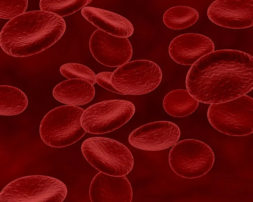
All You Need to Know About Sickle Cell Anaemia
What is Sickle Cell Anaemia? In this condition, haemoglobin within the red blood cells is affected. Haemoglobin is an essential component of the blood and is responsible for carrying oxygen from your blood to other body parts. Sickle cell anaemia is an inherited blood disorder affecting red blood cells. Normal haemoglobin cells are flexible and round, which helps them move even through smaller blood vessels. However, in the case of sickle cell anaemia, the haemoglobin cells get a crescent shape known as haemoglobin S, which makes the cells rigid and lack flexibility, due to which the blood flow is restricted. This prevents vital oxygen from reaching vital organs and tissues throughout the body, which can lead to severe complications like pain, infection, and organ damage. Sickle cell anaemia is a lifelong condition, but there are treatment options that can help reduce symptoms of sickle cell anaemia. What are the Symptoms of Sickle Cell Anaemia? Sickle cell anaemia symptoms may appear in childhood by the age of 5 or 6 months. These sickle cell anaemia symptoms may vary, with some displaying mild symptoms while others showing more severe complications. Common sickle cell anaemia symptoms include: Frequent episodes of pain Fatigue Weakness Paleness Jaundice (yellow tint on the skin and whites of the eyes) Swelling of the hands and feet What are the Types of Sickle Cell Disease? The sickle cell anaemia types depend on the genes a person inherits from their parents. For instance: Haemoglobin SS This is the most severe form of sickle cell anaemia and can affect 65% of people with the condition. Anyone with this form of sickle cell anaemia usually inherits one gene with haemoglobin S from each parent. Most of their haemoglobin is abnormal, which causes chronic anaemia. Haemoglobin SC This mild to moderate sickle cell anaemia type can affect 25% of individuals suffering from sickle cell anaemia. In this case, you may have inherited only one haemoglobin S gene from one parent and another abnormal type haemoglobin C gene from the other parent. Haemoglobin (HbS) beta thalassemia People with beta thalassemia, a form of sickle cell anaemia, inherit only one haemoglobin S gene from one parent and another beta thalassemia gene from the other. This sickle cell anaemia type is divided into two forms: HbS beta+, which is very mild and can affect about 8% of individuals with sickle cell anaemia, and HbS beta 0, which affects 2% of individuals with sickle cell anaemia. The second form is much more severe and shows sickle cell anaemia symptoms similar to haemoglobin SS. There are a few other forms of sickle cell anaemia, such as haemoglobin SD, SE, and which are very rare. What Causes Sickle Cell Anaemia? The primary sickle cell anaemia cause is a genetic mutation in the HBB gene. The function of this gene is the creation of part of the haemoglobin, and any mutation in the gene codes for abnormal haemoglobin. Those who inherit this disease in an autosomal recessive manner (i.e., parents do not have this condition) do not display sickle cell anaemia symptoms or signs. In this case, each child's parents carry one copy of the mutated gene, which can cause sickle cell anaemia. What are the Risk Factors of Sickle Cell Anaemia? Some ethnic groups are more likely to develop sickle cell anaemia as compared to others. These include: People of Middle Eastern, Asian, Indian, and Mediterranean descent Hispanic Americans who hail from South or Central America African descendants, among whom 1 in 12 individuals carry the sickle cell anaemia gene What are the Complications of Sickle Cell Anaemia? Sickle cell anaemia can affect different parts of the body. Some effects may be acute, meaning they start suddenly, while others are chronic and last a long time. Sickle cell anaemia symptoms begin early and remain lifelong. Pain This is one of the most common complications of sickle cell anaemia. As the sickled cells start to get stuck and block blood flow, this can cause an acute pain crisis, also known as a sickle cell crisis, Vaso-Occlusive Episode (VOE), or Vaso-Occlusive Crisis. Anaemia Sickle cell anaemia causes red blood cells to die early, leading to acute cases of anaemia. In this case, there will not be enough healthy red blood cells to carry oxygen to the body parody, causing the abovementioned symptoms. Acute Chest Syndrome This is a life-threatening medical condition that can cause injury in the lungs, difficulty breathing, and low levels of oxygen in the rest of the body. Blood Clots If you have sickle cell anaemia, your cells are more likely to clot, which increases the chances of developing deep vein thrombosis, leading to pulmonary embolisms. Vision Problems If the blood flow to your eyes gets blocked due to sickle cell anaemia, it can lead to vision loss and, in some cases, permanent blindness. Strokes If the blood flow to your brain is hampered, your brain does not get the required amount of oxygen to function correctly, which in turn can lead to a stroke. Organ Damage Individuals with sickle cell anaemia run the risk of issues related to the heart, lungs, kidneys, and other vital organs. How is Sickle Cell Anaemia Diagnosed? A sickle cell anaemia diagnosis can usually be given to newborn babies after a routine screening test. This test takes a small blood sample and checks for various conditions, including sickle cell anaemia. Prenatal testing may also give a positive or negative diagnosis. How is Sickle Cell Anaemia Treated? Sickle cell anaemia treatment can include medication, blood and marrow transplants, transfusions, and gene therapy. This treatment starts with antibiotics. Other medications like Voxelotor, Crizanlizumab, Hydroxyurea, and L-glutamine help reduce the symptoms of this condition. Your healthcare provider may suggest sickle cell anaemia treatments like acute transfusion or red blood cell transfusion to treat sickle cell anaemia and prevent complications. A stem cell transplant from a suitable donor can help cure sickle cell anaemia. How Can I Prevent Sickle Cell Anaemia? Sickle cell anaemia prevention is not always possible as it is a genetic disease. If you or your partner suffer from it, it is best to talk to your healthcare provider about genetic testing or counselling to understand the probability of passing the mutated gene to your children. Conclusion Sickle cell anaemia is a lifelong condition, but recent advances in healthcare have made huge strides in helping with its prevention and treatment. Regular diagnostic tests and genetic testing for babies and adults enable you to understand if you are at risk of developing this condition. Metropolis Labs provides world-class facilities and best-in-class diagnostic services in the comfort of your home so you can keep track of your health. Contact us today for more details.
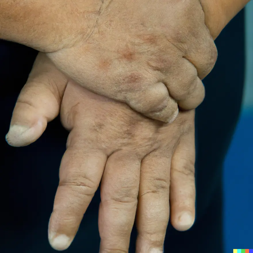
Acromegaly: A Comprehensive Guide Explaining a Growth Hormone Disorder
What is acromegaly? Acromegaly is an uncommon hormonal condition characterised by an overproduction of growth hormone, resulting in abnormal growth, particularly in the extremities such as the hands and feet. Who does acromegaly affect? Acromegaly can impact individuals of any age, though it is typically identified more frequently in middle-aged adults, with an average age of diagnosis around 40 years. How common is acromegaly? Acromegaly is a rare condition, occurring in 3 to 14 individuals per 100,000, or approximately 50 to 70 individuals per million. How does acromegaly affect my body? Acromegaly causes the accelerated growth of body tissues and bones, resulting in the gradual emergence of disproportionately large hands and feet, accompanied by a range of additional symptoms over time. What causes acromegaly? Acromegaly causes include overproduction of growth hormone (GH) for an extended period. The primary acromegaly cause in adults is usually a benign pituitary tumour (adenoma) that produces excess GH. Symptoms such as headaches and vision problems may arise due to the tumour pressing on nearby brain tissues. In rare cases, non pituitary tumours elsewhere in the body, such as the lungs or pancreas, can cause acromegaly by secreting GH or growth hormone-releasing hormone (GH-RH). What are the symptoms of acromegaly? Common acromegaly symptoms include enlarged hands and feet, leading to difficulties in wearing previously fitting rings and shoes. Gradual alterations in facial shape may occur, such as a protruding lower jaw and brow bone, enlarged nose, thickened lips, and widened spacing between teeth. Acromegaly symptoms can vary among individuals and may encompass: Enlargement of hands and feet. Increased size of facial features like bones, lips, nose, and tongue. Coarse, oily, thickened skin. Excessive sweating and body odour. Presence of skin tags. Fatigue, muscle weakness, and joint pain. Deepened voice due to enlarged vocal cords and sinuses. Severe snoring from upper airway obstruction. Vision difficulties and persistent or severe headaches. Irregular menstrual cycles in women and erectile dysfunction in men. Decreased libido. How is acromegaly diagnosed? Without relying on specific tests, here's how acromegaly diagnosis is done: Patient History: The healthcare provider reviews medical background, focusing on symptoms like changes in appearance, joint discomfort, or vision issues. Physical Examination: Conducted to identify signs such as enlarged extremities, facial alterations, and skin abnormalities. Symptom Observation: Diagnosis inferred from symptoms and physical characteristics, although they may be subtle or overlap with other conditions. Clinical Assessment: Diagnosis based on the healthcare provider's judgement, considering characteristic features and suggestive symptoms of acromegaly. What tests will be done to diagnose acromegaly? Acromegaly diagnosis involves various assessments and tests: Blood tests to measure growth hormone (GH) levels, often supplemented with a glucose tolerance test. Testing for insulin-like growth factor 1 (IGF-1) to assess abnormal growth. Imaging studies such as X-rays, MRI scans, and sonograms to identify excess bone growth and detect tumours. Following diagnosis, MRI and CT scans aid in locating and determining the size of pituitary tumours. If no pituitary tumour is found, doctors may search for tumours elsewhere in the body contributing to excessive GH production. Despite estimated prevalence rates, acromegaly often goes undiagnosed, leading to potential underestimation of affected individuals. How is acromegaly treated? Acromegaly treatment typically involves a combination of surgical intervention and medications. Surgery for Acromegaly: Transsphenoidal surgery: This common approach involves removing the pituitary tumour through the nasal passage to extract as much of the tumour as possible while preserving normal pituitary function. Medications for Acromegaly: Somatostatin analogues: Drugs such as octreotide and lanreotide are administered via injections to reduce the secretion of growth hormone from the pituitary gland. Dopamine agonists: Medications like cabergoline may be used, particularly if the tumour secretes both growth hormone and prolactin. Growth hormone receptor antagonists: Injectable drugs like pegvisomant block the action of growth hormone, lowering IGF-1 levels and managing symptoms. Other medications: In some cases, combinations of drugs like cabergoline or pegvisomant with somatostatin analogues may be prescribed to achieve better control over growth hormone and IGF-1 levels. Other acromegaly treatments include Radiation Therapy: Radiation therapy may be considered if surgery is not feasible or if the tumour persists after surgery. It involves targeted radiation to shrink or destroy tumour cells. Stereotactic radiosurgery (gamma knife) delivers focused radiation to the tumour, minimising damage to surrounding tissues. Is acromegaly curable? In certain cases, acromegaly can be cured, although not in all instances. Surgical removal of a pituitary tumour causing acromegaly yields approximately an 85% success rate for small tumours and around 40% to 50% for large tumours. What is the outlook for acromegaly? The outlook for acromegaly varies based on factors such as treatment effectiveness and the presence of complications. Surgical removal of pituitary tumours, especially small ones, can result in a favourable outcome for many patients. Long-term management through medications or radiation therapy helps control symptoms and prevent complications. What are the complications of acromegaly? Untreated acromegaly can result in various complications, including: Cardiovascular issues Arthritis Diabetes and impaired glucose tolerance Hypertension Vision impairments Additionally, the condition increases the likelihood of developing colon polyps, which are small growths on the colon lining and can potentially lead to colorectal cancer. When to see a doctor? If you notice any of the following symptoms associated with acromegaly, it's recommended to consult a doctor: Observable alterations in physical appearance like enlarged hands, feet, or facial features. Ongoing joint pain or discomfort. Vision difficulties, including changes in peripheral vision or trouble focusing. Symptoms indicative of hormonal imbalances, such as fatigue, excessive sweating, or irregular menstrual cycles. Conclusion Acromegaly, a rare hormonal disorder, leads to symptoms like enlarged extremities, facial features, joint discomfort, and vision issues. Diagnosis involves a medical history review, physical examination, and tests. Acromegaly treatment includes surgery and medication, with long-term management often necessary. Untreated acromegaly can cause complications like cardiovascular problems, arthritis, and increased cancer risk. We at Metropolis Labs provide accurate health check-ups and testing facility. Our labs are successfully functioning all across India. You can avail our at-home testing facilities, if you are not willing to step out of your house. Schedule your test today with us.
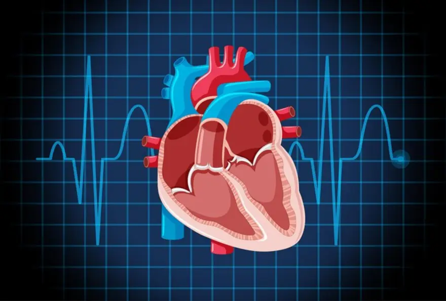
Understanding Tetralogy of Fallot (TOF): Symptoms, Treatment & Causes
Tetralogy Of Fallot (TOF) is a rare congenital heart defect. TOF develops when a baby's heart fails to develop normally during pregnancy. It is important for you to know about this condition and understand its causes, potential complications and treatment options. So let's delve deeper into the biology of tetralogy of fallot to get a better understanding. What is Tetralogy of Fallot? Tetralogy of Fallot is a congenital (present since birth) heart defect characterised by four anatomical abnormalities in a heart. This condition affects blood flow to lungs and your body. Four abnormalities of tetralogy of Fallot Tetralogy of Fallot involves four main heart problems: Ventricular Septal Defect: A hole between your heart's lower chambers. Pulmonary Stenosis: Narrowing of the valve controlling blood flow to your lungs. Overriding Aorta: The aorta, which carries oxygen-rich blood, is shifted toward your right ventricle. Right Ventricular Hypertrophy: Thickening of your heart's right lower chamber. What are the Symptoms of Tetralogy of Fallot? Some of the common tetralogy of fallot symptoms are: Cyanosis (bluish tint to the skin, lips, and nails) Shortness of breath, especially during feeding Rapid breathing Poor weight gain and growth Clubbing of fingers and toes due to chronic lack of oxygen Tet spells Tet spells are sudden episodes of worsened cyanosis and breathlessness in a tetralogy of fallot baby (2-6 months age). They occur when the narrowing of the pulmonary artery suddenly restricts blood flow to the lungs. These spells can be triggered by crying, feeding, dehydration or waking up from a nap and require immediate medical attention. Other Tetralogy of Fallot Symptoms and Signs Some other tetralogy of fallot symptoms include: Difficulty feeding or excessive fussiness during feeding Heart murmur detected during a physical examination of tetralogy of fallot Developmental delays or failure to meet developmental milestones Increased risk of infections, particularly respiratory infections, due to reduced oxygen levels in the blood Tetralogy of Fallot symptoms in Adults The symptoms of tetralogy of fallot in adults may include: Fatigue and breathlessness during physical activity Fainting spells (syncope) Cyanosis may worsen over time Heart palpitations or irregular heartbeats is a common tetralogy of fallot symptom in adults What are the Complications of Tetralogy of Fallot? The complications of tetralogy of fallot may include: Arrhythmias: Irregular heart rhythms due to structural abnormalities associated with tetralogy of fallot. Heart failure: Weakening of your heart muscle over time is a common complication associated with tetralogy of fallot. Infective endocarditis: Bacterial infection of your heart's inner lining. Delayed growth and development: Due to chronic oxygen deficiency caused by tetralogy of fallot. Pulmonary hypertension: Increased blood pressure in your lungs. Stroke: Increased risk due to blood clots or arrhythmias. Paradoxical embolism: Blood clots traveling from your veins to your arteries through a hole in your heart. Neurological complications: Such as developmental delay, seizures, brain abscess etc. What Causes Tetralogy of Fallot? The exact tetralogy of fallot causes are often unknown, but several factors may be involved. For instance: Genetic factors: Mutations in certain genes can increase the risk of tetralogy of fallot. These genes may include NKX2-5, GATA 4, CITED 2 etc. Environmental factors: Exposure to certain environmental toxins, such as chemicals or pollutants, during pregnancy could potentially disrupt normal heart development and increase the risk of tetralogy of fallot. Additionally, maternal infections, particularly during the first trimester, have been linked to a higher incidence of congenital heart defects. Maternal factors: Poor maternal nutrition, inadequate prenatal care, maternal diabetes, and substance abuse (including alcohol and drugs) during pregnancy can influence foetal development and increase the risk of tetralogy of fallot. Chromosomal abnormalities: Conditions involving chromosomal abnormalities, such as Down syndrome (Trisomy 21), are associated with a higher prevalence of congenital heart defects, including tetralogy of fallot. These chromosomal anomalies disrupt normal foetal development, including cardiac formation. How is Tetralogy of Fallot Diagnosed? The diagnosis of tetralogy of fallot is done through a combination of physical examination, imaging tests like echocardiography, blood tests and sometimes genetic testing to determine if certain mutations are associated with tetralogy of fallot. Tests before birth Prenatal screening tests like foetal echocardiography can detect tetralogy of fallot. Foetal echocardiography allows visualisation of the foetal heart to identify structural abnormalities, including tetralogy of fallot. Maternal blood tests, such as Maternal Serum Alpha-Fetoprotein (MSAFP), may indicate an increased risk of congenital heart defects, including tetralogy of fallot. Tests in infancy Newborn screening: Routine physical examination after birth may detect signs suggestive of Tetralogy of Fallot. Echocardiogram: This specialised ultrasound of the heart confirms tetralogy of fallot diagnosis by visualising structural abnormalities. Electrocardiogram (ECG): Detects abnormal heart rhythms or patterns suggestive of tetralogy of fallot. Chest X-ray: Helps assess heart size and lung congestion caused by tetralogy of fallot. Oxygen saturation monitoring: Measures the level of oxygen in the blood, aiding in assessing the severity of cyanosis. Genetic testing: Identifies underlying genetic mutations associated with tetralogy of fallot, especially in syndromic cases. Tests in Childhood or Adulthood In addition to the above tests, other tests for tetralogy of fallot for children and adults include Exercise stress testing: Evaluates heart function during physical activity with the help of an ECG stress test machine, assessing exercise tolerance and identifying any limitations caused by tetralogy of fallot. Cardiac MRI or CT scan: Provides detailed imaging of your heart and blood vessels, aiding in assessment of tetralogy of fallot and planning for potential interventions. How is Tetralogy of Fallot Treated? TOF treatment depends on its severity and may involve: Surgical repair: Complete repair for tetralogy of fallot typically involves closing the ventricular septal defect, relieving pulmonary stenosis, and repositioning the aorta. This is usually performed in infancy. Palliative procedures: Temporary tetralogy of fallot surgery may be needed to improve blood flow to your lungs before complete repair, such as a shunt placement. Medications: Drugs may be prescribed to manage symptoms of tetralogy of fallot, prevent complications, or prepare for surgery in TOF treatment. These may include prostaglandins, diuretics, anticoagulants etc. What are the Risk Factors of Tetralogy of Fallot? Some of the factors that makes one vulnerable to tetralogy of fallot are: Family history( if present in parent or sibling) Poor maternal nutrition during pregnancy Exposure to toxins or viral infections during pregnancy Older maternal age increases the risk of tetralogy of fallot How Can I Reduce my Risk? There are several ways by which you can reduce your risk of tetralogy of fallot: Keep blood sugar levels under control before and during pregnancy Avoid exposure to infections of any kind and fully treat your tetralogy of fallot if you have it. Discuss genetic counselling if you have a family history of tetralogy of fallot or other congenital heart defects Follow prenatal care guidelines and consult with doctors regularly What Can I Expect if I Have Tetralogy of Fallot? If you have Tetralogy of Fallot and have undergone treatment for complete repair of the heart, you can expect to do any daily activity that is done by a normal healthy human with a normal life expectancy, provided you regularly keep an eye on your heart health and manage symptoms associated with tetralogy of fallot. If you leave it untreated, you might face cardiovascular problems throughout your life and it can impact the survival rate. Conclusion Tetralogy of fallot is a congenital heart defect that requires lifelong management. Although it presents unique challenges, tetralogy of fallot can be effectively managed with early diagnosis, appropriate medical care, and surgical interventions. If your family has a history of congenital heart defects, consider getting a blood test done to check for tetralogy of fallot in your baby. Metropolis Healthcare is at your service with hassle-free, reliable and comprehensive blood test facilities, both at home and within our advanced labs. Book your test today!

Understanding Tourette Syndrome: Symptoms, Treatment & Causes
What is Tourette syndrome? Tourette Syndrome (TS) is a neurological condition characterised by repetitive movements or involuntary sounds known as tics. Tourette syndrome symptoms arise as sudden twitches, movements, or vocalisations that individuals experience uncontrollably. These tics can significantly impact their daily lives. How common is Tourette syndrome? Tourette syndrome, a prevalent neurodevelopmental disorder, impacts approximately 1 per cent of the population around the world. Is Tourette’s the only tic disorder? No, Tourette syndrome is not the only tic disorder. Other recognised tic disorders are:- Persistent (Chronic) Motor or Vocal Tic Disorder: Involves motor or vocal tics that last over a year, but not both. Provisional Tic Disorder: Characterised by one or more motor or vocal tics lasting less than a year. Other Specified Tic Disorder: This is applied when symptoms do not meet a specific tic disorder criteria but still cause distress or impairment. Unspecified Tic Disorder: This is used when symptoms do not align with any specific Tic disorder or when adequate information is lacking for a precise diagnosis. What are the symptoms of Tourette syndrome? The primary Tourette syndrome symptom is tics, which can vary in severity from mild and hardly noticeable to frequent and conspicuous. Factors like stress, excitement, illness, or fatigue can exacerbate them. Severe tics may impact social interactions or work performance. There are two types of Tourette syndrome symptoms: Motor tics involve physical movements, such as arm or head jerking, blinking, facial expressions, shoulder shrugging, or mouth twitching. Vocal tics encompass sounds or verbalisations, including barking, throat clearing, coughing, grunting, echolalia (repeating others' words), shouting, sniffing, or swearing. Tics can be categorised as simple, affecting only one or a few body parts like eye blinking or facial expressions, or complex Tourette syndrome symptoms like coordinated movements or vocalisations. What causes Tourette syndrome? Although the exact Tourette syndrome causes remain unknown, studies indicate that disruptions in neural communication within specific brain regions and imbalances in neurotransmitters, which relay nerve signals, may contribute to its onset. Tourette syndrome may also result from gene alterations inherited from parents or occurring during prenatal development. What are the risk factors for Tourette syndrome? Risk factors for Tourette syndrome: Genetic predisposition: Family history increases susceptibility Gender: Males are more commonly affected Age of onset: Symptoms typically emerge in childhood or adolescence. Prenatal and perinatal influences: Complications during pregnancy or childbirth may heighten the risk. Neurological co-morbidities: Conditions like ADHD or OCD often coexist. Environmental exposures: Toxins or stressors during critical brain development periods can contribute. Immune system involvement: Dysfunctions or abnormalities may play a role. How is Tourette syndrome diagnosed? Diagnosing Tourette syndrome typically involves a comprehensive assessment by a healthcare professional. While there is no specific test for Tourette syndrome, diagnosis is based on clinical evaluation and criteria established by medical guidelines, which may include: Medical History: The healthcare provider will review the individual's medical history, including symptoms, family history of tic disorders, and any other relevant medical conditions or medications. Physical Examination: A physical examination may be conducted to rule out other medical conditions that could be causing the symptoms. Diagnostic Criteria: Tourette syndrome is diagnosed based on specific diagnostic criteria, which typically include the presence of both motor and vocal tics, frequent tics lasting over a year, starting before the age of 18, and not being caused by medications, substances, or other medical conditions. Tics must also evolve in location, frequency, or severity. Exclusion of Other Conditions: The healthcare provider may order additional tests, such as blood tests or imaging studies like MRI scans, to rule out other medical conditions that could be causing the symptoms. Consultation with Specialists: In some cases, consultation with specialists such as neurologists or psychiatrists may be recommended to confirm the diagnosis and develop a comprehensive Tourette syndrome treatment plan. What is the treatment for Tourette syndrome? Tourette syndrome has no cure, but Tourette syndrome treatment aims to manage disruptive tics. The options include: Medication: Dopamine-blocking medicines may control tics, although with possible side effects. Botulinum injections can ease specific tics, like motor and phonic. Attention deficit hyperactivity disorder (ADHD) meds might aid focus, but they can worsen tics in some cases. Central adrenergic inhibitors (adrenaline controllers) or antidepressants may help manage behavioural and emotional symptoms. Antiseizure meds may be effective for some. Tourette syndrome therapies: Behaviour therapy, such as Cognitive Behavioural Interventions for Tics, can help monitor and manage tics. Psychotherapy addresses associated issues like ADHD, OCD, or anxiety. Deep brain stimulation (DBS) is an effective Tourette syndrome therapy option for severe, treatment-resistant tics, but it is still under research to determine safety and efficacy. How can medication help Tourette syndrome? Medication addresses Tourette syndrome symptoms by balancing brain neurotransmitters, notably dopamine: Dopamine blockers (e.g., haloperidol, risperidone) reduce tics by blocking dopamine receptors. Stimulant ADHD meds (e.g., methylphenidate) enhance focus but may exacerbate tics. Central adrenergic inhibitors (e.g., clonidine) manage behavioural symptoms such as impulse control and rage. SSRIs (Serotonin reuptake inhibitors) like fluoxetine help with sadness, anxiety, and OCD, often linked with Tourette syndrome. Antiseizure meds like topiramate can also lessen tic severity. Medication aims to improve symptoms and quality of life. It is tailored to each individual's needs and side-effect tolerance. How can behavioural therapy help Tourette Syndrome? Behavioural therapy, like Cognitive Behavioural Interventions for Tics (CBIT), aids Tourette syndrome management by teaching tic monitoring, habit-reversal training (HRT), and relaxation techniques to reduce tic frequency and severity. It increases awareness, offers environmental modifications, and provides education and support to empower individuals and their families in coping with Tourette syndrome challenges effectively. Combined with medication and other treatments, it forms a comprehensive approach to improve tic control and overall quality of life. Conclusion Tourette syndrome poses challenges for those affected, impacting daily life with its characteristic tics and associated symptoms. Lately, understanding and management of TS have improved significantly. While no cure exists, various treatments aim to relieve symptoms and enhance quality of life. Medications offer relief, although with potential side effects. While behavioural therapies empower individuals to manage tics effectively, deep brain stimulation has shown promise in severe cases. For an accurate diagnosis, book your consultation, MRI and blood tests at Metropolis Labs and contact your doctor for immediate treatment. Schedule your tests today.


 WhatsApp