Preventive Healthcare
Omentum: Meaning, Types, Functions, Clinical Importance and Tests
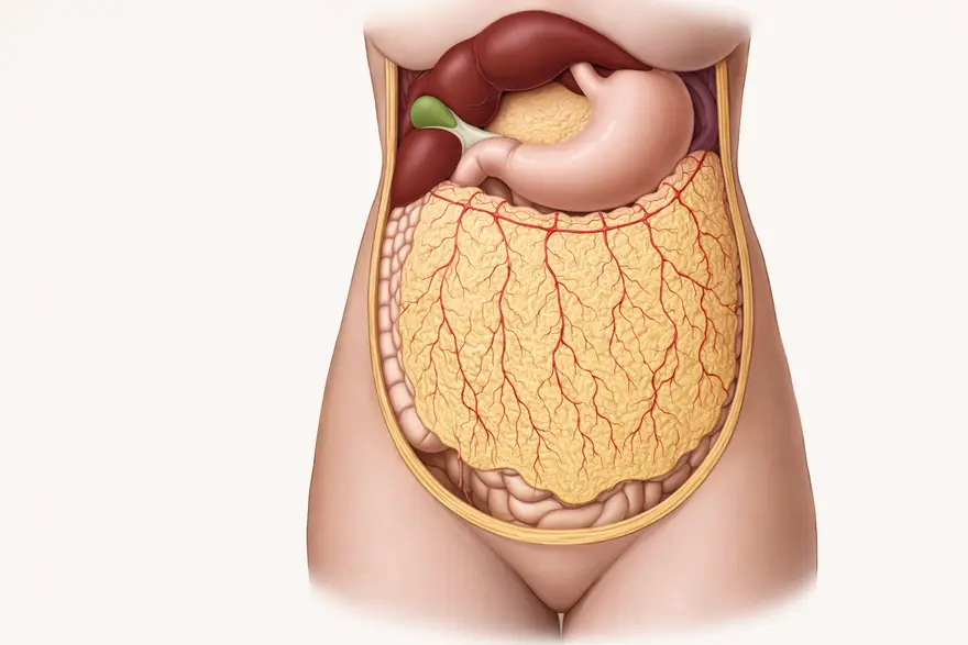
Table of Contents
The omentum is a large, apron-like fold of the abdominal lining that hangs from the stomach and lies over the intestines. It is made up of visceral peritoneum and contains fatty tissue, blood vessels, lymphatic channels, and immune cells.
Most people only hear about the omentum when it appears on a scan report or during an explanation of abdominal pain, infection, or cancer spread within the abdomen. Understanding what it is and what it does can make medical discussions and test results much easier to follow.
This article is for general information. If you have severe or persistent abdominal symptoms, speak to a doctor promptly.
What is Omentum?
Inside the abdomen, a thin, smooth membrane called the peritoneum lines the abdominal wall and covers many organs. The peritoneum has two layers: the parietal peritoneum (lining the abdominal wall) and the visceral peritoneum (covering abdominal organs). Between these layers is the peritoneal cavity, a potential space that allows organs to move smoothly rather than rubbing against each other.
The omentum is formed when the visceral peritoneum folds and fuses into a double-layered sheet. It is not simply a passive covering. It is living tissue with a strong blood supply and active immune components. Because it sits in the middle of the abdomen and can move, it often plays a role in how the body responds to inflammation and infection inside the abdominal cavity.
Types of Omentum
There are two main parts of the omentum. Both connect to the stomach, but they extend to different organs and have different clinical significance.
Greater omentum
The greater omentum is the larger and more familiar portion. It attaches to the greater curvature of the stomach and drapes downward over the small intestines and the transverse colon. Many descriptions compare it to a curtain because it hangs loosely and can shift position.
The greater omentum often contains a noticeable amount of fat, which can vary greatly between individuals. This fat is not just storage tissue. It supports blood vessels, immune cell clusters, and healing responses.
In clinical language, the greater omentum is sometimes called the “policeman of the abdomen” because it can move towards inflamed areas and stick to them, helping isolate the problem and limit spread within the abdomen.
Lesser omentum
The lesser omentum is smaller and runs between the lesser curvature of the stomach (and the first part of the duodenum) and the liver. It forms a thin sheet that is important for both anatomy and surgery.
The lesser omentum is commonly described in two parts:
- The hepatogastric ligament, between the liver and the stomach
- The hepatoduodenal ligament, between the liver and the duodenum
The hepatoduodenal ligament is particularly important because it contains the portal triad, the main vein, artery, and bile duct travelling to and from the liver. For this reason, the lesser omentum is frequently discussed in abdominal surgery and when interpreting certain scan findings.
Omentum Function
The omentum has several roles that are relevant to everyday health and to clinical care. These functions overlap, and they often become more obvious when the abdomen is inflamed or injured.
Mechanical protection and cushioning
The omentum provides a soft layer over the intestines and other abdominal organs. This can reduce friction as organs move during digestion and normal activity.
Helping prevent surfaces from sticking
By sitting between organs and the abdominal wall, the omentum can help reduce direct contact. This contributes to smoother movement inside the abdomen. Adhesions can still form after surgery or infection, but the omentum is part of the system that helps maintain mobility.
Immune defence
The omentum contains immune cell collections often called milky spots. These are areas where immune activity is concentrated and can support responses to pathogens and irritants within the peritoneal cavity.
Containing inflammation and infection
Because the omentum is mobile, it may migrate towards a site of inflammation and adhere to it. This can help wall off infection or inflammation, reducing the chance of it spreading widely within the abdomen.
Fat storage and metabolic activity
The omentum stores visceral fat. Visceral fat is metabolically active, meaning it interacts with hormones and inflammatory pathways. This is one reason abdominal fat distribution is linked to broader metabolic health.
Supporting tissue repair
The omentum has a strong blood supply and biological factors associated with healing. In selected situations, this regenerative potential is used in surgical practice.
Omentum in Digestion and Immunity
The omentum sits close to the stomach, intestines, and major digestive blood vessels, which places it in an ideal position to support both digestion-related movement and immune surveillance in the peritoneal cavity.
From an immunity perspective, the milky spots allow immune cells to detect antigens and respond to threats within the abdomen. This becomes particularly relevant during infections or inflammatory conditions where the body needs to limit spread. In practical terms, this is one reason the omentum may be found adhering to an inflamed appendix or another inflamed organ during surgery.
It is important to remember that abdominal symptoms are often non-specific. Pain, tenderness, nausea, or fever can have many causes, and the omentum is just one part of a larger clinical picture. Doctors typically rely on history, examination, blood tests, and imaging to determine the cause.
Clinical Importance of Omentum
Doctors pay attention to the omentum because it can be directly affected by disease or because it reflects disease elsewhere in the abdomen. The omentum can be involved in:
- acute abdominal pain syndromes
- inflammatory and infectious processes
- surgical repair and healing
- spread of disease within the abdomen, including certain cancers
If you have severe abdominal pain, pain that worsens over time, fever, persistent vomiting, fainting, or blood in stool or vomit, seek urgent medical care. These symptoms can be caused by many conditions, and early assessment matters.
Omentum in Abdominal Diseases
Omental infarction
Omental infarction occurs when part of the omentum loses its blood supply. This is uncommon, but it can cause sudden localised abdominal pain and can mimic appendicitis or gallbladder inflammation. Imaging, especially CT, is often used to distinguish it from other urgent causes of abdominal pain.
Omental torsion
Torsion means twisting. If the omentum twists, blood flow may be compromised, leading to inflammation and pain. Because the symptoms overlap with other abdominal conditions, imaging is usually needed to reach a diagnosis.
Inflammation related to nearby organs
Inflammation from nearby organs can involve the omentum. In these cases, the omentum may adhere to an inflamed area, which can be protective, but it can also contribute to tenderness and inflammatory changes seen on scans.
Omental thickening and “omental cake”
Radiology reports may mention omental thickening. In some cases, more extensive thickening is described as “omental cake”. These are descriptive terms, not diagnoses. Depending on the clinical situation, they may be associated with inflammation, infection, or malignant disease. Doctors interpret these findings alongside symptoms, blood tests, and sometimes tissue sampling.
Adhesions after surgery or inflammation
Adhesions are bands of scar tissue that can form after abdominal surgery or significant inflammation. The omentum can be part of adhesion formation because it tends to move towards inflamed tissue and may stick to it during healing.
Imaging and Diagnostic Tests to Identify the Omentum
Omental conditions are rarely diagnosed from symptoms alone, because many abdominal problems feel similar at first. Doctors often use imaging and laboratory tests to understand what is happening.
Imaging can help identify:
- inflammation and fat stranding within the omentum
- focal masses or nodules
- fluid collections within the abdomen
- patterns that suggest infarction, torsion, infection, or spread of malignant disease
Ultrasound (abdomen)
Ultrasound is commonly used as an initial test for abdominal symptoms. It is safe and widely available, and it helps assess many abdominal organs. However, it can be limited for detailed omental evaluation, especially if bowel gas obscures the view or if the area of concern is deep.
CT scan (abdomen and pelvis)
CT Scan is often the most informative imaging test for omental pathology. It can show inflammatory changes, focal infarction, torsion-related patterns, omental thickening, and deposits. CT also helps doctors look for alternative causes of pain and assess the wider abdomen in a single examination.
MRI
MRI can be useful when more soft tissue characterisation is needed or when CT is not suitable. The decision depends on the clinical question and what the doctor is trying to confirm.
PET CT
PET CT may be used in oncology pathways to assess metabolic activity and the extent of disease spread, when a doctor considers it appropriate.
Laboratory tests
Blood tests do not identify the omentum directly, but they support the overall assessment. Depending on symptoms and suspected cause, a doctor may recommend tests such as a full blood count and markers of inflammation. Further tests are selected based on individual risk factors, examination findings, and imaging results.
If your doctor recommends blood tests as part of an abdominal assessment, Metropolis Healthcare can perform the requested laboratory investigations and provide reports for your doctor to review and interpret in conjunction with imaging and clinical findings.
Biopsy or fluid analysis
If imaging suggests malignancy, chronic infection, or an unclear cause, the doctor may recommend tissue sampling or analysis of abdominal fluid. This helps confirm diagnosis and guide treatment planning.
Surgical Considerations with Omentum
The omentum is important in surgery because it is mobile, well supplied with blood, and biologically active. In selected cases, surgeons use it to support healing, protect surgical joins within the bowel, or cover defects. This approach is based on the omentum’s ability to bring blood supply and healing factors to an area that needs support.
Surgical decisions involving the omentum depend on the patient’s underlying condition, anatomy, prior operations, and the goals of treatment. Your surgeon will explain the reasoning if an omental procedure is being considered.
Omentum and Its Role in Cancer
In some abdominal cancers, the omentum is a common site for spread within the peritoneal cavity. This happens because the omentum contains blood vessels, lymphatic channels, and immune structures that can trap cells circulating within abdominal fluid. On imaging, doctors may look for omental nodules, thickening, or more extensive involvement.
Scan reports may use terms such as omental deposits or peritoneal disease. These findings require careful interpretation, and confirmation may involve further imaging, tumour marker testing, or biopsy depending on the clinical situation.
Omentectomy
An omentectomy is the surgical removal of part or all of the omentum. It may be performed in certain cancers when the omentum is involved or when removal supports staging and management. The decision is individual and usually made by a multidisciplinary team, taking into account imaging, surgical findings, and pathology.
FAQs
What is the function of the omentum?
The omentum helps protect abdominal organs by cushioning them and reducing friction during movement. It also supports immune defence through specialised immune cell clusters, and it can move towards inflamed areas to help contain infection and inflammation. In addition, it stores visceral fat and contributes to tissue repair through its blood supply and healing-related factors.
What diseases affect the omentum?
The omentum can be affected by conditions such as omental infarction and omental torsion, both of which can cause acute abdominal pain. It can also become involved in inflammation or infection related to nearby organs, and it may show thickening or nodularity on imaging. In some cancers, the omentum can be a site of spread within the abdomen, which is why it is assessed during staging and follow-up.
Can the omentum be removed surgically?
Yes. Doctors may recommend removal of part or all of the omentum for specific reasons, including selected cancer surgeries or when the omentum itself is diseased. Many people can live without a portion of the omentum, but the impact and the approach depend on the underlying condition and the overall surgical plan.
What is the greater omentum?
The greater omentum is the larger fold of the visceral peritoneum that attaches to the greater curvature of the stomach and drapes over the intestines. It contains fat, blood vessels, lymphatic channels, and immune tissue. It helps cushion organs and may move towards inflamed areas to limit spread of infection or inflammation.
References
- Matricardi P. M., Kleine-Tebbe J., Hoffmann H. J., et al. (2016). Molecular diagnosis of allergic diseases: Current status and future perspectives. Allergy, 71(11), 1473–1488. PMID: 27159990
- Scala E., Villalta D., Giani M., et al. (2018). Molecular allergology and clinical relevance: The allergen microarray approach. Clinical Chemistry and Laboratory Medicine, 56(9), 1416–1424. PMID: 29701458
- Standring S. (Ed.). (2021). Gray’s Anatomy: The Anatomical Basis of Clinical Practice. 42nd Edition. Elsevier.
- Drake R. L., Vogl A. W., Mitchell A. W. M. (2019). Gray’s Anatomy for Students. 4th Edition. Elsevier.
- Moore K. L., Dalley A. F., Agur A. M. R. (2018). Clinically Oriented Anatomy. 8th Edition. Wolters Kluwer.
- Sadler T. W. (2019). Langman’s Medical Embryology. 14th Edition. Wolters Kluwer.
- Meza-Perez S., Randall T. D. (2017). Immunological Functions of the Omentum. Trends in Immunology, 38(7), 526–536. PMID: 28579319
- Rangel-Moreno J., Moyron-Quiroz J. E., Carragher D. M., et al. (2009). Omental milky spots develop in the absence of lymphoid tissue-inducer cells and support B and T cell responses to peritoneal antigens. Immunity, 30(5), 731–743. PMID: 19427241
- Di Nicola V. (2019). Omentum a powerful biological source in regenerative surgery. Regenerative Therapy, 11, 182–191. PMID: 31453273
- Naffaa L. N., Shabb N. S., Haddad M. C. (2003). CT findings of omental torsion and infarction: case report and review of the literature. Clinical Imaging, 27(2), 116–118. PMID: 12639779
- Puylaert J. B. (1992). Right-sided segmental infarction of the omentum: clinical, US, and CT findings. Radiology, 185(1), 169–172. PMID: 1523302
- Liebermann-Meffert D. (2000). The greater omentum. Anatomy, embryology, and surgical applications. Surgical Clinics of North America, 80(1), 275–293. PMID: 10685153
- Wang A. W., Prieto J. M., Cauvi D. M., et al. (2020). The Greater Omentum-A Vibrant and Enigmatic Immunologic Organ Involved in Injury and Infection Resolution. Shock, 53(4), 384–390. PMID: 31389904
- McLachlin A. D., Denton D. W. (1973). Omental protection of intestinal anastomoses. American Journal of Surgery, 125(1), 134–140. PMID: 4683467