Preventive Healthcare
Sternum (Breastbone): Anatomy, Function, and Common Conditions
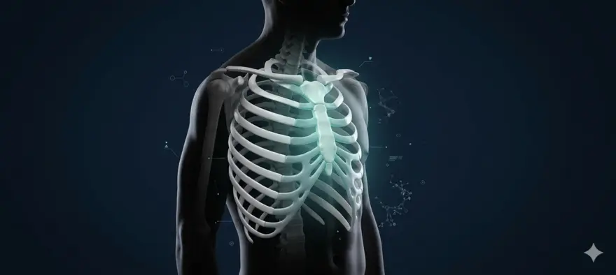
Table of Contents
- What Is a Sternum (Breastbone)?
- Where Is the Sternum Bone Located?
- Sternum Anatomy: Structure and Sternum Parts
- Sternum Diagram With Labelled Parts
- How the Sternum Connects With Ribs (Sternum Ribs Structure)
- What Is the Function of the Sternum?
- Development and Growth of the Sternum
- Common Sternum Conditions and Structural Disorders
- How Is the Sternum Examined or Diagnosed?
- Diagnostic Tests for Identifying Sternum Issues
- Treatment Approaches for Sternum Conditions
- Can You Live Without a Sternum?
- Sternum vs Ribcage: What Is the Difference?
- Interesting Facts About the Sternum Bone
- When Should You Seek Medical Advice?
- Supporting Your Bone Health and Overall Wellbeing
- Frequently Asked Questions
- References
You may not think about it often, but the bone at the centre of your chest is doing a remarkable job every single day. It shields your most vital organs, anchors your ribcage, and supports the muscles that help you breathe. This bone is your sternum, more commonly known as your breastbone. Understanding its structure and what can go wrong with it can help you make sense of chest pain and know when to seek medical care.
What Is a Sternum (Breastbone)?
The sternum is a long, flat bone located at the front and centre of your chest. It runs vertically from just below your collarbone to the upper abdomen. Its primary role is to protect the heart, lungs, and major blood vessels that lie behind it. It also forms the front anchor of the ribcage and serves as an attachment point for key muscles. Another widely used name for it is the breastbone.
Where Is the Sternum Bone Located?
The sternum sits in the midline of your chest, right at the front. You can feel it by placing your hand flat against the centre of your chest. It connects to structures on both sides of your body and runs from top to bottom in the following position:
- At the top, it meets your clavicles (collarbones) at the sternoclavicular joints
- On either side, it connects to the ribs via cartilage
- At the back, it faces the thymus gland, the heart, and large blood vessels
- At the bottom, it tapers into a small, pointed projection that sits just above the upper abdomen
Sternum Anatomy: Structure and Sternum Parts
The sternum is not a single uniform bone. It is made up of three distinct sections that gradually fuse together as you grow into adulthood. Each section has its own shape, function, and connections to the rest of the skeleton.
Manubrium
The manubrium is the broad, upper portion of the sternum. It is roughly trapezoid in shape and is the widest part of the bone.
- It articulates with the clavicles on either side at the sternoclavicular joints
- It connects with the first pair of ribs directly and the second pair of ribs at its lower border
- It houses a small notch at the top centre called the jugular notch, which you can feel at the base of your throat
- It meets the body of the sternum at a slight angle, forming a small ridge called the sternal angle (or angle of Louis), which is a useful anatomical landmark in clinical examinations
Body of the Sternum (Gladiolus)
The body, also called the gladiolus, is the longest part of the sternum. It makes up the central bulk of the bone.
- It is flat, narrow, and elongated
- It connects with the second through seventh pairs of ribs via costal cartilage
- It runs from the sternal angle down to the xiphoid process
- It provides the primary surface area for rib attachment along the sides of the chest
Xiphoid Process
The xiphoid process is the smallest and lowest section of the sternum. Its name comes from the Greek word for sword.
- It is made largely of cartilage at birth and in childhood
- It gradually calcifies and begins turning to bone from early adulthood, typically completing this process around the age of 40
- Its shape varies considerably from person to person. It may be straight, curved, bifurcated (forked), or deviated to one side
- It serves as an attachment point for the diaphragm and some abdominal muscles
- In CPR training, the xiphoid process is used as a reference point to identify the correct position for chest compressions, just above it on the lower half of the sternum
Sternum Diagram With Labelled Parts
Visualising the sternum helps make its anatomy clearer. Picture an upside-down sword. The wide, handle-like top is the manubrium. The long flat middle section is the body. And the narrow pointed tip at the bottom is the xiphoid process. The ribs attach along the sides of the body like rungs branching outward from a central column. Together, they form the front wall of the ribcage.
How the Sternum Connects With Ribs (Sternum Ribs Structure)
The sternum does not connect directly to the ribs in the way two bones typically meet at a joint. Instead, it connects through costal cartilage, which is a flexible, elastic type of cartilage that allows the chest to expand and contract during breathing.
- The first rib attaches directly to the manubrium
- The second rib connects at the junction between the manubrium and the body
- Ribs three through seven attach along the body of the sternum via individual costal cartilage segments
- Ribs eight through ten do not attach directly to the sternum. They connect to the cartilage of the rib above them
- Ribs eleven and twelve are called floating ribs because they do not attach to the sternum at all
How Many Ribs Attach to the Sternum?
Seven pairs of ribs attach directly to the sternum. These are known as the true ribs. The remaining five pairs on each side (ribs eight to twelve) either attach indirectly through cartilage or not at all, and are referred to as false ribs and floating ribs respectively.
What Is the Function of the Sternum?
Protection of Vital Organs
The sternum sits directly in front of the chest cavity, forming a bony shield over some of your most vital structures. These include:
- The heart and its major vessels
- The lungs
- The thymus gland, which plays an important role in your immune system
- The oesophagus and trachea (partially)
Structural Support of the Ribcage
The sternum is the front anchor of the ribcage. Without it, the ribs on either side would have no stable midline structure to connect to. It effectively holds the two halves of the ribcage together at the front, maintaining the shape and integrity of the thoracic cavity.
Muscle Attachment and Movement
Several important muscles attach to the sternum, including:
- The pectoralis major, a large chest muscle involved in arm movement and upper body strength
- The sternocleidomastoid muscle, which runs from the sternum to the skull and assists in neck movement
- The diaphragm, which attaches to the xiphoid process and is the primary muscle of breathing
- The rectus abdominis, which runs from the xiphoid process down the front of the abdomen
- The transversus thoracis, a muscle that assists in exhalation by compressing the ribcage
Development and Growth of the Sternum
The sternum begins forming in the foetal stage, developing from two parallel cartilaginous bands that fuse together along the midline. At birth, the sternum is almost entirely cartilaginous. Over time, this cartilage is gradually replaced by bone through a process called ossification.
- The manubrium is usually fully ossified by early childhood
- The body fuses from several segments progressively through childhood and adolescence
- The sternal angle fully forms during this fusion process
- The xiphoid process remains largely cartilaginous well into adulthood and completes ossification last
How the Sternum Changes With Age
- At birth and in infancy, the sternum is soft, flexible, and predominantly cartilaginous
- During childhood, ossification begins and the bone gradually becomes harder
- During adolescence and early adulthood, the separate segments of the body fuse together
- By around age 25, the manubrium and body are usually fully fused
- The xiphoid process calcifies more slowly, often completing this process between the ages of 40 and 60
- In older adults, the fully ossified sternum may be more susceptible to fracture from falls or trauma
Common Sternum Conditions and Structural Disorders
Sternum Fracture
A sternal fracture is a break in the breastbone, most commonly caused by blunt force trauma to the chest. It frequently occurs in road accidents, particularly when the chest strikes the steering wheel or seatbelt. Sports injuries and falls can also cause it.
Costochondritis
Costochondritis is one of the most common causes of chest pain that is not related to the heart. It involves inflammation of the costal cartilage, which connects the ribs to the sternum.
Pectus Excavatum
Pectus excavatum, also known as sunken chest or funnel chest, is a congenital condition where the sternum and front ribs grow inward rather than outward, creating a concave appearance.
Pectus Carinatum
Pectus carinatum, or pigeon chest, is the opposite of pectus excavatum.
Sternal Infections (Osteomyelitis)
Infection of the sternum, known as sternal osteomyelitis, is relatively rare but serious.
How Is the Sternum Examined or Diagnosed?
- A physical examination to assess tenderness, swelling, and the exact location of pain
- A review of your medical history, including any recent trauma, illness, or surgery
- Palpation of the sternoclavicular joint, the costochondral junctions, and the xiphoid area
- Assessment of your breathing pattern and any pain that worsens with inspiration
- Referral for imaging if bone injury, infection, or structural abnormality is suspected
Diagnostic Tests for Identifying Sternum Issues
- Chest X-ray: A standard first-line test to assess for fractures, structural deformities, or lung involvement
- CT scan: Provides detailed cross-sectional images and is more sensitive than X-ray for detecting sternal fractures, infections, and masses
- MRI: Useful for assessing soft tissue involvement, including infection, tumour infiltration, or bone marrow changes
- Bone scan: May be used to detect infection, stress fractures, or cancer spread to the sternum
- Blood tests: Including inflammatory markers (CRP, ESR) and a complete blood count to detect signs of infection or systemic illness
- Electrocardiogram (ECG): Often performed when chest pain is present to rule out cardiac causes
Treatment Approaches for Sternum Conditions
- Rest and activity modification for fractures and costochondritis
- Over-the-counter anti-inflammatory medications such as ibuprofen for pain and swelling
- Physical therapy to restore movement and strengthen surrounding muscles after injury
- Bracing for structural conditions such as pectus carinatum in growing children
- Corticosteroid injections for persistent costochondritis or inflammation
- Antibiotics or antifungal therapy for sternal infections
- Surgical intervention for displaced fractures, severe deformities, deep sternal wound infections, or post-cardiac surgery complications
- Management of underlying conditions such as acid reflux, pleurisy, or bronchitis when these are causing substernal pain
Can You Live Without a Sternum?
In rare cases, the sternum can be partially or fully removed.
Sternum vs Ribcage: What Is the Difference?
|
Feature |
Sternum |
Ribcage |
|
What it is |
A single flat bone |
A bony structure made up of 12 pairs of ribs, the sternum, and the thoracic vertebrae |
|
Location |
Centre front of the chest |
Surrounds the entire thoracic cavity |
|
Primary role |
Protects organs and anchors the ribcage at the front |
Encloses and protects the heart, lungs, and other thoracic organs |
|
Composition |
Three fused sections (manubrium, body, xiphoid process) |
24 individual rib bones plus the sternum and spine |
|
Movement |
Largely stationary |
Expands and contracts with every breath |
|
Connections |
Connects to clavicles and ribs via cartilage |
Connects to the sternum at the front and thoracic spine at the back |
Interesting Facts About the Sternum Bone
- The word sternum comes from the Greek word sternon, meaning chest
- The sternum is approximately 15 to 17 centimetres long in adults
- It is one of the main sites used for bone marrow biopsy, as it is relatively accessible and contains active marrow
- The sternal angle (where the manubrium meets the body) is used by doctors to count rib levels and locate important anatomical structures during examination
- During CPR, chest compressions are delivered to the lower half of the sternum, just above the xiphoid process
- The thymus gland, which sits directly behind the sternum, is largest in childhood and gradually shrinks after puberty
- Congenital absence of the sternum, though extremely rare, has been documented
When Should You Seek Medical Advice?
- Your pain follows a direct blow, fall, or accident
- The pain is persistent or worsening over time
- You have difficulty breathing or pain that worsens significantly with each breath
- You notice swelling, bruising, or warmth over the sternum
- You have a fever alongside chest pain, which may suggest infection
- You experience sudden, severe chest pain accompanied by sweating, nausea, shortness of breath, or pain radiating to the arm or jaw
Supporting Your Bone Health and Overall Wellbeing
Your skeletal system, including the sternum and the broader Bones that make up your chest wall, relies on adequate nutrition, physical activity, and regular health monitoring to stay strong throughout your life.
Frequently Asked Questions
Is the Sternum the Same as the Breastbone?
Yes. The sternum and the breastbone refer to the same bone.
Where Is the Sternum Located in the Body?
The sternum is located at the front and centre of the chest.
What Organs Does the Sternum Protect?
The sternum protects the heart, lungs, major blood vessels, the thymus gland, and partially the oesophagus and trachea.
At What Age Does the Sternum Fully Fuse?
The sternum fuses in stages.
Why Is the Sternum Important in CPR?
During cardiopulmonary resuscitation (CPR), compressions are delivered to the lower half of the sternum.
References
- Drake RL, Vogl AW, Mitchell AWM. Gray's Anatomy for Students. 4th ed. Philadelphia: Elsevier; 2019.
- Moore KL, Dalley AF, Agur AMR. Clinically Oriented Anatomy. 8th ed. Philadelphia: Wolters Kluwer; 2018.
- Knipe H, Murphy A, et al. Sternum anatomy. Radiopaedia. 2023.
- Mollen KP, Kane TD. Pectus excavatum and carinatum. Curr Opin Pediatr. 2008;20(3):328-332. PMID: 18475100.
- Harston A, Roberts C. Classification of sternum fractures and associated injuries. Injury. 2011;42(11):1170-1174. PMID: 21689826.
- Pikkarainen E, Räsänen P, Roine RP, et al. Sternal wound infections. Eur J Cardiothorac Surg. 2010;37(1):184-190.
- Slipman CW, Derby R, Simeone FA, Mayer TG. Interventional Spine: An Algorithmic Approach. Philadelphia: Saunders Elsevier; 2008.
- World Health Organization. Cardiovascular diseases (CVDs). Geneva: WHO; 2023.