Preventive Healthcare
Basal Cell Carcinoma: Spotting It Early & Treatment Approaches
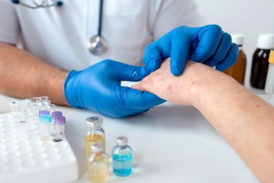
Table of Contents
- What is Basal Cell Carcinoma (BCC)?
- Who is at Risk?
- Causes of Basal Cell Carcinoma
- Early Signs & Symptoms of Basal Cell Carcinoma
- Types of Basal Cell Carcinoma
- How Basal Cell Carcinoma is Diagnosed
- Treatment Options for Basal Cell Carcinoma
- Recovery and Follow-Up
- Prevention Tips
- Conclusion
- References
What is Basal Cell Carcinoma (BCC)?
Basal cell carcinoma (BCC) is the most common type of skin cancer, accounting for nearly 80% of all cases. It develops from the basal cells — the deepest layer of the epidermis (outer skin). These cells normally replace old skin cells, but when damaged (most often by ultraviolet (UV) light), they can begin to grow abnormally and form cancerous lesions.
Though basal cell carcinoma grows slowly and rarely spreads (metastasises), untreated cases can cause significant local tissue destruction, scarring, and disfigurement.
Early detection and prompt basal cell carcinoma treatment lead to excellent outcomes, often with a complete cure. With timely diagnosis and modern treatments, nearly all basal cell carcinomas are completely curable.
Who is at Risk?
Anyone can develop basal cell carcinoma, but certain factors make some people more vulnerable. Awareness of these risks can help in early detection and prevention.
- Fair skin, light eyes, or blond/red hair: Less melanin means less natural protection against UV damage.
- Prolonged or intense sun exposure: Outdoor work or frequent sunbathing increases risk.
- Living in sunny or high-altitude areas: Stronger UV radiation damages skin cells over time.
- Use of tanning beds: Artificial UV light accelerates DNA injury in basal cells.
- Personal or family history of skin cancer: Indicates higher genetic susceptibility.
- Older adults: Cumulative UV exposure over decades raises risk.
- Weakened immune system: Long-term immunosuppressive therapy after transplants or for chronic illness.
- Occupational exposure: Jobs involving arsenic, radiation, or tar-based chemicals.
- Inherited conditions: Rare syndromes such as Gorlin (Basal Cell Nevus) Syndrome increase lifelong risk.
Causes of Basal Cell Carcinoma
According to the National Institutes of Health (NIH), the main causes of basal cell carcinoma stem from cumulative DNA damage in the skin’s basal cells — most often due to prolonged exposure to ultraviolet (UV) radiation. Over time, this damage disrupts normal cell repair, leading to uncontrolled growth and cancer formation.
Common contributing factors include:
- Chronic UV exposure: Long-term sunlight or tanning bed use causes DNA mutations that trigger abnormal cell growth.
- Intermittent intense exposure: Severe sunburns, particularly in childhood or adolescence, increase lifetime risk.
- Ionising radiation: Prior radiotherapy or occupational radiation exposure can damage skin cells.
- Contact with carcinogens: Substances like arsenic, coal tar, and certain industrial solvents raise cancer risk.
- Weakened immune system: Seen in organ transplant recipients, HIV-positive individuals, or those on prolonged corticosteroids.
- Genetic predisposition: Inherited DNA-repair defects, such as in Gorlin (Basal Cell Nevus) Syndrome, heighten susceptibility.
- Chronic skin injury or scars: Areas of long-standing inflammation, burns, or scars may develop cellular changes over time.
Early Signs & Symptoms of Basal Cell Carcinoma
Recognising basal cell carcinoma symptoms early ensures simpler, less invasive treatment.
Lesions often develop on sun-exposed areas such as the face, ears, neck, scalp, shoulders, and back.
Common basal cell carcinoma symptoms include:
- A pearly or waxy bump that may bleed easily
- A flat, flesh-coloured or brown scar-like lesion
- A sore that doesn’t heal or heals and returns
- Tiny visible blood vessels (telangiectasia) on the lesion surface
- Crusting or ulceration in the centre of a raised lesion
- Itching, pain, or oozing in advanced stages
Although basal cell carcinoma rarely spreads, neglecting these early signs can cause deep tissue invasion and disfigurement, especially around the nose, eyes, or ears. If you notice any of these symptoms, consult a dermatologist promptly.
Types of Basal Cell Carcinoma
Dermatologists identify several BCC types based on how the tumour looks under the skin and how aggressively it grows. Recognising the type helps guide the most effective treatment plan.
- Nodular Basal Cell Carcinoma:
The most common form. Appears as a shiny, dome-shaped bump with visible blood vessels and may bleed or form a crust. - Superficial Basal Cell Carcinoma:
Presents as a flat, red, or scaly patch — often on the trunk, shoulders, or back. It tends to grow slowly and may resemble eczema or psoriasis. - Morpheaform (Sclerosing) Basal Cell Carcinoma:
Appears as a firm, scar-like lesion with poorly defined borders. This type grows more deeply and can be harder to remove completely. - Pigmented Basal Cell Carcinoma:
Contains melanin, giving it a brown, blue, or black colour. It can look like melanoma, so a biopsy is essential for diagnosis. - Infiltrative Basal Cell Carcinoma:
Grows deeper into the surrounding skin and sometimes nerves, carrying a higher risk of recurrence after treatment.
How Basal Cell Carcinoma is Diagnosed
Diagnosis of BCC involves careful clinical evaluation and laboratory testing to confirm the cancer type, depth, and extent of spread. Dermatologists rely on both visual inspection and microscopic confirmation to plan the right treatment.
Steps in basal cell carcinoma diagnosis include:
- Clinical Examination:
The dermatologist inspects the lesion’s size, colour, shape, and surface pattern — often using a dermatoscope for magnified evaluation. - Skin Biopsy:
A small tissue sample is taken under local anaesthesia and sent for histopathological examination. This is the gold standard for confirming basal cell carcinoma. - Histological Analysis:
Pathologists identify the characteristic abnormal basal cells, determine the specific subtype, and assess margins and invasion depth. - Imaging (CT or MRI):
Used in advanced or recurrent cases to evaluate tissue, bone, or nerve involvement, particularly around the face or scalp. - Molecular or Genetic Tests (rare):
Performed when aggressive or inherited variants are suspected to guide targeted therapy.
Treatment Options for Basal Cell Carcinoma
The goal of basal cell carcinoma treatment is to remove the cancer completely while preserving skin function and appearance.
Treatment choice depends on the tumour’s size, depth, location, and Basal Cell Carcinoma type.
1. Surgical Treatments
Surgery remains the most effective and commonly recommended approach for basal cell carcinoma treatment.
- Excisional Surgery:
The tumour and a margin of surrounding healthy tissue are surgically removed to ensure complete clearance. - Curettage and Electrodessication:
Suitable for small or superficial lesions; involves scraping the tumour and using an electric current to destroy remaining cancer cells. - Mohs Surgery:
The gold standard for high-risk, recurrent, or facial BCCs. The tumour is removed layer by layer, with each layer examined under a microscope until all cancer cells are gone, maximising cure rates and minimising scarring. - Cryosurgery:
Uses liquid nitrogen to freeze and destroy abnormal cells. It is often chosen for small, superficial tumours or for patients unfit for surgery.
2. Non-Surgical Treatments
These therapies are used when surgery is not suitable or for specific superficial basal cell carcinoma types.
- Topical Medications:
Prescription creams such as Imiquimod or 5-Fluorouracil (5-FU) stimulate the immune system or destroy abnormal cells. - Photodynamic Therapy (PDT):
Combines a light-sensitising cream with controlled light exposure to selectively kill cancer cells. - Radiation Therapy:
Used for patients who cannot undergo surgery or for tumours located near delicate structures (e.g., eyes, nose, or ears).
3. Advanced or Rare Cases
For locally advanced or metastatic basal cell carcinoma, newer targeted and immune-based therapies are effective.
- Targeted Therapy:
Drugs like Vismodegib and Sonidegib block abnormal cell-signalling pathways (Hedgehog pathway inhibitors) that drive tumour growth. - Immunotherapy:
Used for resistant cases where the immune system is activated to fight residual cancer cells. - Multidisciplinary Management:
Plastic or reconstructive surgeons may be involved after large excisions to restore appearance and function.
Recovery and Follow-Up
Recovery after treatment is typically excellent, with most wounds healing within 2–4 weeks, depending on the size and type of procedure performed. Surgical treatments like excision or Mohs surgery usually leave minimal scarring when properly cared for.
Because basal cell carcinoma can recur, even after successful treatment, ongoing monitoring is crucial. Studies show that up to 10% of patients may develop recurrence within five years, often in nearby or new skin areas.
Doctors usually recommend:
- Monthly skin self-checks: Examine your entire body, including scalp and back, for any new growths or non-healing sores.
- Annual dermatology exams: Regular professional evaluations help detect early recurrence or new lesions.
- Prompt medical review: Report any suspicious, changing, or scar-like patches immediately for assessment.
Prevention Tips
Preventing basal cell carcinoma revolves around consistent sun protection, early detection, and healthy skin habits. Since most basal cell carcinoma causes are linked to long-term ultraviolet (UV) exposure, prevention is both simple and effective when followed daily.
To reduce your risk of basal cell carcinoma:
- Use broad-spectrum sunscreen (SPF 30 or higher) every day, even on cloudy days. Reapply every 2–3 hours when outdoors.
- Avoid peak sunlight hours (10 a.m. – 4 p.m.), when UV radiation is strongest.
- Wear protective clothing, such as long sleeves, wide-brimmed hats, and UV-blocking sunglasses.
- Avoid tanning beds, which emit harmful artificial UV rays.
- Treat precancerous skin lesions (like actinic keratoses) early to prevent progression.
- Schedule regular skin checks, especially if you’ve had basal cell carcinoma before or have fair skin.
Conclusion
Basal cell carcinoma (BCC) is a slow-growing but potentially destructive form of skin cancer. It arises mainly from long-term UV exposure but is highly curable when recognised early. Prompt consultation with a dermatologist, protective sun habits, and regular skin checks are the best defences.
If you’re advised to undergo diagnostic or follow-up tests for skin or systemic cancers, you can book them easily with Metropolis Healthcare. We offer over 4,000 tests, home collection in 10,000+ locations, and reliable reports via the Metropolis Healthcare App, website, phone, or WhatsApp.
FAQs
1. How fast does basal cell carcinoma grow?
Basal cell carcinoma typically grows slowly — over months or years — but can cause local tissue destruction if untreated.
2. Can basal cell carcinoma be cured?
Yes. When diagnosed early and treated effectively, basal cell carcinoma has a cure rate exceeding 95%, especially after Mohs surgery or complete excision.
3. Does basal cell carcinoma come back after treatment?
Recurrence is possible, particularly for aggressive subtypes or lesions near the nose, ears, or lips. Regular follow-up reduces the risk.
4. How can I spot basal cell carcinoma early?
- Watch for new or changing skin growths that bleed, crust, or don’t heal.
- Look for shiny, scar-like, or pearly bumps.
- Check sun-exposed areas monthly.
- Seek a dermatologist’s opinion for persistent lesions.
5. Is basal cell carcinoma dangerous?
Although basal cell carcinoma rarely spreads, ignoring it can lead to deep invasion and damage to skin, cartilage, or bone. Early medical evaluation ensures simple and successful treatment.
References
- https://www.mayoclinic.org/diseases-conditions/basal-cell-carcinoma/symptoms-causes/syc-20354187
- https://my.clevelandclinic.org/health/diseases/4581-basal-cell-carcinoma
- https://www.icmr.gov.in/icmrobject/uploads/STWs/1725952384_dermatology_skin_cancers.pdf
- https://www.who.int/news-room/fact-sheets/detail/ultraviolet-(uv)-radiation
- https://pmc.ncbi.nlm.nih.gov/articles/PMC8339433/