Preventive Healthcare
What Is Ureteral Obstruction? Causes, Symptoms, And Treatment Options
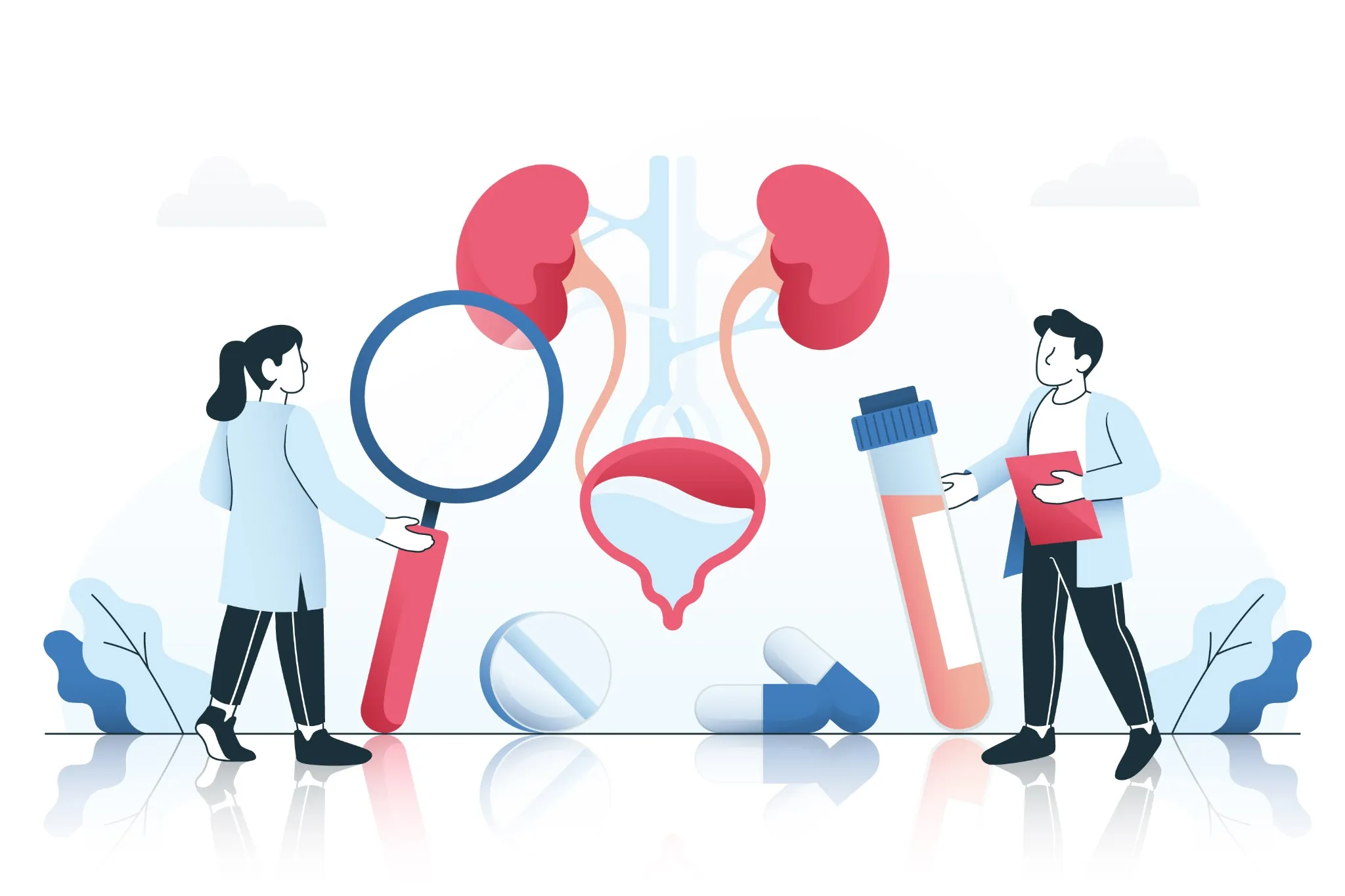
Table of Contents
- What Is a Ureteral Obstruction?
- Types of Ureteral Obstruction
- What Causes Ureteral Obstruction?
- Symptoms of Ureteral Obstruction
- Complications of Untreated Ureteral Obstruction
- When to Seek Emergency Care
- How Ureteral Obstruction Is Diagnosed
- Treatment Options for Ureteral Obstruction
- Surgical Options
- Ureteral Stents: Purpose & What to Expect
- Nephrostomy Tube: When It's Needed
- Recovery After Treatment
- Lifestyle & Home Care After Treatment
- How to Prevent Ureteral Obstruction
- When to Follow Up with a Urologist
- Conclusion
- FAQs
What Is a Ureteral Obstruction?
A ureteral obstruction occurs when something blocks one or both ureters, the narrow tubes that carry urine from your kidneys to your bladder. This blockage prevents normal urine flow, causing urine to back up toward the kidneys. The resulting pressure increase leads to kidney swelling, medically known as hydronephrosis.
Ureteral obstruction can develop suddenly, such as when a kidney stone lodges in the ureter, or gradually over months from scar tissue formation. Without proper treatment, this condition can cause permanent kidney damage, infections, and, in severe cases, kidney failure.
Types of Ureteral Obstruction
By Location:
- Ureteropelvic junction (UPJ) obstruction: A blockage where the ureter meets the kidney
- Ureterovesical junction (UVJ) obstruction: Blockage where the ureter enters the bladder
- Mid-ureteral obstruction: Blockage along the middle portion of the ureter
By Cause:
- Intrinsic obstruction: Caused by problems within the ureter itself, such as stones or tumors
- Extrinsic obstruction: Results from external pressure on the ureter from surrounding structures
By Onset:
- Acute obstruction: Develops suddenly, often causing severe pain
- Chronic obstruction: Develops slowly over time, sometimes with minimal symptoms
Acute vs. Chronic Ureteral Obstruction
Acute ureteral obstruction typically occurs suddenly when a kidney stone becomes lodged in the ureter. This creates intense, cramping pain in your side or back, often accompanied by nausea and vomiting.
Chronic ureteral obstruction develops gradually from causes like scar tissue, tumours, or congenital abnormalities. You may experience vague back discomfort, fatigue, or recurrent urinary tract infections.
What Causes Ureteral Obstruction?
• Kidney stones: The most frequent cause; stones form in your kidney and lodge in the ureter.
• Ureteral strictures: Scar tissue from previous surgery, radiation, stones, infections, or injury, causing permanent narrowing.
• Tumours within the urinary tract: Cancerous or benign growths affecting your ureter, kidney, bladder, or surrounding tissues.
• Blood clots: Clots from bleeding in your kidney or urinary tract, blocking the narrow ureteral channel.
• Abdominal or pelvic cancers: Colorectal, gynecologic, prostate, or lymphatic cancers that press on the ureter externally.
• Enlarged prostate: Benign enlargement or prostate cancer affecting bladder outflow and ureter function.
• Retroperitoneal fibrosis: Abnormal fibrous tissue growth behind your abdomen that encases and constricts the ureters.
Other Medical Conditions That Can Lead to Obstruction
• Endometriosis: Endometrial tissue outside your uterus, involving the ureter and causing external compression.
• Severe pelvic organ prolapse: Advanced cases occasionally kink or compress the ureters.
• Large uterine fibroids: Significant fibroid growth potentially affecting the ureter position.
• Congenital abnormalities: Birth defects like duplicated ureters or ureteroceles affecting normal urine flow.
• Inflammatory disorders: Conditions like tuberculosis or schistosomiasis, causing ureter wall thickening.
• Surgical complications: Post-operative scarring from abdominal, pelvic, or urinary tract procedures.
Symptoms of Ureteral Obstruction
• Severe flank or side pain
• Sudden, wave-like pain
• Abdominal pain or fullness
• Blood in the urine (hematuria)
• Frequent urination or urgency
• Pain or burning during urination
• Reduced urine output
• Cloudy or foul-smelling urine
• Fever and chills
Symptoms in Children vs. Adults
|
Age Group |
Common Symptoms |
Key Differences |
|
Children |
Abdominal pain, fever, vomiting, reduced appetite |
May not localise pain well; symptoms are often non-specific. |
|
Adults |
Severe flank pain, blood in urine, nausea |
Can better describe pain location and intensity |
Complications of Untreated Ureteral Obstruction
• Progressive kidney damage: Continued pressure causes irreversible kidney tissue death.
• Chronic kidney disease: Permanent reduction in kidney function requiring ongoing management.
• Kidney infections (pyelonephritis): Occur when bacteria multiply in stagnant urine, causing a serious infection.
• Sepsis: A life-threatening infection spreading throughout your bloodstream.
• Complete kidney failure: Total loss of kidney function requiring dialysis or transplantation.
• Kidney stone formation: Stagnant urine promotes additional stone development.
When to Seek Emergency Care
• Severe, unbearable flank or abdominal pain that doesn't respond to pain medication
• High fever (above 38.3 °C (101 °F)) with chills combined with urinary symptoms
• Complete inability to urinate for more than 12 hours
• Signs of severe infection such as confusion, rapid heartbeat, or difficulty breathing
• Persistent vomiting, preventing fluid intake and medication
• Blood clots in urine or heavy bleeding
How Ureteral Obstruction Is Diagnosed
- Medical history review: Discussing symptoms, previous kidney problems, and medications.
- Physical examination: Checking for tenderness, swelling, or masses.
- Urine tests: Detecting blood, infection, or abnormal cells.
- Blood tests: Assessing kidney function and infection markers.
- Imaging studies: Visualising the urinary tract structure and blockage location.
- Specialised tests: If initial tests are inconclusive.
Imaging Tests for Ureteral Obstruction
• Ultrasound: A non-invasive test showing kidney swelling and potential blockages
• CT scan: Detailed cross-sectional images revealing stones, tumours, or other obstructions
• Intravenous pyelogram (IVP): Contrast dye injection highlighting urinary tract abnormalities, according to MedlinePlus
• MRI (magnetic resonance imaging): Provides detailed soft-tissue imaging for complex cases
• Nuclear medicine scans: Functional studies showing how well your kidneys process urine
Laboratory Tests
• CBC (Complete Blood Count) Test: Checking for signs of infection or anaemia
• Comprehensive Metabolic Panel (CMP): Assesses kidney function and electrolyte balance.
• Urine Routine Test (Urine R/M Test): Examining urine for blood cells, bacteria, and crystals
• Urine Culture Test: Identifying specific bacteria causing infection
• Electrolyte Test: Evaluates mineral balance and kidney function
Treatment Options for Ureteral Obstruction
Treatment for ureteral obstruction depends on the underlying cause, its severity, and your overall health. Treatment goals include relieving the urinary tract blockage, preserving kidney function, and preventing complications.
For acute cases with severe pain, immediate pain relief and hydration are priorities. Chronic cases may require more complex interventions to address underlying causes and prevent further kidney damage.
Treatment for Tumour-Related Obstructions
The approach depends on whether the tumour is benign or malignant, its location, and your overall health status. Treatment may involve surgical removal, chemotherapy, radiation therapy, or a combination of approaches. In some cases, temporary drainage procedures, such as ureteral stents or nephrostomy tubes, help preserve kidney function while addressing the underlying cancer.
Surgical Options
• Ureteroscopy with stone removal: Inserting a thin scope through your bladder to remove or break up stones.
• Shock wave lithotripsy: Using sound waves to break kidney stones into smaller pieces.
• Ureteral stent placement: Inserting a small tube to keep your ureter open.
• Pyeloplasty: Surgical repair of ureteropelvic junction obstruction.
• Ureteroplasty: Reconstruction of damaged or narrowed ureter segments.
• Nephrostomy tube insertion: Creating temporary drainage directly from your kidney.
Ureteral Stents: Purpose & What to Expect
Ureteral stents are thin, flexible tubes placed inside your ureter to maintain urine flow around blockages. During stent placement, you will receive local anesthesia or sedation while the doctor inserts the stent through your bladder using a cystoscope. Most patients experience some discomfort, urinary frequency, and mild blood in the urine for several days after placement. While stents effectively restore urine flow, they require regular replacement every few months to prevent complications like infection or blockage.
Nephrostomy Tube: When It's Needed
A nephrostomy tube provides an alternative drainage route when ureteral stenting isn't possible or effective. This tube is inserted directly through your back into the kidney, allowing urine to drain into an external collection bag. While more invasive than stents, nephrostomy tubes effectively preserve kidney function in severe cases.
Recovery After Treatment
Recovery from ureteral obstruction treatment varies depending on the procedure performed and the underlying condition. Most patients experience gradual improvement in symptoms over several days to weeks following successful treatment. During recovery, you'll need regular follow-up appointments to monitor kidney function, ensure proper healing, and prevent recurrence.
Lifestyle & Home Care After Treatment
• Stay well-hydrated: Drink 8–10 glasses of water daily unless otherwise advised by your doctor.
• Follow prescribed medications: Complete antibiotic courses and take pain medications as directed.
• Monitor symptoms: Track urine output, colour, and any pain or fever.
• Maintain good hygiene: Especially important if you have stents or drainage tubes.
• Attend follow-up appointments: Regular monitoring ensures proper healing and early detection of complications.
• Avoid heavy lifting: Protect surgical sites and prevent tube displacement.
How to Prevent Ureteral Obstruction
• Drinking sufficient water helps prevent kidney stone formation.
• Limit sodium, oxalate-rich foods, and excessive protein intake according to your stone type.
• Regular medical check-ups for early detection and treatment of underlying conditions.
• Prompt treatment of urinary tract infections.
• Following recommended guidelines for early detection of pelvic and abdominal cancers.
• Managing underlying conditions such as controlling diabetes, high blood pressure, and other chronic diseases.
When to Follow Up with a Urologist
• Recurrent kidney stones
• Persistent or worsening symptoms after initial treatment
• Changes in kidney function detected through blood tests
• Complications from stents or drainage tubes
• New onset of blood in urine or urinary symptoms
• Family history of kidney disease or recurrent obstructions
Conclusion
Understanding ureteral obstruction empowers you to recognise symptoms early and seek appropriate medical care. Whether caused by kidney stones, strictures, or other conditions, prompt diagnosis and treatment prevent serious complications and preserve kidney function.
The most important step you can take is listening to your body and seeking medical evaluation when experiencing persistent flank pain, blood in urine, or changes in urination patterns. Early intervention significantly improves outcomes and reduces the risk of permanent kidney damage.
At Metropolis Healthcare, we support your journey towards better urological health through comprehensive diagnostic services. Our extensive portfolio of more than 4,000 tests helps identify ureteral obstruction early. With our convenient home sample collection service across more than 10,000 touchpoints across India, you can access accurate diagnostics from the comfort of your home, enabling timely detection and monitoring of urinary tract conditions.
FAQs
What is ureteral obstruction?
Ureteral obstruction occurs when something blocks the tubes carrying urine from the kidneys to the bladder, causing urine backup, kidney swelling, and potential complications if untreated.
What causes a ureter to become blocked?
- Kidney stones
- Scar tissue from previous surgery or injury
- Tumours pressing on or growing within the ureters
- Blood clots blocking the narrow ureteral channel.
- Congenital abnormalities affecting the ureter structure
Can ureteral obstruction go away on its own?
Small kidney stones may sometimes pass naturally with increased fluid intake, but most obstructions require medical intervention to prevent permanent kidney damage.
How painful is ureteral obstruction?
Pain intensity varies but can be excruciating, particularly with kidney stones. The pain typically occurs in waves, radiating from the side to the groin.
What tests diagnose ureteral obstruction?
- CT scans to visualize urinary tract blockages.
- Ultrasound to detect kidney swelling.
- Blood tests measuring kidney function
- Urine tests checking for infection or blood
How is ureteral obstruction treated?
Treatment depends on the cause but may include pain management, stent placement, stone removal procedures, surgical repair, or addressing underlying conditions like tumours.
Can a blocked ureter cause kidney failure?
Yes, untreated ureteral obstruction can lead to permanent kidney damage and failure, especially if both kidneys are affected or if treatment is delayed.
How long is the recovery after ureteral stent placement?
Most people adapt to stents within a few days, though some discomfort may persist. Complete recovery typically occurs within weeks of stent removal.
What foods help prevent ureteral blockages?
- Citrus fruits contain natural citrate.
- Low-sodium foods to reduce stone formation
- Moderate calcium intake from dietary sources
- Limited oxalate-rich foods if prone to stones