Preventive Healthcare
What is the Talus Bone? Functions, Common Injuries, and Treatment
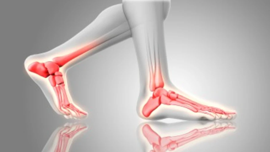
Table of Contents
- What is the Talus Bone?
- Functions of the Talus Bone
- Anatomy of the Talus Bone
- Common Injuries of the Talus Bone
- Symptoms of Talus Bone Injuries
- Diagnosis of Talus Bone Injuries
- Diagnostic Tests to Identify a Talus Bone Fracture
- Treatment Options for Talus Bone Injuries
- How Long Does It Take to Recover from Talus Bone Injuries?
- Preventing Talus Bone Injuries
- Conclusion
- FAQs
What is the Talus Bone?
The talus bone, or ankle bone (talus), is one of the seven tarsal bones in the hindfoot. Sitting between the tibia, fibula, and heel bone (calcaneus), it forms the key link that allows the foot to move smoothly in multiple directions. Unlike most bones, no muscles attach to the talus; it is mostly covered by articular cartilage and relies on surrounding ligaments and joint surfaces for movement and stability.
Despite its small size, the talus is clinically very important. A report published in StatPearls (2023) notes that talus fractures make up only about 1% of all foot and ankle fractures. However, up to 70% of severe ankle injuries may involve cartilage or osteochondral damage to the talus. Its deep position and relatively poor blood supply increase the risk of complications after injury, making early diagnosis and proper treatment essential.
Functions of the Talus Bone
The talus bone performs several critical functions that enable normal foot and ankle movement:
- Weight transmission: Transfers body weight from the tibia and fibula down to the foot.
- Ankle joint formation: Creates the primary hinge joint, allowing up-and-down foot movements.
- Subtalar joint participation: Enables inversion and eversion movements, allowing adaptation to uneven surfaces.
- Arch support: Contributes to maintaining the foot's natural arch structure.
- Balance maintenance: Provides stability during standing, walking, and dynamic activities.
- Shock absorption: Helps distribute forces during impact activities like jumping or running.
Anatomy of the Talus Bone
The talus bone anatomy comprises three distinct regions: the head, neck, and body. Each section plays a specific role in foot mechanics and joint function.
The head forms the front portion, articulating with the navicular bone and contributing to the complex midfoot joint system. The neck is the narrowed midsection that contains crucial ligament attachments stabilizing both the subtalar and ankle joints. The body constitutes the largest portion, featuring the dome-shaped surface that forms the main ankle joint with the tibia.
Approximately two-thirds of the talus bone surface is covered with articular cartilage—more than most other bones in the body. This extensive cartilage coverage facilitates smooth joint movement but also limits the bone's entry points for blood vessels, contributing to healing challenges when fractures occur.
Common Injuries of the Talus Bone
Several types of injuries commonly affect the talus bone, ranging from minor stress fractures to severe trauma:
- Talus bone fractures: Typically result from high-energy trauma such as motor vehicle accidents or falls from height.
- Osteochondral lesions: Combined cartilage and bone injury resulting from severe ankle sprains or repetitive stress.
- Stress fractures: Small cracks developing from repetitive loading in athletes or active individuals.
- Avascular necrosis: Loss of blood supply leading to bone tissue death, typically following severe fractures.
- Post-traumatic arthritis: Degeneration of the ankle joint that develops months or years after the original injury.
Symptoms of Talus Bone Injuries
Recognising talus bone injury symptoms enables prompt medical evaluation and treatment.
Common signs include:
- Deep ankle pain that worsens with standing or weight-bearing activities
- Significant swelling around the ankle and hindfoot area
- Difficulty walking or complete inability to bear weight
- Limited ankle movement in all directions
- Tenderness when touching the ankle joint area
- Ankle instability or the feeling that the joint might give way
- Clicking or catching sensations during foot movement
- Persistent aching that continues even at rest
Diagnosis of Talus Bone Injuries
Healthcare providers use a systematic approach to diagnose talus bone injuries:
- History: How the injury happened and how symptoms started/worsened
- Physical exam: Swelling, tenderness, deformity, and ankle movement
- X-ray: First-line test to look for obvious fractures and joint alignment
- CT scan: Detailed view of complex or subtle fracture patterns
- MRI: Detects subtle stress fractures, cartilage or ligament injuries, and early signs of avascular necrosis (AVN)
- Bone health tests: Such as the Calcium Profile, Vitamin D Plus Profile, Bone Screening Profile, Bone Formation and Resorption Marker Profiles, Osteocalcin, and Osteomon Profile—help assess bone strength and healing capacity
Diagnostic Tests to Identify a Talus Bone Fracture
Several diagnostic approaches help identify and evaluate talus bone fractures:
- Standard ankle X-rays provide initial fracture detection and alignment assessment.
- CT scans offer detailed three-dimensional views for surgical planning.
- MRI studies detect subtle fractures and assess bone viability.
- Bone Profile evaluates overall bone metabolism and healing capacity.
- Arthritis Profile identifies inflammatory conditions affecting healing.
- Osteomon profile assesses bone formation and resorption markers.
Treatment Options for Talus Bone Injuries
Treatment approaches vary significantly based on injury type, severity, and patient factors:
- Non-surgical management: Immobilisation, protected weight-bearing, and physical therapy for stable fractures.
- Surgical intervention: Internal fixation with screws or plates for displaced fractures.
- Bone grafting: The placement of bone tissue to stimulate healing in complex or non-union fractures.
- Joint replacement: Consideration for severe arthritis or avascular necrosis cases.
- Rehabilitation programmes: Structured physical therapy to restore function and prevent complications.
How Long Does It Take to Recover from Talus Bone Injuries?
Recovery depends on injury severity and whether surgery is needed. Simple, non-displaced talus fractures generally heal within 8–12 weeks, while complex fractures can take 3–6 months or longer. Because of the talus’s limited blood supply, healing may be delayed, and regular follow-up imaging is essential. Patients may also undergo metabolic evaluations, such as the Bone Resorption Marker Test or Arthritis Profile, to ensure there are no underlying bone conditions delaying recovery.
Preventing Talus Bone Injuries
While not all talus bone injuries are preventable, several strategies can reduce your risk:
- Proper footwear: Wear supportive, well-fitted shoes suitable for your specific activities.
- Gradual training progression: Avoid sudden increases in activity intensity or duration.
- Ankle strengthening exercises: Build supporting muscle strength around the joint.
- Balance training: Improve proprioception to prevent falls and awkward landings.
- Surface awareness: Be cautious on uneven or slippery surfaces.
- Bone health maintenance: Ensure adequate calcium and vitamin D intake.
Conclusion
Talus bone injuries need early diagnosis, proper treatment, and regular follow-up, as even small fractures can affect ankle movement and long-term joint health. Using targeted imaging and bone health tests helps detect complications early and supports better recovery.
Metropolis Healthcare offers strong support on this journey with 4,000+ tests, full-body checkups, and specialty bone and joint testing, backed by over 10,000 home sample collection touchpoints. Patients receive quick, accurate reports and can conveniently book through the website, app, WhatsApp, or a phone call, so they can focus on healing while their diagnostic needs are handled smoothly.
FAQs
How is a talus bone fracture treated?
Treatment depends on whether the bone is displaced. Non-displaced fractures are managed with immobilisation and restricted weight-bearing, while displaced fractures typically require surgery. Regular imaging and Bone and Joint Tests help monitor healing.
What causes talus bone pain?
Common causes include fractures, sprains, cartilage injuries, arthritis, Osteomalacia, or metabolic bone disorders. Persistent pain should be evaluated with imaging and tests like the Bone Profile or Arthritis Profile.
Can you walk with a broken talus bone?
Most people cannot walk comfortably with a talus bone fracture due to severe pain and instability. Walking on a broken talus can worsen the injury, so immediate medical assessment is essential.
What are the signs of a talus bone injury?
Key signs include ankle pain, swelling, bruising, difficulty walking, and reduced ankle range of motion. Severe injuries may also cause deformity or locking of the ankle joint.