Preventive Healthcare
Choroid Plexus Tumor: Symptoms, Diagnosis & Treatment
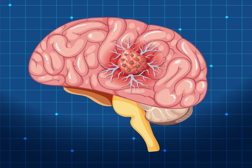
Table of Contents
- What is a Choroid Plexus Tumor?
- Choroid Plexus Tumor Causes
- Choroid Plexus Tumor Symptoms
- Diagnosis of Choroid Plexus Tumors
- Imaging and Diagnostic Methods for Choroid Plexus Tumors
- Risk Factors for Choroid Plexus Tumors
- Treatment Options for Choroid Plexus Tumors
- Prognosis of Choroid Plexus Tumors
- Choroid Plexus Tumors in Children
- Choroid Plexus Tumors in Adults
- Conclusion
- FAQs
What is a Choroid Plexus Tumor?
A choroid plexus tumor is a rare intraventricular neoplasm that arises from the choroid plexus epithelium — the tissue lining the brain’s ventricles responsible for cerebrospinal fluid (CSF) production. The choroid plexus is located within all four ventricles of the brain — lateral, third, and fourth — where it produces and regulates cerebrospinal fluid within your brain, where this specialised tissue continuously produces cerebrospinal fluid. This protective fluid cushions your brain and spinal cord whilst delivering essential nutrients.
These Tumors can be either benign (non-cancerous) or malignant (cancerous). Choroid plexus papilloma (WHO Grade I) is benign, whereas atypical choroid plexus papilloma (Grade II) and choroid plexus carcinoma (Grade III) are progressively more aggressive forms. The choroid plexus serves as a critical barrier between your blood and brain tissue, making Tumors in this location particularly significant.
As these tumors enlarge, they may obstruct CSF pathways, leading to hydrocephalus and increased intracranial pressure — the main cause of early symptoms, leading to increased pressure within your brain. This pressure buildup often triggers the first noticeable symptoms.
Choroid Plexus Tumor Causes
The exact cause of choroid plexus Tumor development remains largely unknown, though researchers have identified several contributing factors. Somatic DNA mutations in choroid plexus epithelial cells can trigger uncontrolled proliferation, disrupting normal CSF homeostasis, disrupting normal cellular function and death cycles.
Genetic predisposition, particularly germline TP53 mutations seen in Li-Fraumeni syndrome, is the most recognized risk factor for choroid plexus carcinoma. Families with Li-Fraumeni syndrome, characterised by TP53 mutations, show increased risk for developing choroid plexus carcinoma. These genetic alterations affect how cells regulate growth, division, and programmed death.
Choroid Plexus Tumor Symptoms
Symptoms largely reflect increased intracranial pressure or hydrocephalus, resulting from CSF flow obstruction or overproduction due to blocked cerebrospinal fluid drainage.
• Persistent headaches that worsen over time
• Nausea and vomiting, particularly in the morning
• Irritability or sudden behavioural changes in children
• Developmental delays or regression in milestones
• Balance problems and coordination difficulties
• Vision changes, including blurred or double vision
• Seizures that may be new or increasing in frequency
• Excessive drowsiness or unusual lethargy
• Bulging fontanelle (soft spot) in infants
Diagnosis of Choroid Plexus Tumors
- Comprehensive clinical evaluation: Your doctor assesses symptoms and medical history, and performs neurological examinations
- MRI: With contrast is the imaging modality of choice, providing detailed information about tumor margins, vascularity, and ventricular anatomy to visualise the Tumor's size, location, and characteristics
- CT Scan: May reveal coarse calcifications, hyperdense intraventricular masses, or associated hydrocephalus and can detect calcifications within the Tumor
- Lumbar puncture: May be used selectively to evaluate CSF cytology, especially in suspected choroid plexus carcinoma, but is avoided preoperatively in hydrocephalus due to herniation risk
- Surgical biopsy: Tissue sample collection during surgery enables precise Tumor classification
- Neuropathological analysis: Expert microscopic examination determines the exact Tumor grade and type
- Genetic testing: Analysis for TP53 mutations and other genetic abnormalities that influence treatment planning
Imaging and Diagnostic Methods for Choroid Plexus Tumors
• MRI typically shows a lobulated, cauliflower-like enhancing intraventricular mass, often attached to the choroid plexus
• CT scan shows well-defined masses with distinctive density patterns
• Contrast-enhanced imaging highlights blood vessel patterns within Tumors
• Diffusion-weighted imaging helps distinguish between benign and malignant types
• Spinal imaging checks for Tumor spread throughout the nervous system
Risk Factors for Choroid Plexus Tumors
• CPC most frequently occurs in children under five, while CPP is more evenly distributed across age groups
• Genetic predisposition, including TP53 gene mutations and Li-Fraumeni syndrome
• Family history of brain Tumors or hereditary cancer syndromes
• Gender variations with slight male predominance in certain age groups
Treatment Options for Choroid Plexus Tumors
• Gross total surgical resection: It is the cornerstone of treatment and offers the best prognosis, particularly for choroid plexus papilloma and obtain diagnostic samples
• Chemotherapy protocols: Chemotherapy (often using cisplatin, etoposide, or vincristine-based regimens) targets residual or disseminated disease, particularly in CPC.
• Radiation therapy: Radiation therapy is generally reserved for older children and adults with residual or recurrent CPC, as it is avoided in infants due to neurodevelopmental risks
• Clinical trials: Access to emerging treatments, including targeted therapy and immunotherapy
• Supportive care: Management of hydrocephalus and symptom control throughout treatment
• Multimodal therapy: Combination treatments based on Tumor grade and surgical outcomes
Surgical Treatment for Choroid Plexus Tumors
Surgery remains the cornerstone of choroid plexus Tumor treatment. The primary goal involves removing as much Tumor as possible whilst preserving surrounding healthy brain tissue. Complete surgical resection significantly improves outcomes for both benign and malignant Tumors.
Modern neurosurgical techniques utilise advanced imaging guidance and microscopic precision. When post-operative scans reveal residual Tumor tissue, a second surgery may be recommended, particularly for choroid plexus carcinoma cases. Surgical success depends heavily on Tumor location within the choroid plexus and surrounding anatomy.
Chemotherapy and Radiation Therapy
• Chemotherapy regimens: Administered after surgery for grade 2 and grade 3 Tumors to target remaining cancer cells
• Radiation planning: Recommended for choroid plexus carcinoma and cases with incomplete surgical removal
• Treatment timing: Usually begins several weeks after surgical recovery to allow proper healing
• Combination protocols: Often used together for aggressive Tumors to improve long-term outcomes
• Clinical trial participation: Patients may access newer agents through research studies
• Individualised planning: Treatment decisions consider patient age, Tumor grade, and overall health status
Prognosis of Choroid Plexus Tumors
Prognosis depends on tumor grade, completeness of resection, and patient age. Five-year survival exceeds 90% for CPP but ranges from 40–60% for CPC and treatment completeness. Benign choroid plexus papillomas generally carry excellent outcomes when completely removed. However, choroid plexus carcinoma presents more challenges due to its aggressive nature and tendency to spread.
Complete surgical resection represents the strongest predictor of favourable outcomes. Patients achieving total Tumor removal experience significantly better long-term survival rates. Age at diagnosis also influences prognosis, with younger patients often showing remarkable resilience during treatment.
Choroid Plexus Tumors in Children
Children account for up to 70% of choroid plexus tumor cases, with CPP being more common than CPC, with unique considerations affecting their care. The choroid plexus location in developing brains makes early symptoms particularly concerning for parents.
Children typically present with symptoms related to increased brain pressure, including persistent headaches, developmental delays, and coordination problems. Treatment approaches in paediatric cases emphasise balancing aggressive Tumor control with minimising long-term developmental and neurological side effects from chemotherapy and radiation therapy.
Choroid Plexus Tumors in Adults
While choroid plexus Tumor cases predominantly affect children, adults can also develop these rare brain Tumors. Adult presentations often involve more subtle symptoms that may develop gradually over time. The choroid plexus Tumor symptoms in adults might include persistent headaches, cognitive changes, or subtle neurological deficits.
In adults, choroid plexus tumors are rare but may be treated more aggressively, as the risks of radiation and chemotherapy-related neurotoxicity are lower, as the developing brain concerns present in children are not applicable. However, the rarity of these Tumors in adults means treatment protocols often adapt paediatric approaches to adult patients.
Conclusion
A choroid plexus tumor diagnosis can feel overwhelming, but early detection, multidisciplinary care, and advances in neurosurgical and molecular oncology have greatly improved outcomes, but understanding the condition empowers you to navigate treatment decisions confidently. Early recognition of choroid plexus Tumor symptoms, prompt diagnostic evaluation with MRI scan and biopsy, and comprehensive choroid plexus Tumor treatment at specialised centres offer the best outcomes for patients and families.
Working closely with experienced neuro-oncology teams ensures access to the latest therapeutic advances whilst maintaining focus on your overall wellbeing and quality of life.
At Metropolis Healthcare, we support your diagnostic needs with comprehensive testing services spanning over 4,000 advanced tests. Our extensive network of 10,000+ touchpoints ensures convenient access to precise diagnostics when you need them most. From routine screenings to specialised neurological assessments, our home sample collection service brings expert care directly to your doorstep.
FAQs
Can choroid plexus Tumors be cancerous?
Yes, choroid plexus carcinoma represents the malignant form, though benign papillomas are more common. Proper biopsy and pathological examination determine the exact Tumor type.
How is a choroid plexus Tumor diagnosed?
Diagnosis involves MRI scan imaging, CT scan protocols, cerebrospinal fluid analysis, and surgical biopsy. These comprehensive tests confirm the diagnosis and guide treatment planning.
What are the symptoms of choroid plexus Tumors?
• Persistent headaches and nausea
• Vision problems and balance difficulties
• Seizures and behavioural changes
• Developmental delays in children
Is surgery the only treatment for choroid plexus Tumors?
Surgery remains the primary treatment, but choroid plexus carcinoma often requires additional chemotherapy or radiation therapy. Treatment plans depend on Tumor grade and individual factors.
References
1. https://www.ncbi.nlm.nih.gov/books/NBK539749/
2. https://my.clevelandclinic.org/health/diseases/choroid-plexus-carcinoma
3. https://www.cancer.gov/rare-brain-spine-tumor/tumors/choroid-plexus-tumors
4. https://www.sciencedirect.com/science/article/pii/S2772610X25000133