Preventive Healthcare
Mammography Explained: What to Expect, Results, and Why It Matters
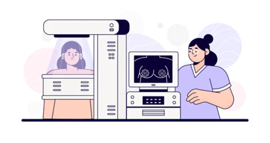
Table of Contents
- What Is Mammography?
- Why Mammograms Are Important?
- Types of Mammograms
- Who Should Get a Mammogram?
- Preparing for a Mammogram: Step-by-Step Guide
- Mammogram Procedure: What to Expect
- Is Mammography Painful?
- Understanding Mammogram Results
- Risks and Limitations of Mammography
- Mammogram Safety During Pregnancy
- Tips for a Comfortable Mammogram Experience
- Cost of Mammogram in India
- Conclusion
- FAQs
- References
What Is Mammography?
Mammography is a specialised medical imaging technique that uses low-dose X-rays to create detailed images of the breast, known as mammograms. The primary goal of mammography is to detect breast cancer and other breast diseases at an early stage, often before symptoms develop or a lump can be felt, improving the chances of successful treatment. During the exam, the breast is compressed between two plates to spread the breast tissue, allowing for a clearer image with a minimal dose of radiation.
As a woman, understanding what a mammogram is, why it's important, and what to expect from different mammogram types can help you make informed decisions about your breast health.
Why Mammograms Are Important?
According to the World Health Organisation (WHO), five-year survival rates for breast cancer surpass 90% in high-income countries. In contrast, survival rates are lower in other regions, with India reporting around 66%. These differences are largely due to unequal access to early detection, timely diagnosis, and effective treatment. Mammograms play a crucial role in this process because they are currently the most effective screening tool for detecting breast cancer early, when it is most treatable.
Early detection through regular mammography has been shown to lower the risk of fatality from breast cancer by enabling treatment before the cancer spreads. Mammograms can reveal tumours too small to be felt and identify abnormal growths or calcifications that may signal early cancer, leading to prompt intervention.
Types of Mammograms
Screening Mammogram
A screening mammogram is performed on women who do not have any symptoms of breast disease. It typically involves taking two or more X-ray images of each breast to look for hidden cancers or changes that cannot be felt. Regular screening mammograms are recommended for women to find cancers at an early, more treatable stage and to reduce breast cancer mortality.
Diagnostic Mammogram
A diagnostic mammogram procedure is done when there are signs or symptoms of breast problems, such as a lump, pain, nipple discharge, or a change in breast shape. It involves more images and takes longer than a screening mammogram, allowing for a closer look at areas of concern. It is also used if a screening mammogram detects an abnormality or if the patient has breast implants, which can make imaging more challenging.
Digital Mammography & 3D Mammogram (Tomosynthesis)
Digital mammography, also called full-field digital mammography (FFDM), replaces traditional X-ray film with electronic detectors, resulting in images that are stored and viewed on a computer. This technology offers greater image quality, easier storage, and potentially lower radiation doses.
3D mammography, or breast tomosynthesis, takes multiple images from different angles to create a three-dimensional view of the breast. It improves the detection of small cancers and reduces the false positives, especially in women with dense breast tissue.
Who Should Get a Mammogram?
- For women at average risk, most major guidelines (including ACR and USPSTF) recommend mammographic screening every 1–2 years starting at age 40.
- Screening may continue until around 74, depending on overall health and life expectancy.
- For women with a family history of breast cancer or other risk factors, annual mammograms or annual MRI screenings may be recommended starting at a younger age.
- Women who notice changes in their breasts, such as lumps, pain, or nipple discharge, must get a mammogram procedure done immediately.
- Women with a personal history of breast cancer or certain benign breast conditions should have regular mammograms, as the purpose of a mammogram is early detection.
Preparing for a Mammogram: Step-by-Step Guide
- Schedule your appointment for a time when your breasts are least likely to be tender (usually a week after your period).
- Bring previous mammogram images or records if available.
- Remove jewellery and clothing from the waist up; you will be given a gown to wear.
Avoid deodorants, powders, or lotions on test day.
Do not apply deodorant, antiperspirant, powders, or lotions under your arms or on your breasts on the day of the exam. These substances can appear as white spots on the X-ray and interfere with the image quality.
Wear comfortable, two-piece clothing.
Opt for a two-piece outfit so you only need to remove your top for the exam, making the experience easier and more comfortable.
Inform the technician about breast implants or pregnancy.
Notify the technologist if you have breast implants, as special techniques are needed for accurate imaging, or if you are or might be pregnant, so that safety precautions can be taken.
Mammogram Procedure: What to Expect
Step 1: Positioning
You will stand in front of the mammography machine, and a technologist will position your breast on a flat support plate. Your arm, shoulder, and head may need to be adjusted to get a clear image.
Step 2: Compression
Another plate is gently pressed down on your breast to flatten it. Compression is essential to spread the tissue for a clearer image, reduce motion, and minimise radiation dose. The compression lasts only a few seconds for each image.
Step 3: Imaging
X-rays are taken from different angles, usually two per breast (top-to-bottom and side-to-side). The process is repeated for the other breast. The entire mammogram procedure typically takes about 20 minutes.
Is Mammography Painful?
Most women experience only mild discomfort or pressure during the compression phase, which lasts just a few seconds per image. While some may find it briefly uncomfortable, severe pain is uncommon. Discomfort can be minimised by scheduling the mammogram when breasts are less likely to be tender and by communicating with the technologist.
For example, if you have sensitive breasts, you can take an over-the-counter pain reliever before your appointment or apply a numbing gel patch to reduce discomfort.
Understanding Mammogram Results
Mammogram results are categorised to guide next steps, helping interpret findings from different mammogram types and determine if further testing or follow-up is needed.
Category 0: Incomplete (Needs more testing)
The images are not clear, or additional views are necessary. Further imaging, such as additional mammogram pictures or an ultrasound, is needed before a final result can be given.
Category 1: Negative (No signs of cancer)
No abnormalities are seen. The breast tissue appears normal, and no further testing is needed until the next routine screening.
Category 2: Benign Finding
The images show a non-cancerous condition, such as cysts or fibroadenomas. No action is required other than routine screening.
Category 3: Probably Benign (Follow-up needed)
A finding is likely non-cancerous, but a short-term follow-up mammography (typically in six months) is recommended to monitor for any changes.
Category 4: Suspicious Abnormality (Biopsy may be required)
There is a suspicious area that is not clearly cancer but requires further investigation, typically a breast biopsy, to determine if cancer is present.
Category 5: Highly Suggestive of Malignancy
The findings are very likely to be cancer, and a breast biopsy is strongly recommended to confirm the diagnosis.
Risks and Limitations of Mammography
- Exposure to a small amount of ionising radiation.
- False positives can lead to unnecessary anxiety and additional tests.
- False negatives, where some cancers may be missed, especially in women with dense breast tissue.
- Overdiagnosis and overtreatment of cancers that may not have become life-threatening.
- Mammograms may be less accurate in younger women or those with dense breasts.
Mammogram Safety During Pregnancy
Mammograms are generally avoided during pregnancy unless necessary. It is important to confirm pregnancy status before undergoing a mammogram, and a pregnancy test is recommended if there is any uncertainty.
The estimated uterine radiation dose from a standard bilateral two-view mammogram is approximately 0.03 µGy (3 × 10⁻⁸ Gy), which is negligible compared to natural background exposure. This makes mammography relatively safe if absolutely necessary during pregnancy, especially with abdominal shielding, which approximates the dose to a foetus during the first trimester. Wearing a lead apron (shielding the abdomen) can cut foetal exposure by at least half, making mammography relatively safe if necessary during pregnancy.
Tips for a Comfortable Mammogram Experience
- Schedule your mammogram for when your breasts are least tender.
- Take an over-the-counter pain reliever before the exam if you are concerned about discomfort.
- Communicate with the technologist if you feel pain or need to reposition.
- Practise relaxation techniques, such as deep breathing, to minimise anxiety.
- Bring a previous mammogram for comparison if done at a different site.
Cost of Mammogram in India
In India, the cost of a mammogram varies depending on the type of test and the imaging technology used.
- Screening mammography, performed as a routine preventive check, typically ranges from ₹ 1,000 to ₹ 3,500.
- Diagnostic mammography, which involves additional views and detailed imaging to investigate specific concerns, usually costs between ₹ 2,000 and ₹ 5,000.
- Digital mammography, which provides enhanced imaging especially for women with dense breast tissue, can range from ₹ 2,500 to ₹ 6,000.
- Many government and charitable hospitals may also offer subsidised or free mammograms for women from lower-income groups.
Conclusion
Mammography is a powerful tool in the fight against breast cancer. By understanding the mammogram procedure, mammogram types, and how to interpret mammogram results, you can take charge of your breast health and make informed decisions. Remember, early detection is key, and regular screening mammograms can save lives.
If you have concerns about your breast health or are due for regular checkups, it’s important to consult a trusted healthcare provider. Metropolis Healthcare, a leading chain of diagnostic labs across India, offers over 4,000+ tests to support your health journey, including gene panels, blood tests, ultrasounds, biopsies, and other diagnostic investigations. With advanced NABL- and CAP-certified lab facilities, experienced specialists, and personalised care, Metropolis is committed to delivering accurate results and empowering you to make informed decisions about your well-being.
FAQs
1. At what age should you get your first mammogram?
Most guidelines recommend beginning routine screening at age 40, though some, like the WHO and certain European programs, begin at 45–50. Women with higher risk should consult their doctor about starting earlier based on individual risk factors and discussions with your healthcare provider. Women with a family history or higher risk may need to start earlier.
2. How often should you get a mammogram?
Screening frequency varies between every 1–2 years depending on age, personal risk, and regional guidelines. Women at high risk (family history, BRCA mutation, prior chest radiation) may need annual screening and supplemental MRI, age, and medical advice.
3. Can mammograms detect all types of breast cancer?
While mammograms are highly effective, they cannot detect all cancers. Some cancers may be hidden, especially in women with dense breast tissue, and additional tests like ultrasound or MRI may be necessary.
4. Is a mammogram safe for women with implants?
Yes, mammograms are safe for women with breast implants. However, additional images (called implant displacement views) are needed to see the breast tissue more clearly. Ensure you inform the technologist about your implants before the exam.
5. What happens if my mammogram is abnormal?
If your mammogram shows an abnormality, don't panic. Most abnormal findings are not cancer. You may need additional imaging, such as a diagnostic mammogram, ultrasound, or MRI. In some cases, a breast biopsy may be recommended to determine if the abnormality is cancerous. Your doctor will guide you through the next steps and discuss any necessary treatment options.
References
- https://www.who.int/news-room/events/detail/2025/10/01/default-calendar/breast-cancer-awareness-month-2025
- https://pmc.ncbi.nlm.nih.gov/articles/PMC9142711/
- https://ijbi.in/quality-assurance-guidelines-for-breast-imaging-breast-imaging-society-india/
- https://asianheartinstitute.org/blog/mammography-test-in-mumbai-india-procedure-costs-and-what-to-expect/#
- https://www.starimaging.in/blog/how-much-does-a-mammography-test-cost-in-delhi-average-prices-and-factors.htm
- https://my.clevelandclinic.org/health/diagnostics/4877-mammogram