Preventive Healthcare
Astrocytomas Explained: Types, Diagnosis & Management
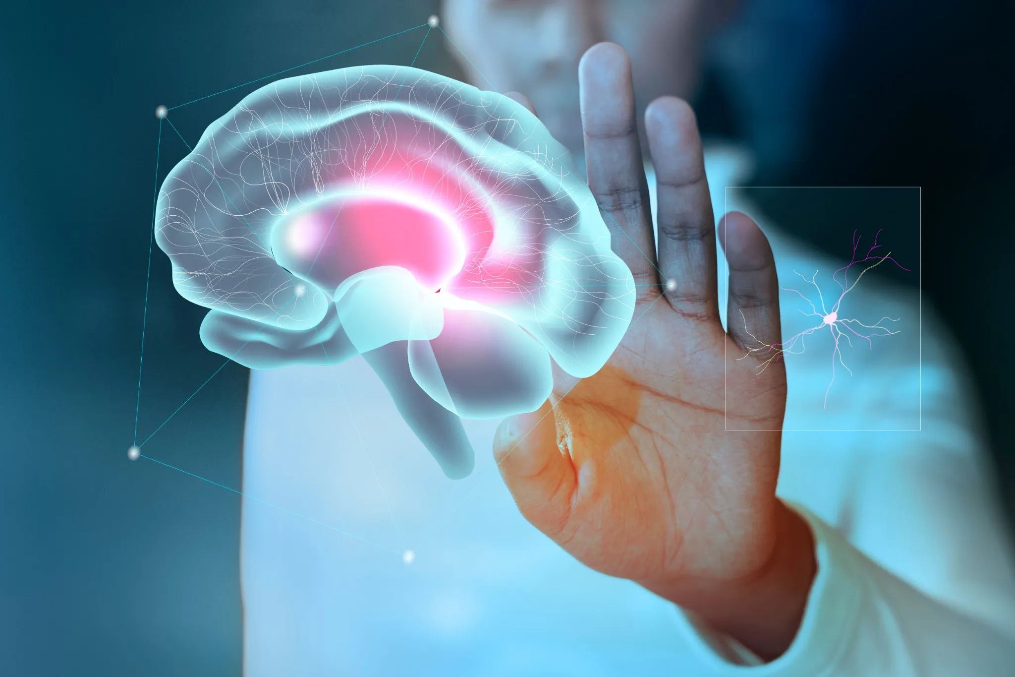
Table of Contents
- What Are Astrocytomas?
- Types of Astrocytomas
- Causes and Risk Factors for Astrocytomas
- Symptoms of Astrocytomas
- How Astrocytomas Are Diagnosed
- Diagnostic and Imaging Methods for Detecting Astrocytomas
- Treatment Options for Astrocytomas
- Managing Life with an Astrocytoma Diagnosis
- Prognosis and Survival Rates for Astrocytomas
- Conclusion
- FAQs
- References
What Are Astrocytomas?
An astrocytoma is a type of primary brain tumor that develops from astrocytes—supportive glial cells that help nourish and protect neurons. These are among the most common primary brain tumours, accounting for approximately 60% of primary adult brain tumors, and unlike secondary tumours, they originate within brain tissue itself.
Astrocytes normally regulate the blood-brain barrier, neurotransmitters, and nutrient flow, but when they become cancerous, they form tumours that disrupt normal brain function. Their behaviour varies according to tumour grade and molecular features.
Modern WHO classification integrates histopathological findings with molecular profiling, enabling more accurate diagnosis and targeted treatment. Astrocytomas can develop anywhere in the brain or spinal cord, but most often appear in the cerebral hemispheres, brainstem, or cerebellum, influencing both symptoms and treatment choices.
Types of Astrocytomas
Astrocytoma grading follows the World Health Organization (WHO) classification system, which categorises these brain tumours into four distinct grades based on their appearance under a microscope and growth characteristics:
- Grade 1 Astrocytomas (Pilocytic Astrocytoma): These slow-growing, benign tumours typically occur in children and young adults. Pilocytic astrocytomas are typically localized and seldom infiltrate surrounding brain tissue, making them the most treatable form.
- Grade 2 Astrocytomas (Low-grade Diffuse Astrocytomas): These tumours grow slowly but tend to infiltrate surrounding brain tissue. They commonly affect adults between the ages of 20 and 50 and may progress to higher grades over time if left untreated.
- Grade 3 Astrocytomas (Anaplastic Astrocytomas): These intermediate-grade tumours grow more rapidly and spread aggressively through brain tissue. They account for roughly 2% of all primary brain tumors and typically occur in adults aged 30–50.
- Grade 4 Astrocytomas (Glioblastoma): The most aggressive form, glioblastoma grows rapidly and spreads extensively throughout brain tissue. Glioblastomas comprise about 60% of all astrocytomas and may evolve from lower-grade tumors (secondary glioblastoma) or arise de novo (primary glioblastoma).
Causes and Risk Factors for Astrocytomas
The exact causes of astrocytomas remain largely unknown, as most cases develop spontaneously without identifiable triggers. Unlike many other cancers, astrocytomas lack well-established preventable risk factors, making prevention strategies limited. However, researchers have identified several potential contributing factors that may increase your risk of developing these astrocytoma brain tumours.
Age represents one of the most significant risk factors, with different astrocytoma types affecting specific age groups. While pilocytic astrocytomas predominantly occur in children and young adults, higher-grade astrocytomas typically develop in older adults. Gender also plays a role, with men having a slightly higher risk of developing certain types of astrocytomas than women.
As per a 2024 StatPearls review, astrocytomas are the most common gliomas and part of the glial tumours that constitute about 60% of all brain tumours, with Ionizing radiation remains the only well-established environmental risk factor for their development.
Radiation Exposure and Astrocytomas
Radiation can influence the risk of developing astrocytomas, but the level of risk depends on the dose, duration, and context of exposure:
- Previous head or neck radiation, especially in childhood, can increase the risk of secondary astrocytomas years later.
- Occupational exposure to ionising radiation may play a role, but evidence is still inconclusive.
- Medical imaging that uses radiation carries minimal risk when used appropriately.
- Atomic bomb survivors and nuclear accident victims show higher rates of brain tumour development.
Genetics and Astrocytomas
• Hereditary syndromes: Certain genetic conditions like Inherited syndromes such as Li-Fraumeni syndrome, Neurofibromatosis type 1 (NF1), and Tuberous Sclerosis Complex (TSC) significantly increase astrocytoma risk.
• Family history: Having a first-degree relative with a brain tumour slightly increases your risk, though most astrocytomas occur without family history.
• Molecular markers: Such as IDH1/IDH2 mutations, CDKN2A/B deletions, and TERT promoter mutations are key determinants in diagnosis, classification, and prognosis in astrocytoma development and classification.
• Chromosomal abnormalities: Specific genetic changes like EGFR amplification and chromosome alterations contribute to tumour formation.
• DNA repair defects: Inherited problems with DNA repair mechanisms may predispose individuals to developing astrocytomas.
Symptoms of Astrocytomas
Astrocytoma symptoms vary considerably depending on the tumour's size, location, and growth rate. Many people initially dismiss early symptoms as stress, fatigue, or normal ageing, which can delay diagnosis. Recognising these warning signs early enables prompt medical evaluation and treatment.
The symptoms typically develop gradually as the tumour grows and increases pressure within your skull or affects specific brain regions.
Here are the most common astrocytoma symptoms to watch for:
- Persistent headaches that worsen over time, especially morning headaches that improve throughout the day.
- Seizures or convulsions that occur for the first time in adults without a previous seizure history.
- Vision problems, including blurred vision, double vision, or loss of peripheral vision.
- Nausea and vomiting that occur without other illness, particularly morning vomiting.
- Balance and coordination difficulties leading to falls or clumsiness.
- Weakness or numbness in arms, legs, or face, often affecting one side of the body.
- Speech difficulties, including trouble finding words, slurred speech, or language comprehension problems.
- Memory problems and difficulty concentrating or making decisions.
- Personality changes, including mood swings, irritability, or behavioural alterations.
- Fatigue and drowsiness that don't improve with rest.
How Astrocytomas Are Diagnosed
Diagnosing astrocytomas requires a comprehensive approach combining clinical evaluation, advanced imaging, and tissue analysis. The diagnostic process typically begins when you or your doctor notices concerning symptoms that warrant further investigation. Your healthcare team will conduct a thorough neurological examination to assess cognitive function, motor skills, sensory perception, and reflexes.
The diagnostic journey can feel daunting, but understanding each step helps reduce anxiety and ensures you're prepared for what lies ahead. Your doctor will review your medical history, including any previous radiation exposure, family history of brain tumours, or genetic conditions like Li-Fraumeni syndrome that might increase your risk.
Modern astrocytoma diagnosis relies heavily on molecular testing to determine specific genetic characteristics that influence treatment decisions and prognosis. This personalised approach ensures you receive the most appropriate therapy based on your tumour's unique biological features rather than appearance alone.
The integration of clinical findings, imaging results, and molecular diagnostics provides a complete picture that guides your treatment team in developing an optimal care plan tailored to your specific situation.
Diagnostic and Imaging Methods for Detecting Astrocytomas
Several sophisticated diagnostic tools help doctors detect and characterise astrocytomas accurately. Each method provides unique information that contributes to a comprehensive diagnosis:
- MRI Scan: Gold standard for brain tumour imaging, showing precise tumour size, location, and spread.
- CT Scan: Often used first in emergencies to detect mass effect, bleeding, or calcifications.
- PET Scan: Evaluates tumor metabolism to differentiate active tumor from post-treatment changes or necrosis.
- Biopsy procedures: Provide definitive diagnosis via tissue analysis, e.g. Histopathological examination (via stereotactic or open biopsy), with possible core or intraoperative (‘rush’) analysis, and selected molecular or microbiology tests on biopsy tissue.
- Lumbar puncture (Spinal Tap): Occasionally used to study cerebrospinal fluid when spinal or leptomeningeal spread is suspected.
- Neuropsychological testing: Evaluates memory, speech, and thinking to map the functional impact of the tumour.
Treatment Options for Astrocytomas
Treatment for astrocytomas requires a multidisciplinary approach tailored to your specific tumour type, grade, location, and overall health status. Your treatment team typically includes neurosurgeons, neuro-oncologists, radiation oncologists, and supportive care specialists working together to optimise your outcomes.
• Surgical resection: The primary treatment for most astrocytomas, aiming to remove as much tumour tissue as possible while preserving neurological function.
• Radiation therapy: External beam radiation targets residual tumor cells after surgery to reduce recurrence risk and reduces recurrence risk, particularly important for higher-grade tumours.
• Chemotherapy: Temozolomide, the most commonly used chemotherapy for astrocytomas, is often combined with radiation therapy for Grade III and IV tumors for optimal results.
• Targeted therapy: Targeted therapies that act on specific molecular pathways within tumor cells, such as EGFR or BRAF alterations, offering hope for more effective and less toxic treatments.
• Clinical trials: Experimental treatments, including immunotherapy and novel drug combinations, may be available for eligible patients.
• Supportive care: Medications to control seizures, reduce brain swelling, and manage symptoms improve quality of life throughout treatment.
Managing Life with an Astrocytoma Diagnosis
Living with an astrocytoma means adjusting to a new reality while focusing on quality of life and hope. Many people manage their condition for years with the right treatment and support. Strong coping strategies, a reliable support system, and active involvement in care decisions are crucial.
Regular follow-ups with MRI scans become routine to monitor tumour changes or recurrence. Though scan days can be stressful, early detection makes treatment more effective, and managing “scanxiety” through relaxation, counselling, or support groups can help.
Rehabilitation services, such as cognitive therapy for memory or attention problems, and physical or occupational therapy for mobility and daily activities, often improve functioning. Gentle, regular exercise within your limits can also boost mood and energy.
Work life may need adjustment; some continue working with reasonable accommodations, while others require time off or disability support. Honest communication with employers usually helps find practical solutions.
Prognosis and Survival Rates for Astrocytomas
Understanding prognosis can guide treatment and life planning, but outcomes vary widely between individuals. Survival depends mainly on tumour grade, age, molecular profile, and how much of the tumour can be safely removed.
Broadly, Grade 1 pilocytic astrocytomas have excellent long-term survival after complete surgery. Grade 2 astrocytomas often have a median survival is approximately 6–8 years for Grade II, 2–3 years for Grade III, and 12–15 months for Grade IV (glioblastoma) Younger patients and those with favorable molecular profiles (such as IDH-mutant or MGMT-methylated tumors) and more complete surgical resection are all linked with better outcomes. Modern molecular testing help personalize both prognosis and treatment, marking a shift toward precision neuro-oncology.
Conclusion
Astrocytomas are complex brain tumours that demand early diagnosis, personalised treatment, and regular follow-up. Prognosis depends on tumour grade, age, molecular markers, and how much of the tumour can be safely removed. However, advances in neuro-oncology and molecular testing are steadily improving outcomes and quality of life. Staying informed about your tumour type and closely engaging with your care team are key to making confident decisions.
For accurate and reliable diagnostics, Metropolis Healthcare offers 4,000+ tests, including advanced molecular and speciality panels, along with comprehensive full body checkups. Patients benefit from home sample collection backed by 10,000+ touchpoints, quick turnaround times, and strict quality controls. Booking is simple and convenient via website, app, WhatsApp, or phone, ensuring you get timely, high-quality reports that support effective management of conditions like astrocytomas.
FAQs
What are the causes of astrocytomas?
Astrocytomas develop from mutations in astrocyte DNA, influenced by genetic conditions such as Li-Fraumeni syndrome, radiation exposure, and environmental factors. In some cases, no clear cause is identified.
What are the symptoms of an astrocytoma?
Symptoms include persistent headaches, seizures, vision changes, memory problems, and loss of coordination. These symptoms occur due to pressure on specific brain regions.
How is an astrocytoma diagnosed?
Diagnosis involves neurological exams, an MRI scan, a CT scan, and biopsies to determine tumour type and grade. Astrocytoma radiology imaging helps identify tumour boundaries and structure.
Can astrocytomas be treated successfully?
Yes, treatments like surgery, radiation, chemotherapy, and targeted therapy can effectively manage many types of astrocytomas. Early diagnosis improves outcomes significantly.
Are astrocytomas hereditary?
Most astrocytomas are not hereditary, but genetic conditions like Li-Fraumeni syndrome and neurofibromatosis can increase risk.
What is the life expectancy for astrocytoma patients?
Life expectancy varies by tumour grade. Pilocytic astrocytoma has excellent survival, while glioblastoma has a significantly lower prognosis. Early intervention improves outcomes.
References
- https://my.clevelandclinic.org/health/diseases/17863-astrocytoma
- https://www.mayoclinic.org/diseases-conditions/astrocytoma/symptoms-causes/syc-20576675
- https://www.aans.org/patients/conditions-treatments/astrocytoma-tumors/
- https://www.ncbi.nlm.nih.gov/books/NBK559042/