Preventive Healthcare
Angiosarcoma: Understanding an Aggressive Vascular Cancer
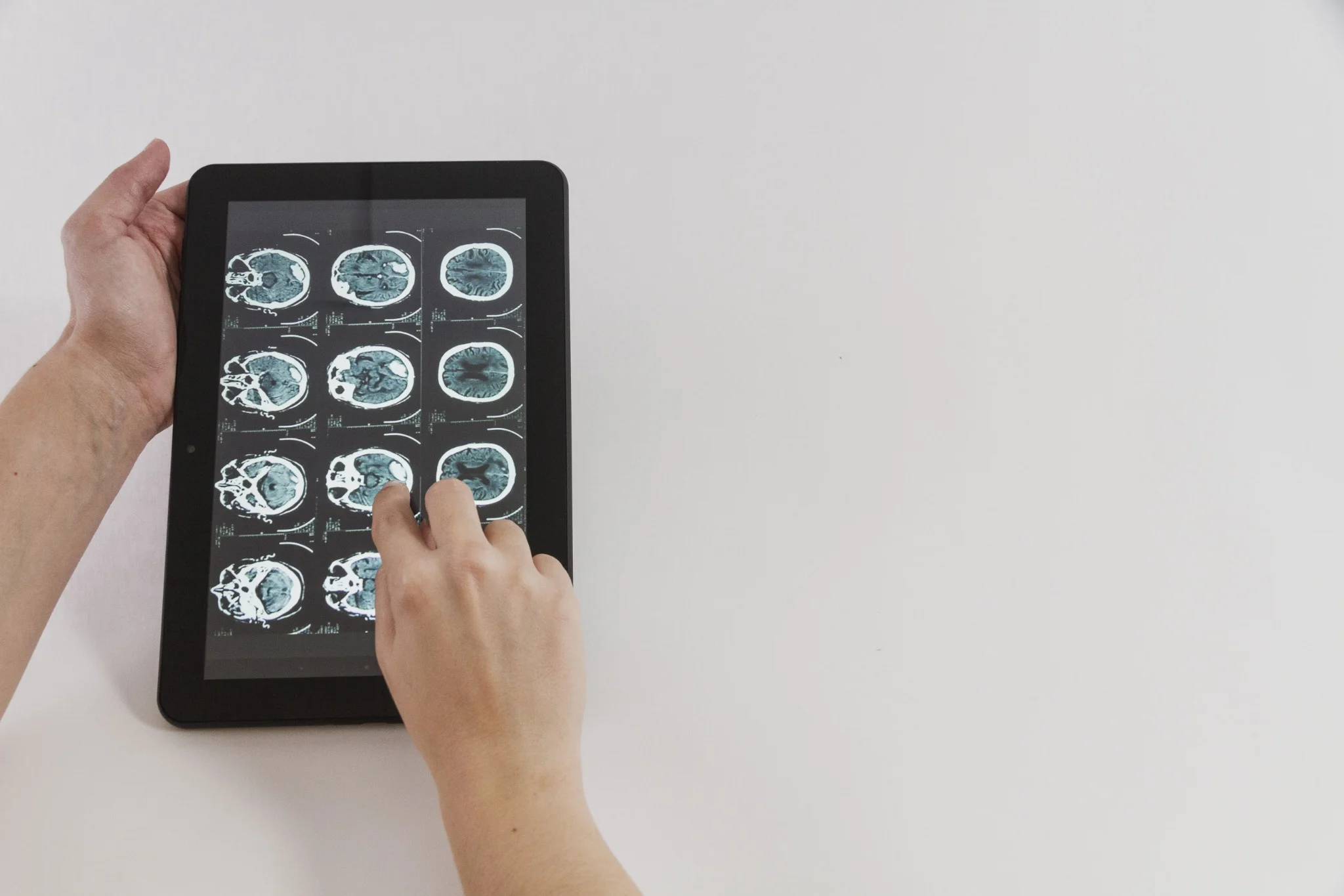
Table of Contents
What is Angiosarcoma?
Angiosarcoma is a rare, aggressive malignant tumour arising from endothelial cells that line blood vessels or lymphatic vessels. Because these vessels are found throughout the body, angiosarcoma can develop in many different locations, most commonly in the skin, soft tissues, breast, liver, and heart. Angiosarcoma is classified as a type of soft tissue sarcoma, characterised by malignant vascular differentiation, a sarcoma known for its aggressive nature and tendency to spread quickly to other parts of the body.
According to the National Cancer Institute, angiosarcoma can affect people of any age but is most often diagnosed in people over the age of 70. Early detection is challenging because symptoms may be vague or mimic other conditions, contributing to the generally poor prognosis associated with angiosarcoma.
Types of Angiosarcoma
Angiosarcomas can be classified into several types based on their location and underlying risk factors:
- Primary cutaneous angiosarcoma: Typically arises on the scalp or face of elderly individuals and may occur without identifiable risk factors.
- Lymphoedema-Associated Angiosarcoma (Stewart-Treves Syndrome): This develops in areas with chronic lymphoedema, commonly after a mastectomy for breast cancer.
- Radiation-Induced Angiosarcoma: This type arises after radiation therapy, often for breast cancer treatment.
- Primary visceral angiosarcoma: Arises within internal organs such as the liver, breast, spleen, or heart.
- Deep Soft Tissue Angiosarcoma: This type is found in muscles, ligaments, or adipose tissue.
Causes and Risk Factors of Angiosarcoma
- Radical mastectomy: Surgical removal of the entire breast and nearby lymph nodes can lead to chronic lymphoedema, a risk factor for angiosarcoma.
- Radiotherapy: Previous radiation treatment, particularly for breast cancer or lymphoma, raises the risk of secondary angiosarcoma.
- Foreign materials: In rare cases, implanted medical devices and synthetic grafts have been associated with angiosarcoma development.
- Environmental carcinogens: Occupational or environmental exposure to chemicals such as vinyl chloride, thorium dioxide (Thorotrast), and arsenic is associated particularly with hepatic angiosarcoma.
- Immunosuppression: Severe immune suppression, such as in HIV/AIDS, increases susceptibility to vascular tumours; however, Kaposi sarcoma is far more common than angiosarcoma.
- Preexisting benign lesions: Rarely, chronic bone infarcts, Paget's disease of bone, and chronic osteomyelitis may give rise to angiosarcoma.
- Genetic predisposition: Germline mutations in tumour suppressor genes such as POT1 or TP53 (Li-Fraumeni syndrome) may increase susceptibility to angiosarcoma, including cardiac subtypes.
Symptoms of Angiosarcoma
The signs and symptoms of angiosarcoma vary depending on the tumour's location. Some common symptoms include:
- Angiosarcoma that affects the skin:
- A bruise-like, purple or red patch that grows and may resemble a rash or skin infection
- Swelling and pain near the affected area
- A lump or nodule that may bleed or ulcerate
- Thickening of the skin, which may become tender or swollen
- Angiosarcoma that affects organs:
- Symptoms differ based on the involved organ:
- Heart: Chest pain, shortness of breath, abnormal heart rhythms
- Liver: Abdominal pain, swelling, yellowing of the skin and eyes (jaundice)
- Breast: A lump, swelling, or skin discolouration that can indicate angiosarcoma of breast.
- General: Unexplained weight loss, fatigue, or symptoms related to metastatic spread
- Symptoms differ based on the involved organ:
Diagnosis of Angiosarcoma
Diagnosing angiosarcoma can be challenging due to its rarity and often non-specific symptoms. The diagnostic process typically begins with a thorough physical examination and a detailed review of your medical history, with a focus on any factors that may increase your risk for this cancer.
If your doctor suspects angiosarcoma, they will likely order imaging tests such as MRI, CT scans, or ultrasound to assess the size, depth, and extent of the tumour. However, a biopsy is necessary to confirm the diagnosis and differentiate angiosarcoma from other types of tumours. Additional molecular and immunohistochemical tests may be performed on the biopsy sample to further characterise the cancer cells and guide treatment planning.
Diagnostic Tests for Angiosarcoma
- Tumour/Cancer Marker Profile Test
- Cancer Profile Test
- Female Cancer Detection Profile Test
- CD31 IHC
- CD34 IHC
- Tumor Marker Panel (270 Genes), Comprehensive, FFPE
- Cancer marker profile (neuroendocrine tumours)
- Cancer marker profile (Brain and Pituitary)
Treatment Options for Angiosarcoma
Treatment for angiosarcoma depends on various factors, including the tumour's size, location, and stage, as well as your overall health.
The main treatment approaches include:
- Surgery: The primary treatment for localised angiosarcoma is surgical removal of the tumour with wide margins to reduce the risk of recurrence.
- Radiation therapy: Often used after surgery to target any remaining cancer cells or as the main treatment if surgery is not feasible.
- Chemotherapy: Systemic medicines like paclitaxel, doxorubicin, or ifosfamide may be used, particularly for advanced or metastatic disease.
- Targeted therapy: Some angiosarcomas show sensitivity to targeted agents such as tyrosine kinase inhibitors (e.g., pazopanib) or VEGF pathway inhibitors, though responses vary, though research in this area is ongoing.
- Immunotherapy: Immune checkpoint inhibitors such as pembrolizumab and nivolumab are being evaluated in clinical trials and have shown benefit in select angiosarcoma cases, though not yet established as standard treatment.
- Multimodal approach: Combinations of surgery, radiation, and chemotherapy are frequently necessary to treat aggressive or widespread angiosarcoma.
Prognosis and Survival Rates
Angiosarcoma is known for its poor prognosis, largely due to its aggressive nature and propensity for early spread. Survival rates vary depending on factors such as the tumour's size, location, stage at diagnosis, and the extent to which it can be surgically removed.
Overall, five-year survival rates are typically below 40%, depending on tumour location and stage. Cutaneous angiosarcoma of the scalp and face often has the poorest outcomes, with localised tumours generally having better outcomes than those that have metastasised. Tumours affecting the scalp, face, and deep tissues tend to have worse prognoses, as do those diagnosed at more advanced stages. Early detection and prompt treatment are crucial for improving outcomes and quality of life.
Prevention and Monitoring
- Avoid unnecessary exposure to known carcinogens such as vinyl chloride and arsenic.
- Monitor for lymphoedema after cancer surgery or radiation therapy, and promptly report any persistent swelling to your healthcare provider.
- Attend regular medical check-ups, especially if you have risk factors like chronic lymphoedema or a history of radiation treatment.
- Have any new or changing skin lesions evaluated early, particularly if you have known risk factors for angiosarcoma.
- Consider genetic counselling if you have a family history of inherited syndromes that increase angiosarcoma risk.
- Participate in recommended follow-up imaging and surveillance after initial treatment for angiosarcoma to monitor for recurrence.
By staying vigilant and working closely with your healthcare team, you can improve your chances of detecting angiosarcoma early when it is most treatable.
Metropolis Healthcare offers a comprehensive portfolio of more than 4,000 tests and profiles, ranging from routine diagnostics to highly specialised tests for bladder conditions and cancer, infectious diseases, and genetic conditions. Our team of qualified blood collection technicians can make at-home visits for sample collection, which are then processed at Metropolis' advanced diagnostic labs. Test reports are delivered promptly via email and the user-friendly Metropolis Healthcare App, empowering you with the information you need to make informed health decisions.
FAQs
What causes angiosarcoma?
Angiosarcoma develops when cells lining blood or lymph vessels acquire mutations that cause them to multiply uncontrollably. The risk of these mutations increases after exposure to radiation, chronic lymphoedema, certain chemicals, or in the presence of specific genetic predispositions, though many cases have no clear cause.
How is angiosarcoma diagnosed?
Diagnosing angiosarcoma involves a physical exam, imaging tests (MRI, CT), and a biopsy of the affected tissue. Specialised laboratory tests are then used to confirm the presence of angiosarcoma cells and help guide treatment decisions.
What is the life expectancy with angiosarcoma?
Life expectancy varies, but approximately 41-43% of people with angiosarcoma survive five years after diagnosis. Earlier-stage, localised tumours generally have better outcomes, while advanced or metastatic disease unfortunately carries a poorer prognosis.
Can angiosarcoma be cured?
A cure is possible for some individuals with localised angiosarcoma if the tumour can be completely removed surgically. However, the aggressive nature of this cancer and its high risk of recurrence make achieving long-term remission challenging.
What are the early signs of angiosarcoma?
Early signs may include a rapidly growing bruise-like or purple skin patch, swelling, a persistent lump, or unexplained pain, particularly in those with known risk factors such as chronic swelling or a history of radiation therapy.
How common is angiosarcoma?
Angiosarcoma is extremely rare, comprising only around 1-2% of soft tissue sarcomas.
References
- https://my.clevelandclinic.org/health/diseases/22778-angiosarcoma
- https://www.cancer.gov/pediatric-adult-rare-tumor/rare-tumors/rare-vascular-tumors/angiosarcoma
- https://www.mayoclinic.org/diseases-conditions/angiosarcoma/symptoms-causes/syc-20350244
- https://emedicine.medscape.com/article/276512-overview
- https://www.ncbi.nlm.nih.gov/books/NBK441983/
- https://www.frontiersin.org/journals/surgery/articles/10.3389/fsurg.2022.819099/full