Preventive Healthcare
Osteochondroma: Medical Information & Health Facts
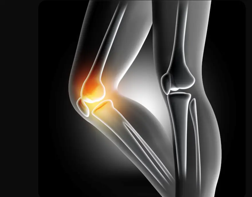
Table of Contents
- What is Osteochondroma?
- Types of Osteochondroma
- Causes of Osteochondroma
- How Does Osteochondroma Develop?
- Symptoms of Osteochondroma
- Diagnosis of Osteochondroma
- Diagnostic and Imaging Methods for Detecting Osteochondroma
- Treatment for Osteochondroma
- Managing Osteochondroma Symptoms
- Osteochondroma and Multiple Hereditary Exostoses (MHE)
- Prevention of Osteochondroma
- Prognosis for Osteochondroma
- Conclusion
- FAQs
- References
What is Osteochondroma?
Osteochondroma is a benign (non-cancerous) bone growth that develops as a cartilage-capped bony projection from the surface of bones. The osteochondroma meaning encompasses both its structural composition—consisting of both bone and cartilage, most often developing near the growth plates of long bones. This condition primarily affects children and teenagers during periods of rapid skeletal development.
They present as firm, immobile lumps that feel hard to the touch. Osteochondroma typically develops at the ends of long bones such as the femur, tibia, humerus, and bones around the knee and shoulder. Each growth consists of either a stalk (pedunculated type) or a broad base (sessile type) attached to the underlying bone, capped with cartilage that continues to grow until skeletal maturity.
Types of Osteochondroma
• Solitary osteochondroma: A single, isolated growth affecting one bone location
• Multiple osteochondromas: Several growths throughout the skeleton, often linked to a genetic condition called Multiple Hereditary Exostosis (MHE)
• Sessile osteochondroma: A broad-based, flat growth directly attached to the bone surface
• Pedunculated osteochondroma: A mushroom-like growth projecting outward on a narrow stalk
Causes of Osteochondroma
The exact osteochondroma causes involve disruptions in normal bone development during childhood. These growths develop when a small portion of the growth plate cartilage becomes displaced and continues growing outward instead of contributing to normal bone lengthening.
Genetic factors play a significant role in osteochondroma development. Mutations in genes called EXT1 and EXT2, which normally regulate cartilage formation, can trigger abnormal bone growth patterns.
Environmental factors may also contribute to osteochondroma causes. Some cases develop following bone trauma, surgical procedures, or radiation exposure, though these triggers are less common than genetic factors.
How Does Osteochondroma Develop?
Osteochondroma development begins when a portion of growth plate cartilage grows outward in an abnormal direction. Instead of contributing to normal bone lengthening, displaced cartilage cells form an outward projection. This process, called endochondral ossification, gradually converts the cartilage base into bone while maintaining a cartilage cap.
The growth continues throughout childhood and adolescence, moving away from the joint as the underlying bone grows normally. Once skeletal maturity is reached, the cartilage cap typically stops growing and may even thin over time.
Symptoms of Osteochondroma
- Painless, hard lump: Most common presentation, typically near joints like knees, shoulders, or hips
- Activity-related pain: Discomfort during movement, especially if the tumour irritates surrounding muscles or tendons
- Joint stiffness: Limited range of motion when the growth impinges on joint structures
- Neurological symptoms: Numbness, tingling, or weakness caused by compression of nearby nerves
- Vascular complications: Rarely, nearby blood vessels may be compressed, leading to reduced circulation or changes in pulse
- Cosmetic concerns: Visible deformity or asymmetry, particularly around prominent bone areas
- Fracture-related pain: Sudden, severe pain that occurs when the stalk of an osteochondroma fractures due to trauma
Diagnosis of Osteochondroma
- Comprehensive medical history: Your doctor will ask about symptom onset, family history of bone diseases, previous injuries, and any changes in the growth's characteristics.
- Physical examination: Careful assessment of the mass's size, location, mobility, and relationship to surrounding structures provides important diagnostic clues.
- Imaging studies: Various techniques help visualise the osteochondroma's structure and assess its impact on nearby tissues and organs.
- Growth monitoring: Serial examinations over time help determine whether the lesion is stable or changing, which influences treatment decisions.
- Genetic testing: For individuals with multiple lesions or strong family histories, genetic analysis may identify specific mutations associated with hereditary conditions.
The diagnostic process aims to distinguish osteochondroma from other bone diseases and determine appropriate management strategies based on individual circumstances.
Diagnostic and Imaging Methods for Detecting Osteochondroma
• X-rays: First-line imaging showing bone continuity, cortical and medullary involvement, and overall tumour morphology
• Magnetic Resonance Imaging (MRI): Superior soft tissue contrast revealing Thickness of the cartilage cap (typically less than 1–2 cm in benign cases), neurovascular relationships, and potential complications
• Computed Tomography (CT): Detailed bone architecture assessment, particularly valuable for complex anatomical regions like pelvis or spine
• Ultrasound imaging: Non-invasive evaluation of Thickness of the cartilage cap (typically less than 1–2 cm in benign cases), especially useful in paediatric patients
• Bone scintigraphy: Occasionally employed to assess metabolic activity and identify multiple lesions in hereditary cases
Osteochondroma radiology findings typically demonstrate characteristic features, including bone continuity, cartilage cap presence, and absence of aggressive features. Radiologists assess key features such as continuity of the cortex and medulla, and cap thickness to distinguish benign osteochondromas from other bone lesions or possible malignant change.
Treatment for Osteochondroma
Osteochondroma treatment approaches vary based on symptoms, location, size, and individual patient factors:
• Active surveillance: Most asymptomatic osteochondromas are managed with periodic monitoring with clinical examinations and imaging studies to ensure stability and detect any concerning changes.
• Surgical excision: Osteochondroma treatment through surgical removal becomes necessary when growths cause pain, restrict movement, compress nearby nerves or vessels, cause functional limitations, or show signs of malignant transformation.
• Physical therapy: Targeted exercises after surgery or during conservative management help maintain joint flexibility, restore strength, and prevent stiffness.
• Pain management: For mild discomfort, nonsteroidal anti-inflammatory drugs (NSAIDs) can provide symptom relief while avoiding more invasive interventions.
Managing Osteochondroma Symptoms
• Regular monitoring: Schedule routine follow-up appointments to track any changes in size, symptoms, or complications that might require treatment adjustments.
• Activity modification: Protect affected areas from trauma while maintaining normal activities, using appropriate padding or supports when participating in contact sports.
• Symptom relief: Apply ice packs and use over-the-counter anti-inflammatory medications for minor pain or swelling related to irritation or bursitis.
• Physical therapy: Engage in prescribed exercises to maintain joint flexibility and muscle strength, particularly important after surgical treatment.
• Prompt evaluation: Seek immediate medical attention for new or worsening symptoms such as severe pain, rapid growth, or neurological changes.
Osteochondroma and Multiple Hereditary Exostoses (MHE)
Multiple hereditary exostosis (MHE) is a more complex form of osteochondroma affecting multiple bones simultaneously. According to MedlinePlus, this genetic condition occurs in approximately 1 in 50,000 people and follows an autosomal dominant inheritance pattern, meaning affected parents have a 50% chance of passing the condition to their children.
People with MHE typically develop numerous osteochondromas throughout their skeleton, often leading to bone deformities, shorter stature, and functional limitations. The condition requires lifelong monitoring, as the risk of malignant transformation, while still low, is higher than with solitary osteochondromas.
Families affected by MHE benefit from genetic counseling to understand inheritance patterns and plan for appropriate medical care across generations.
Prevention of Osteochondroma
Unfortunately, there are no known methods to prevent osteochondroma development. Since most cases result from genetic factors or spontaneous developmental changes, prevention strategies are not currently available.
However, early detection and appropriate monitoring can help identify complications before they become serious. Families with a history of multiple osteochondromas should discuss genetic counselling and regular screening with healthcare professionals.
The focus remains on prompt recognition of symptoms and appropriate medical management rather than prevention of the condition itself.
Prognosis for Osteochondroma
The prognosis for osteochondroma is generally excellent, particularly for solitary lesions. Most people with osteochondroma lead normal, active lives without significant limitations or complications.
Solitary osteochondromas rarely cause serious problems and have minimal impact on life expectancy or quality of life. Surgical treatment, when necessary, typically achieves excellent results with low recurrence rates and few complications. Most patients return to full activities within months of surgery.
Multiple hereditary exostoses carries a more variable prognosis depending on the number and location of lesions. While many people manage well with appropriate care, some experience ongoing challenges with joint function, skeletal deformities, or activity limitations. The slightly elevated bone cancer risk requires lifelong monitoring but affects only a small percentage of patients.
Regular follow-up appointments help ensure early detection of any concerning changes while providing reassurance for the vast majority of people whose osteochondromas remain stable and benign.
Conclusion
Understanding osteochondroma empowers you to make informed decisions about your bone health. These benign growths, while initially concerning, rarely cause serious complications and often require only monitoring rather than active treatment. Recognising osteochondroma symptoms enables timely medical evaluation, while understanding the various diagnostic and treatment options helps reduce anxiety about the condition.
The key to successful osteochondroma management lies in regular monitoring, appropriate symptom management, and seeking prompt medical attention when changes occur. Whether dealing with a solitary growth or multiple lesions, working closely with doctors ensures optimal outcomes.
At Metropolis Healthcare, we understand the importance of accurate diagnosis in managing bone diseases like osteochondroma. Our comprehensive portfolio of more than 4,000 tests includes specialized imaging and laboratory tests that support precise evaluation of bone conditions. With our extensive network of over 220 laboratories and 10,000+ touchpoints across India, we provide convenient access to advanced diagnostic services that guide effective treatment decisions.
FAQs
How Is Osteochondroma Treated?
Osteochondroma is usually managed with observation when symptoms are absent. Surgical removal is considered if the lesion causes pain, functional limitation, nerve or vessel compression, or suspicious growth, with complete excision generally offering long-term relief.
Is Osteochondroma Cancerous?
Osteochondroma is a benign bone tumour and not cancerous. Rarely, malignant transformation into chondrosarcoma can occur, especially in adults showing sudden growth, increasing pain, or cartilage cap thickening, warranting prompt medical evaluation to rule out progression.
Can Osteochondroma Recur?
Recurrence after complete surgical removal is rare. However, younger patients with open growth plates and individuals with multiple hereditary exostoses may develop additional lesions over time, making ongoing monitoring important to detect new growths or emerging symptoms early.
What Are the Symptoms of Osteochondroma?
• Painless hard bump near joints
• Pain with movement or activity
• Numbness from nerve compression
• Restricted joint motion
• Sudden pain with stalk fracture
References
- https://www.ncbi.nlm.nih.gov/books/NBK544296/
- https://my.clevelandclinic.org/health/diseases/21982-osteochondroma
- https://orthoinfo.aaos.org/en/diseases--conditions/osteochondroma/
- https://medlineplus.gov/genetics/condition/hereditary-multiple-osteochondromas/#frequency
- https://pmc.ncbi.nlm.nih.gov/articles/PMC11335184/