Preventive Healthcare
IHC Test (ImmunoHistoChemistry): Purpose, Procedure
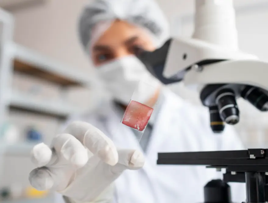
Table of Contents
- What Is an IHC Test?
- How Does IHC Work?
- Step-by-Step Procedure of the IHC Test
- Types of Antibodies Used in IHC Testing
- Common Diseases Diagnosed with IHC
- Types of Cancer Diagnosed Using IHC
- Advantages of IHC in Diagnosing Diseases
- Limitations of IHC Testing
- Risks Associated with IHC
- IHC and Prognosis: What Does It Reveal About the Disease?
- IHC in Treatment Planning and Monitoring
- Preparation for IHC Testing
- Aftercare and Recovery from the IHC Test
- What Are the Costs of IHC Testing?
- Conclusion
- FAQs
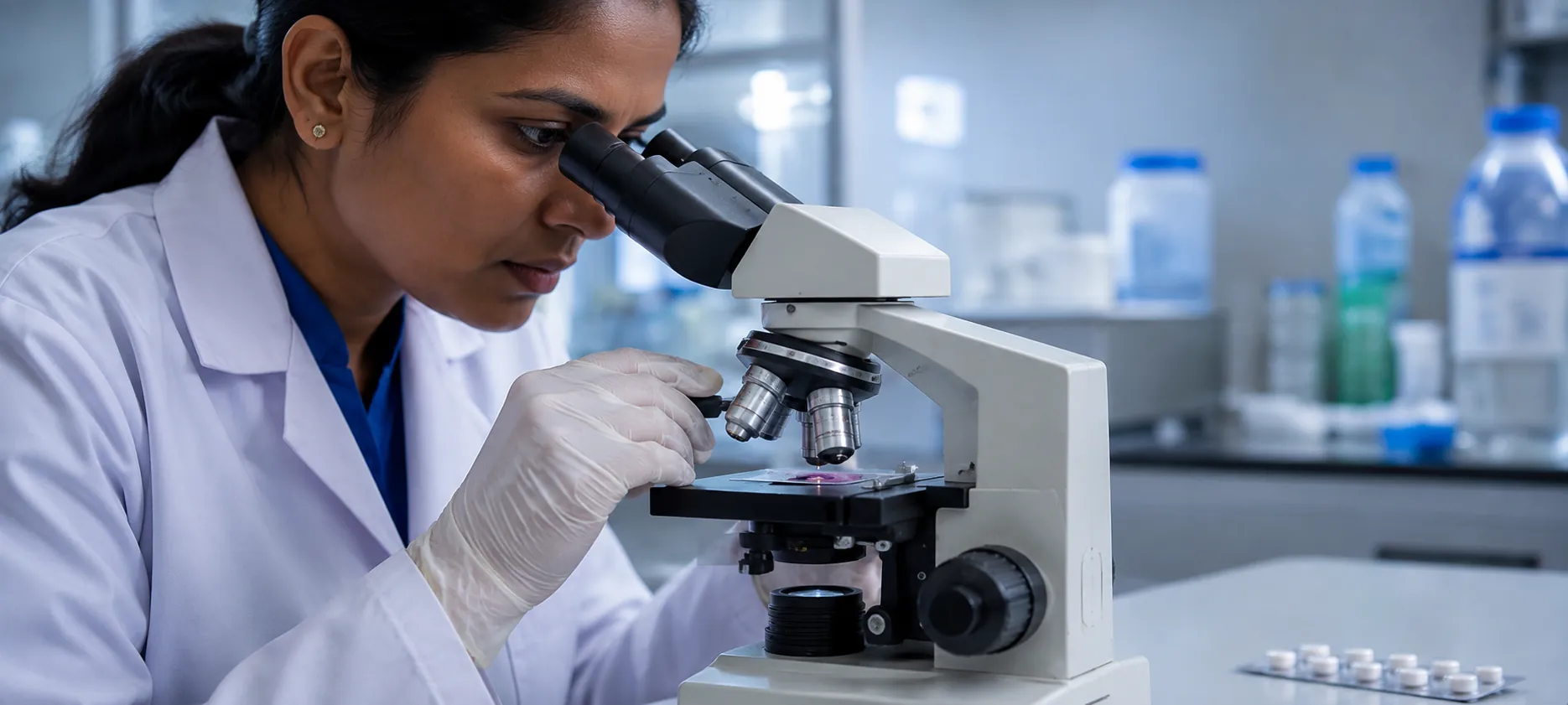
What Is an IHC Test?
The IHC test, or immunohistochemistry, uses antibodies to detect specific proteins in tissue samples, providing crucial information about disease presence and characteristics. This laboratory technique combines principles of immunology and histology to create a detailed picture of cellular activity. During an IHC marker test, pathologists apply special antibodies to your tissue sample, then examine it under a microscope to detect specific target proteins.
How Does IHC Work?
The IHC test functions through a sophisticated process of antibody-antigen binding. Your tissue sample is prepared so that specific antibodies can attach to target proteins if they are present. These antibodies carry markers that create visible signals when viewed under a microscope, allowing pathologists to identify protein locations and concentrations.
Think of it as a highly specific key (antibody) fitting only one type of lock (protein) in your tissue. When the key fits, it creates a visible mark that doctors can interpret. This precision makes the IHC test invaluable for diagnosing conditions like prostate cancer, stomach cancer, and metastatic cancer, where protein identification determines treatment approaches.
Step-by-Step Procedure of the IHC Test
The IHC marker test follows a systematic process to ensure accurate and reproducible results:
- Tissue Collection and Fixation: Your doctor collects tissue through biopsy, then preserves it using formalin to maintain cellular structure and protein integrity.
- Sectioning and Mounting: Laboratory technicians cut extremely thin tissue slices (4-5 microns) and place them on glass slides for examination.
- Antigen Retrieval: Heat treatment or enzymatic processes expose hidden proteins that fixation may have masked, ensuring antibodies can access their targets.
- Primary Antibody Application: Specific antibodies targeting disease-related proteins are applied to tissue sections and allowed to bind.
- Secondary Antibody and Detection: Additional antibodies linked to detection enzymes amplify signals, creating visible markers where target proteins exist.
- Microscopic Examination: Pathologists study the stained tissue slides under a microscope, interpreting staining patterns to determine diagnosis and treatment recommendations.
Types of Antibodies Used in IHC Testing
According to research published in PubMed-indexed journals, antibodies are a core component of immunohistochemistry, with two primary categories commonly used in diagnostic and research settings:
- Monoclonal Antibodies: A uniform population of immunoglobulins that target a single epitope. Generated through hybridoma technology by fusing a single B-cell clone with a myeloma cell line, these antibodies, usually of mouse or rabbit origin, offer high specificity and typically produce less background staining. However, some fixed tissues may mask epitopes, making certain proteins harder to detect with monoclonal antibodies alone.
- Polyclonal Antibodies: A heterogeneous mix of immunoglobulins that bind to multiple epitopes on the same protein. Produced by immunising animals such as rabbits or goats, polyclonal antibodies are available either as whole serum or affinity-purified preparations. Their broader reactivity allows them to detect proteins that may be structurally modified during fixation and, therefore, less accessible to monoclonal antibodies.
Common Diseases Diagnosed with IHC
The IHC test proves invaluable for diagnosing numerous conditions beyond cancer:
- Infectious Diseases: Identifying bacterial, viral, or fungal pathogens within infected tissues helps guide appropriate antimicrobial therapy. For viral infections specifically, an Viral Marker Test can complement IHC by detecting virus-specific proteins or antigens, providing a more targeted diagnostic approach.
- Neurodegenerative Disorders: Detecting protein aggregates associated with Alzheimer's disease or Parkinson's disease supports an accurate neurological diagnosis.
- Muscular Dystrophy: Identifying specific muscle protein deficiencies helps classify dystrophy types and inform treatment strategies.
- Lynch Syndrome: Evaluating DNA mismatch repair proteins helps identify hereditary cancer predisposition syndromes.
Types of Cancer Diagnosed Using IHC
- Breast Cancer: Detecting oestrogen receptors, progesterone receptors, and HER2 proteins guides targeted therapy selection and prognosis assessment.
- Lung Cancer: Identifying specific protein markers helps distinguish between small cell and non-small cell types, informing treatment approaches.
- Lymphoma: Characterising immune cell markers enables accurate lymphoma subtyping, crucial for selecting appropriate chemotherapy regimens.
- Prostate Cancer: Detecting prostate-specific antigen and other markers supports diagnosis and monitoring treatment response.
- Stomach Cancer: Identifying HER2 overexpression and other proteins guides targeted therapy decisions.
- Metastatic Cancer: Determining primary tumour origin through protein profiling helps guide treatment when cancer spreads to distant sites.
Advantages of IHC in Diagnosing Diseases
The IHC test provides precise localisation of proteins within the tissue context, enabling doctors to understand not just whether proteins are present, but exactly where they're located within cells. Unlike blood tests that show general protein levels, the IHC test reveals specific cellular patterns that guide accurate diagnosis. For cancer patients, this precision translates into personalised treatment plans.
Limitations of IHC Testing
Despite its advantages, the IHC test has certain limitations you should be aware of. Technical variability can affect results, particularly during antigen retrieval steps, where inconsistent processing may mask or alter protein detection. Additionally, antibody cross-reactivity sometimes produces false positive results when antibodies bind to unintended targets.
Accurate IHC interpretation requires significant expertise, as staining patterns can sometimes be subtle or ambiguous. Subtle differences in interpretation can occur between pathologists, especially in borderline cases, highlighting the importance of experienced interpretation.
Furthermore, IHC results typically serve as supportive evidence rather than a standalone diagnosis, often requiring correlation with other diagnostic methods for complete accuracy.
Risks Associated with IHC
- Biopsy-related risks: The tissue collection procedure may cause mild pain, minor bleeding, or, rarely, infection at the biopsy site.
- False Results: Technical variations might produce false positive or negative results, potentially affecting diagnosis accuracy.
- Allergic Reactions: Extremely rare reactions to biopsy procedures or local anaesthetics may occur in sensitive individuals.
- Anxiety: Waiting for results can cause emotional stress, though understanding the process often helps reduce uncertainty.
IHC and Prognosis: What Does It Reveal About the Disease?
The IHC test provides crucial prognostic information that shapes your treatment journey and outlook. In breast cancer cases, hormone receptor status determined through IHC testing directly correlates with survival rates and treatment response. Patients with hormone receptor-positive tumours typically experience better outcomes with appropriate therapy.
In lymphoma, specific protein markers identified through IHC testing help predict disease behaviour and treatment responsiveness. This information allows doctors to tailor therapy intensity and monitoring schedules to your specific situation, optimising both effectiveness and quality of life during treatment.
IHC in Treatment Planning and Monitoring
Modern cancer treatment increasingly relies on IHC test results for personalised therapy selection. When treating breast cancer, doctors use HER2 protein levels to determine whether targeted drugs like trastuzumab will be effective. Similarly, lung cancer treatment plans depend heavily on protein profiles revealed through IHC testing.
Beyond initial treatment planning, the IHC marker test helps monitor therapy effectiveness. Repeat biopsies may show changes in protein expression following treatment, indicating whether current approaches are working or require modification.
Preparation for IHC Testing
Preparation for the IHC test mainly involves getting ready for the tissue collection procedure:
- Medication Review: Inform your doctor about blood-thinning medications that might increase bleeding risk during biopsy procedures.
- Fasting: Some biopsy procedures may require fasting, especially if sedation or anaesthesia is used.
- Medical History: Discuss allergies, pregnancy status, or bleeding disorders that might affect the biopsy procedure.
- Consent Process: Understand the biopsy procedure, its risks, and how tissue will be used for diagnosis.
Aftercare and Recovery from the IHC Test
Recovery from an IHC test depends mainly on the biopsy method, as the laboratory analysis itself is non-invasive. Needle biopsies typically require minimal downtime, with most patients resuming normal activities within 24 hours. Surgical biopsies require more extensive recovery, including wound care and activity restrictions as advised by your healthcare team.
What Are the Costs of IHC Testing?
The cost of an IHC test varies depending on how many antibody markers are tested and where the laboratory is located. At Metropolis Healthcare, single markers start from ₹2,300, while complex panels requiring multiple markers cost over ₹10,000.
Conclusion
Understanding what the IHC test is can help you feel more informed and in control during a difficult diagnostic process. It remains a key tool in cancer diagnosis, helping identify tumour-specific markers for accurate treatment planning.
At Metropolis Healthcare, you can access a comprehensive portfolio of 4,000+ tests, including specialised IHC testing for cancer diagnosis. With 10,000+ touchpoints across India and convenient home sample collection, our advanced NABL and CAP accredited laboratory network ensures accurate, timely results when you need them most.
FAQs
How accurate is IHC?
IHC is highly accurate, often above 90–95% for well-established markers. Accuracy depends on antibody quality, tissue processing, and the pathologist's expertise.
What cancers can IHC diagnose?
IHC helps diagnose breast, lung, prostate and stomach cancers, lymphoma, and metastatic tumours by identifying cancer-specific protein markers.
Can IHC be used for non-cancer diagnoses?
Yes. IHC also supports the diagnosis of infections, neurological disorders, muscular dystrophies, and autoimmune diseases by detecting disease-related proteins.
How long does an IHC test take?
IHC usually adds about one extra day to routine processing, with total turnaround typically 2–10 days depending on the complexity of the panel.
What are the benefits of IHC over other tests?
IHC shows protein location within intact tissue, works on archived samples, and helps guide therapy decisions, making it especially useful for cancer subtyping.