Preventive Healthcare
Craniopharyngiomas: A Complete Patient And Caregiver Guide
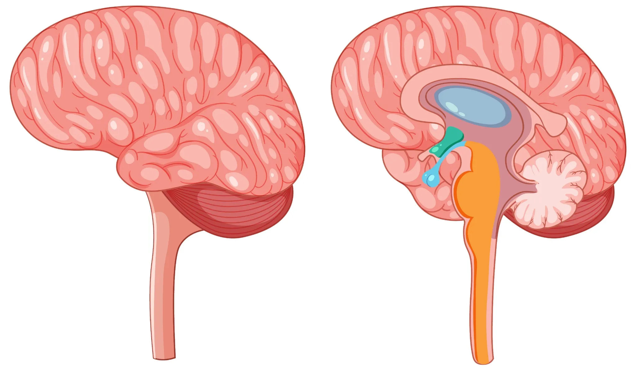
Table of Contents
- What Are Craniopharyngiomas?
- Types of Craniopharyngiomas
- Who Is Affected by Craniopharyngiomas?
- Where Do Craniopharyngiomas Develop in the Brain?
- Symptoms of Craniopharyngiomas
- Causes of Craniopharyngiomas
- How Are Craniopharyngiomas Diagnosed?
- Craniopharyngioma Tumour Growth & Complications
- Craniopharyngioma Treatment Options
- Life After Treatment & Long-Term Monitoring
- Craniopharyngiomas in Children vs. Adults
- Nutrition & Lifestyle Guidance for Recovery
- When to Seek Medical Help
- Outlook & Survival Rate
- Conclusion
- FAQs
- References
What Are Craniopharyngiomas?
Craniopharyngiomas are benign epithelial tumours that arise in the sellar and suprasellar regions of the brain near the pituitary gland. Although benign, these non-pituitary tumours can cause major problems due to their proximity to the pituitary gland, hypothalamus, and optic pathways. Craniopharyngiomas originate from embryonic remnants of Rathke’s pouch, an early structure that gives rise to the anterior pituitary, but instead forms these slow-growing masses.
These tumours typically grow gradually over months or years, pressing against vital structures including the pituitary gland, hypothalamus, optic nerves, and surrounding blood vessels. While craniopharyngiomas don't spread to other parts of your body like cancerous tumours, their proximity to essential brain regions makes them particularly challenging to treat. Early recognition and appropriate medical care are crucial for managing craniopharyngiomas effectively and preventing complications.
Types of Craniopharyngiomas
Medical professionals classify craniopharyngiomas into two distinct types based on their cellular characteristics and typical age of occurrence:
• Adamantinomatous craniopharyngiomas: Most commonly affect children and teenagers, arising from remnant cells of the craniopharyngeal duct during embryonic development.
• Papillary craniopharyngiomas: Predominantly affect adults and arise from metaplastic changes in Rathke’s pouch epithelium rather than anterior pituitary cells.
Who Is Affected by Craniopharyngiomas?
As per a 2025 StatPearls review, craniopharyngiomas occur at an incidence of approximately 0.5–2 cases per million persons per year. They affect both children and adults, showing a bimodal age distribution with peaks at 5–14 years and 45–60 years. This bimodal age distribution means craniopharyngiomas are classified as both a rare childhood brain tumour and an uncommon adult brain tumour.
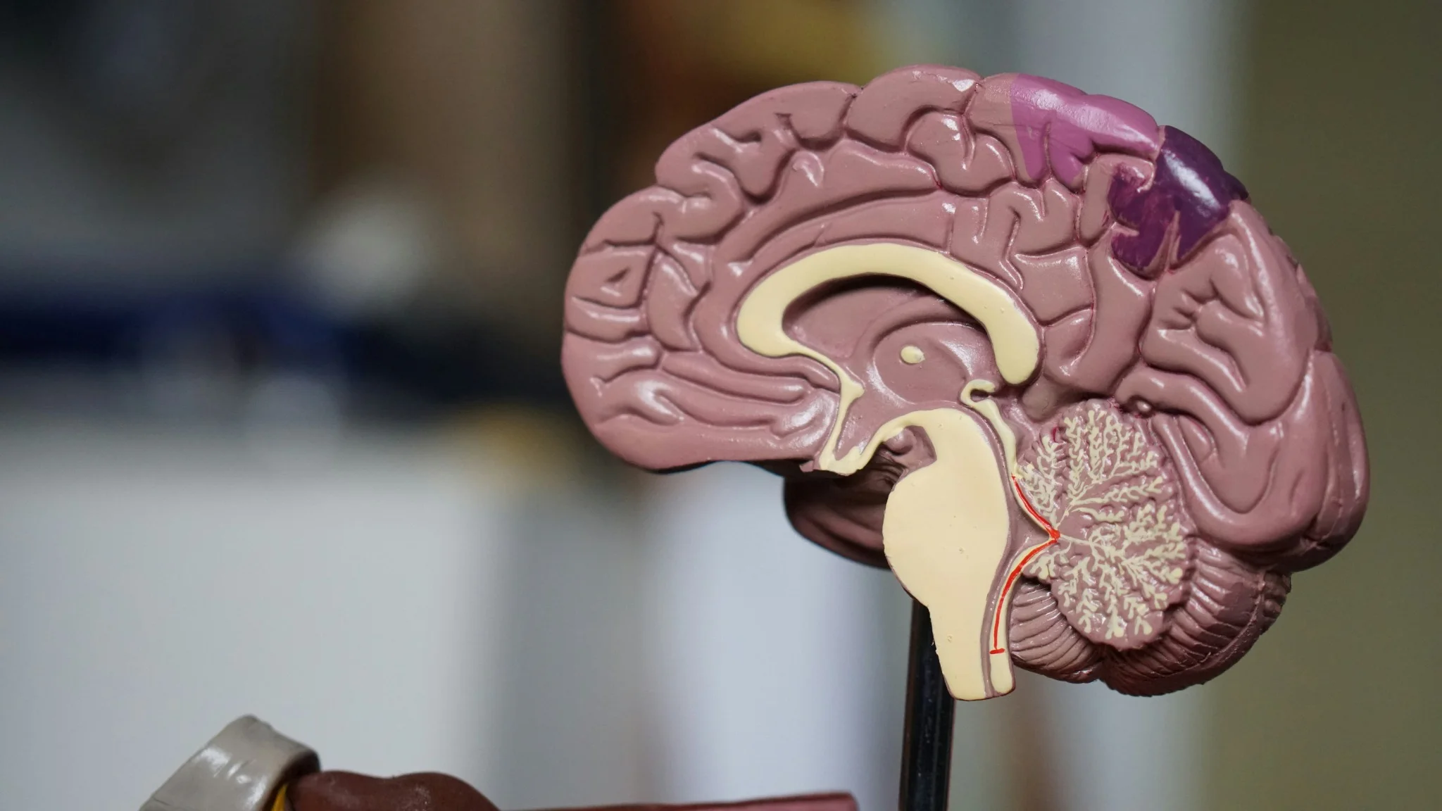
Where Do Craniopharyngiomas Develop in the Brain?
Craniopharyngiomas grow in the sellar and parasellar regions of the brain. This area is around and just above the pituitary gland, which helps control many body functions.
The exact location of the tumour in this region determines which symptoms you may experience. Most craniopharyngiomas originate in the suprasellar cistern and frequently involve the pituitary stalk, sometimes extending into the third ventricle.
They can then grow:
- Upwards toward the third ventricle
- Sideways toward the optic nerves and optic chiasm
Common locations include:
- Suprasellar: Above the pituitary gland; it often affects vision and hormones.
- Intrasellar: In the pituitary area; mainly causes hormone problems.
- Retrosellar or Posteriorly: Extending tumours may compress the brainstem, leading to balance issues, drowsiness, or other neurological symptoms.
Symptoms of Craniopharyngiomas
Craniopharyngioma symptoms develop gradually as the tumour grows and compresses nearby brain structures. The specific symptoms you experience depend on your tumour's size, location, and growth rate.
Common symptoms include:
• Persistent headaches: Often worse in the morning and may worsen over time
• Vision problems: Blurred vision, loss of peripheral sight, or double vision
• Endocrine disturbances: Growth failure in children, menstrual irregularities, fatigue, or delayed puberty.
• Increased urination and thirst: Due to disrupted hormone regulation
• Nausea and vomiting: Particularly common in the morning
• Balance and coordination difficulties: Trouble walking or frequent falls
• Cognitive changes: Memory problems, confusion, or personality alterations
• Sleep disturbances: Excessive daytime sleepiness or disrupted sleep patterns
Causes of Craniopharyngiomas
Craniopharyngiomas are believed to develop from embryonic cells that fail to disappear as the pituitary gland fully forms in the womb. The adamantinomatous subtype (common in children) is linked to CTNNB1 (β-catenin) mutations, while the papillary subtype (in adults) is associated with BRAF V600E mutations, but the exact trigger for this transformation is still unknown.
These tumours are not caused by lifestyle habits, environmental exposures, or typical inherited genetic patterns, and there are currently no known ways to prevent them. Understanding the medical basis of craniopharyngiomas helps patients and families focus on appropriate care and treatment, rather than on feelings of personal responsibility.
How Are Craniopharyngiomas Diagnosed?
Diagnosing craniopharyngiomas typically involves several steps that help your medical team understand your tumour's characteristics and plan appropriate treatment:
- Comprehensive medical history and physical examination: Your doctor will assess symptoms, perform neurological tests, and check for hormonal dysfunction.
- Magnetic Resonance Imaging (MRI): The primary imaging test that provides detailed pictures of your brain and tumour without radiation exposure.
- CT scan: May be used to evaluate bone structures and assist in surgical planning if needed.
- Hormonal blood tests: Assess pituitary and hypothalamic function, including cortisol, ACTH, TSH, FT4, GH, IGF-1, LH, FSH, prolactin, and electrolytes (especially sodium for diabetes insipidus).
- Visual field testing: Formal eye examinations evaluate any vision problems caused by tumour compression.
- Neurosurgical evaluation: If surgery is considered, neurosurgeons review imaging studies and plan treatment approaches.
Craniopharyngioma Tumour Growth & Complications
Craniopharyngiomas are benign but slow-growing tumours. They often enlarge over time, forming both solid and cystic (fluid-filled) parts that can press on nearby brain structures. Even after treatment, they may recur, so long-term follow-up is important.
As they grow, they can cause several complications that affect daily life:
- Hydrocephalus: Buildup of fluid in the brain when the tumour blocks normal cerebrospinal fluid flow.
- Pituitary hormone deficiencies: Reduced hormone levels affecting growth, metabolism, puberty, fertility, and stress response.
- Vision loss: Gradual or sometimes sudden vision problems, especially loss of side (peripheral) vision, which can become permanent without treatment.
- Hypothalamic dysfunction: Problems with appetite, weight, body temperature, thirst, emotions, and sleep.
- Increased intracranial pressure: Severe headaches, nausea, vomiting, drowsiness, and sometimes seizures due to raised pressure inside the skull.
- Memory and learning difficulties: Issues with concentration, memory, and school or work performance affecting overall quality of life.
Craniopharyngioma Treatment Options
Craniopharyngioma treatment approaches depend on your tumour's size, location, symptoms, age, and overall health status. Treatment focuses on the removal or control of the tumour, preserving neurological and hormonal function, and preventing recurrence:
- Surgical resection: Removal of all or part of the tumour using different surgical approaches, chosen based on its position and size.
- Conformal or proton beam radiation therapy: Used to control tumour regrowth when complete surgical resection is unsafe or incomplete.
- Hormone replacement therapy: Long-term medicines to replace hormones that are reduced or lost because of the tumour or its treatment.
- Combination approaches: Surgery followed by radiation to improve local tumour control and lower the chance of regrowth.
- Active surveillance: Careful monitoring with regular MRI scans for small, stable, or asymptomatic tumours where immediate intervention is not required.
Craniopharyngioma Surgery Approaches
Several surgical techniques are available for craniopharyngiomas, each with specific advantages depending on your tumour's characteristics:
- Endoscopic transsphenoidal surgery: A minimally invasive technique through the nasal cavity and sphenoid sinus, preferred for midline or intrasellar/suprasellar tumours.
- Craniotomy approaches: Open surgery through the skull (using different routes) to reach larger, more complex, or laterally extending tumours.
- Endoscopic techniques: Use small cameras and fine instruments through the nose to improve visualisation and allow more precise, targeted tumour removal.
Life After Treatment & Long-Term Monitoring
Recovery after craniopharyngioma treatment usually involves lifelong follow-up, including hormone replacement therapy, regular MRI scans (often every 6–12 months at first), and periodic reviews with your endocrinologist, neurosurgeon, and oncologist.
Many people return to normal or near-normal activities within a few months. Some may still have low energy, memory or concentration problems, or weight changes. In these cases, physiotherapy, occupational therapy, and neuropsychological support can help improve daily functioning, monitor condition, and overall quality of life.
Craniopharyngiomas in Children vs. Adults
Craniopharyngiomas affect children and adults differently, requiring age-specific treatment considerations:
|
Children |
Adults |
|
Primarily of the adamantinomatous type |
Mainly papillary type |
|
Growth and development concerns |
Reproductive and metabolic issues |
|
School performance impacts |
Work and relationship effects |
|
Family support is crucial. |
Self-advocacy is important. |
|
Long-term follow-up is essential. |
Lifetime hormone management |
Nutrition & Lifestyle Guidance for Recovery
Supporting your recovery through proper nutrition and lifestyle modifications can significantly impact your healing process and long-term well-being:
• Balanced nutrition: Focus on whole foods, adequate protein, and essential nutrients to support healing.
• Regular exercise: Gentle physical activity as approved by your medical team to maintain strength and mood.
• Stress management: Techniques like meditation, yoga, or counselling to cope with treatment stress.
• Sleep hygiene: Maintaining regular sleep schedules despite potential hypothalamic dysfunction.
• Social support: Connecting with family, friends, and support groups for emotional well-being.
When to Seek Medical Help
Contact your healthcare provider immediately if you experience any of these warning signs:
• Severe, sudden headaches: Especially if different from your usual pattern
• Rapid vision changes: New visual disturbances or worsening existing problems
• Persistent vomiting: Particularly if accompanied by headache and drowsiness
• Seizures: Any new seizure activity requires immediate medical attention.
• Severe confusion or personality changes: Significant alterations in mental status
• Signs of infection: Fever, neck stiffness, or unusual discharge after surgery
Outlook & Survival Rate
The prognosis for craniopharyngiomas is generally favourable with appropriate treatment. Long-term survival exceeds 90–95%, but morbidity from hormonal, visual, or hypothalamic complications remains common, though long-term quality of life depends on factors like tumour size, treatment approach, and individual response to therapy. Many people with craniopharyngiomas live normal, fulfilling lives with proper medical management and support.
While challenges may persist, particularly regarding hormone management and potential cognitive effects, advances in surgical techniques, radiation therapy, and supportive care continue to improve outcomes. Regular follow-up care and proactive management of treatment-related effects help optimise your long-term prognosis and quality of life.
Conclusion
Craniopharyngiomas are benign but clinically significant tumours that can impact vision, endocrine function, metabolism, and cognition. This makes early diagnosis, timely treatment, and lifelong monitoring essential for both children and adults.
In this journey, reliable diagnostics matter. Metropolis Healthcare offers 4,000+ tests, full body checkups, and speciality testing for both initial evaluation and long-term follow-up. Patients can benefit from home sample collection across 10,000+ touchpoints, quick and accurate reports, and easy booking via website or app, so families can focus on care and recovery, maintaining their diagnostic needs smoothly and professionally.
FAQs
Is craniopharyngioma cancer?
No, craniopharyngiomas are benign brain tumours that don't spread to other parts of your body like cancerous tumours. However, their location near critical brain structures can cause serious health problems, making prompt treatment essential even though they're not cancerous.
Can craniopharyngiomas come back after treatment?
Yes, craniopharyngiomas can recur after treatment, particularly if complete surgical removal wasn't possible. Recurrence rates vary depending on treatment approach, with complete surgical removal offering the lowest risk. Regular monitoring with MRI scans helps detect any regrowth early.
What causes craniopharyngioma in children?
Craniopharyngiomas in children develop from embryonic tissue that should have disappeared during foetal development, but persisted instead. The exact trigger for this transformation isn't known, and these tumours aren't caused by anything parents did or didn't do.
How serious is craniopharyngioma?
While craniopharyngiomas are benign, they can be serious due to their location near vital brain structures. With appropriate treatment, most patients have excellent outcomes, though some may require lifelong hormone replacement therapy and regular monitoring.
How long is the recovery after craniopharyngioma surgery?
Recovery time varies depending on the surgical approach and individual factors. Most patients stay in the hospital for 3-7 days after surgery, with full recovery taking several weeks to months. Some effects, like hormone deficiencies, may require ongoing management.
Can a craniopharyngioma lead to blindness or hormonal problems?
Yes, craniopharyngiomas can cause vision problems if they compress the optic nerves, and hormonal dysfunction is common due to their proximity to the pituitary gland. Early treatment helps prevent permanent damage and manage these complications effectively.
References
- https://my.clevelandclinic.org/health/diseases/22989-craniopharyngioma
- https://www.ncbi.nlm.nih.gov/books/NBK519027/
- https://medlineplus.gov/ency/article/000345.htm