 Home Visit
 Upload


Preventive Healthcare
Diabetic Retinopathy - What It is, Signs, Risk Factors And Diagnosis
3098 Views
0

What is Diabetic Retinopathy?
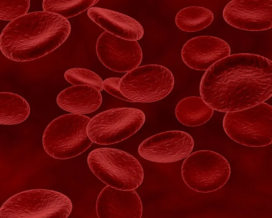
Diabetic Retinopathy is one of the most common complications of long-standing diabetes. Elevated levels of blood sugar cause damage to the retina, which is a light-sensitive tissue of the eyes and their surrounding blood vessels. This causes blurry vision and may lead to total blindness later if left untreated. Diabetic retinopathy is of two types:
- Non-proliferative diabetic retinopathy (NPDR)
- Proliferative diabetic retinopathy (PDR)
Non-Proliferative Diabetic Retinopathy (NPDR): The early stage of diabetic retinopathy is non-proliferative diabetic retinopathy (NPDR). In this case, the blood vessels of the retina swell, putting pressure on the macula of the retina (the central part), resulting in macular edema. This eventually leads to blurred vision.
Proliferative Diabetic Retinopathy (PDR): is a more severe form of diabetic retinopathy. It involves the proliferation or abnormal growth of new blood vessels called neovascularization. These new blood vessels eventually cause a detached retina, which results in total vision loss.
Causes of Diabetic Retinopathy
Prolonged diabetes causes damage to the blood vessels of the whole body. High sugar levels obstruct the small blood arteries leading to your retina, it damages your eyes by causing them to haemorrhage or leak fluid. Your eyes then develop new, poorly functioning blood vessels to make up for these blocked blood vessels. These brand-new blood vessels frequently bleed or leak.
Signs And Symptoms of Diabetic Retinopathy
You can suffer from diabetic retinopathy and not know it as in the early stages it shows no symptoms. As it progresses, the following symptoms begin to appear which include:
- presence of dark floating spots similar to cobwebs in the eyes.
- the feeling of pain and pressure in the eyes.
- microbleeds in the eyes.
- blurry vision.
- poor night and colour vision.
- complete loss of vision
Risk Factors of Diabetic Retinopathy
Diabetes can cause diabetic retinopathy in anyone with the disease. The following factors can raise the chance of developing the eye condition which includes:
- A lengthy history of diabetes.
- having trouble controlling your blood sugar.
- elevated blood pressure.
- high cholesterol level in the blood.
- gestational diabetes during pregnancy.
- Smoking.
What are The Complications of Diabetic Retinopathy?
The development of aberrant blood vessels in the retina is a complication of diabetic retinopathy. If left undiagnosed and untreated, these complications may result in severe eyesight issues which include:
- Vitreous Haemorrhage: The bleeding from the new blood vessels formed in the process of diabetic retinopathy leaks into the vitreous humor which is a gel-like fluid that fills the centre of the eye. Eventually, your retina cannot receive light due to the presence of blood. If there is little bleeding, you could just notice a few dark dots (floaters). In more severe situations, blood can fill the vitreous cavity, completely obstructing your vision.
- Neovascular Glaucoma: New blood vessels of the eye can grow out of the iris, disturbing the fluid balance of the eye. This creates pressure in your eyes which may lead to complete blindness.
- Diabetic Macular Edema (DME): The macula, the area of the retina that controls central vision and is positioned in the centre, is harmed by DME, a consequence of diabetic retinopathy. In this, your retina is unable to absorb fluid from the leaky blood vessels which causes the macula to swell up. It is one of the major causes of vision loss in diabetic retinopathy.
- Retinal Detachment: Sometimes aberrant blood vessels form scar tissue during retinopathy which stretches the retina away from the eyes. This may result in floating dots in your field of vision, bright flashes, or serious vision loss. Retinal detachment is a medical emergency that can cause irreversible blindness.
Diagnosis of Diabetic Retinopathy
To diagnose diabetic retinopathy, your eye doctor may do some tests in addition to conducting a thorough health history and eye examination. A thorough dilated eye exam is the most effective method for diagnosing diabetic retinopathy. To better observe inside your eyes during this examination, drops are put in your eyes to enlarge (dilate) your pupils. Your eye doctor will examine both the inside and outside of your eyes during the examination.
- Visual Acuity Test: It is a basic test that is done to check the degree of vision loss.
- Tonometry: This test is usually done to detect the intraocular pressure of the eye, and to detect the presence of glaucoma.
- Optical Coherence Tomography (OCT): An OCT examination is a type of imaging test that creates retinal images using light waves. Your doctor can measure the thickness of your retina using these photos. How much fluid, if any, has accumulated in the retina can be determined using OCT tests.
- Fluorescein Angiography or Fundus Fluorescein Angiography (FFA): During this test, your doctor will inject a dye into your blood to track the flow of blood in the eye. Photos of the eyes are taken during this whole process to check for broken or leaky blood vessels.
Treatment of Diabetic Retinopathy
The course of treatment of diabetic retinopathy depends upon age, symptoms, and overall general health, especially sugar levels. The good part is that when advanced retinopathy patients receive therapy prior to significant retinal damage, they have a 95% probability of maintaining their eyesight. The treatment of diabetic retinopathy involves:
- Laser treatment: The use of laser therapy, commonly referred to as photocoagulation, can assist stop vision loss. In this kind of surgery, the blood arteries are shrunk or sealed using a laser. To make sure you're comfortable throughout the treatment, your doctor will first provide a local anaesthetic. Additionally, they'll employ medication to widen your pupil. After that, a laser will be used by the doctor to flashlight into your eye.
- Intravitreal injections: A class of medications known as anti-vascular endothelial growth factors (VEGF) is used for diabetic retinopathy to block the certain proteins that cause inflammation and leaky blood vessels in the eyes.
- Vitrectomy: In this operation, scar tissue that is pulling on the retina as well as blood from the vitreous (the middle of the eye) are removed through a small incision in your eye. It is carried out under local or general anaesthesia in a surgery centre or hospital.
Prevention of Diabetic Retinopathy
The key to preventing diabetic retinopathy is to get complete control of your blood sugar profile and get regular eye and physical tests. An eye exam can aid in the diagnosis of eye problems and aware you and your health care practitioner if your diabetes needs to be better monitored. Women with diabetes should have an eye exam before or during pregnancy.


1701259759.webp)


 WhatsApp