 Home Visit
 Upload


Latest Blogs

Exploring Rectal Prolapse: Symptoms, Causes, and Treatment
Rectal prolapse can be uncomfortable to deal with; therefore, understanding the symptoms and complications associated with it is necessary. This guide discusses rectal prolapse in detail, offering insights on treatment options and prevention strategies to help you navigate its challenges effectively. What is rectal prolapse? Rectal prolapse is a condition which occurs when your rectum, the last part of the large intestine, protrudes through your anus. This can lead to discomfort, pain, and difficulty with bowel movements. Rectal prolapse types can include partial, complete, internal, or external, depending on the anatomy of the prolapse. What causes rectal prolapse? Rectal prolapse is primarily caused by weakened pelvic floor muscles and tissues that support your rectum. Chronic straining during bowel movements due to constipation, long-term diarrhoea, ageing, childbirth, or previous pelvic surgery are common rectal prolapse causes. Other factors include neurological conditions, genetic predisposition, and certain connective tissue disorders. What does rectal prolapse look like? Rectal prolapse may look like a reddish or pinkish mass emerging from your anus, typically when exerting pressure during bowel movements. This mass may retract back into the anus or remain visible externally. What are the signs or symptoms of rectal prolapse? Common rectal prolapse symptoms include: The most noticeable rectal prolapse symptom is the protrusion of your rectum through your anus during bowel movements or even when standing or walking. You may experience discomfort, pain, or a feeling of fullness in your rectum or anus. Rectal prolapse can lead to difficulty with your bowel movements, including straining or incomplete evacuation. You may experience leakage of faeces or mucus from the protruding rectum. The exposed rectal tissue can become irritated, leading to itching, burning, or bleeding. You may feel increased pressure or heaviness in your pelvic area, especially when standing or sitting for a long time. How is rectal prolapse diagnosed? Rectal Prolapse diagnosis typically involves: Physical examination: A healthcare provider will perform a physical examination, including a visual inspection of your anus and rectum, to look for signs of prolapse. Digital rectal exam: This involves inserting a gloved, lubricated finger into the rectum to assess the extent of prolapse and check for any abnormalities. Colonoscopy or sigmoidoscopy: These procedures allow for a more detailed examination of the rectum and colon to rule out other potential causes of rectal prolapse symptoms. Imaging tests: In some cases, imaging tests such as MRI or defecography may be ordered to evaluate pelvic floor function and assess the severity of prolapse. Anorectal manometry: This test measures pressure and function in the rectum and anal sphincters, helping to assess bowel function and pelvic floor coordination. Will rectal prolapse go away on its own? Rectal prolapse typically does not resolve on its own and often requires medical intervention, lifestyle changes, pelvic floor exercises, or surgery, depending on the severity of the condition. What happens if rectal prolapse is left untreated? If left untreated, rectal prolapse can cause chronic pain, fecal incontinence, and difficulty passing stool. In severe cases, tissue damage, ulceration and even strangulation of prolapsed tissue can occur, requiring emergency surgery to prevent complications such as tissue necrosis. How do you fix rectal prolapse? Rectal prolapse treatments vary depending on the severity of the condition and individual factors. They include: Abdominal approach (Rectopexy) Rectopexy is a surgical procedure commonly used for rectal prolapse treatment, particularly when the prolapse is significant or associated with other pelvic floor disorders. Rectopexy via the abdominal approach, i.e. through an incision in the belly, is often effective in providing long-term relief from rectal prolapse symptoms. Compared to some other surgical techniques, it lowers the risk of recurrence. Rectal Approach (Perineal) The perineal approach, also known as perineal recto sigmoidectomy or Delorme's procedure, is another surgical option for rectal prolapse therapies. Unlike rectopexy, which is performed through an abdominal incision, the perineal approach involves accessing the rectum through the area between the anus and the genitalia (the perineum). The perineal approach is often preferred for patients who are not candidates for abdominal surgery due to factors such as advanced age, poor overall health, or previous abdominal surgeries. Other options for rectal prolapse therapies include: Conservative management Dietary changes to regulate bowel movements. Pelvic floor exercises to strengthen muscles. Medications to alleviate constipation or diarrhoea. Medical procedures Manual reduction: Non-surgical manipulation to push the prolapse back into place. Rubber band ligation: Placing bands around the prolapsed tissue to cut off its blood supply, causing it to shrink and fall off. Injection sclerotherapy: Injecting a chemical solution into the prolapsed tissue to shrink it. Biofeedback therapy: Training to improve bowel control and pelvic floor function. What are the possible risks or complications of rectal prolapse surgery? Infection: Surgical procedures carry a risk of infection at the incision site or within the pelvic cavity. Bleeding: Post-operative bleeding can occur, either externally from the surgical site or internally within the abdomen or pelvis. Anaesthesia complications: Reactions to anaesthesia, though rare, can include respiratory problems, allergic reactions, or adverse drug interactions. Urinary problems: Surgery near the bladder or urethra can lead to urinary retention, incontinence, or urinary tract infections. Bowel dysfunction: Following rectal prolapse surgery, you may experience changes in bowel habits, including constipation or diarrhoea. Recurrence: Despite surgical intervention, rectal prolapse may recur in some cases, necessitating further treatment. How can I prevent rectal prolapse? Obesity can increase the risk of rectal prolapse, so aim to maintain a healthy weight through a balanced diet and regular exercise Avoid straining during bowel movements Address constipation promptly with dietary changes, fibre supplements, or stool softeners Strengthen pelvic floor muscles with exercises like Kegels to help support the rectum and prevent prolapse If you have a chronic cough, seek treatment to reduce strain on the pelvic floor and rectum Avoid heavy lifting. What is the outlook if I have rectal prolapse? While surgery generally offers favourable outcomes for rectal prolapse, you may experience ongoing symptoms like constipation or incomplete bowel movements. With appropriate medical management or surgery, many people experience significant improvement in symptoms and quality of life, with a low risk of recurrence. Conclusion In conclusion, rectal prolapse is a condition that can significantly impact your daily life. Still, with prompt diagnosis, appropriate treatment and lifestyle modifications, you can find relief and achieve improved long-term outcomes. If you or your family members want to protect yourself from rectal prolapse and its associated complications, get your blood and stool regularly tested at Metropolis Healthcare for timely diagnosis. Metropolis Healthcare strives to provide best-in-class diagnostic service that is not only reliable but also pocket-friendly, executed by the best phlebotomists, and trusted by the best doctors across the country. Hurry up and book your test today!

Understanding Tinea Versicolor: Symptoms, Causes, and Treatment
What is tinea versicolor? Tinea versicolor, a prevalent skin fungal infection, which disrupts natural skin pigmentation, leading to small, discoloured patches. Tinea versicolor commonly affects the torso and upper arms but can also appear on the face and neck, especially in younger children. The tinea versicolor on face can appear to be white, brown, red, or pink in colour or a dry, flaky, or scaly skin with a flat or slightly raised texture. What does tinea versicolor look like? Tinea versicolor spots can manifest as patches that are either lighter or darker than the surrounding skin, presenting in hues of white, pink, or light brown. These patches may exhibit dryness or scaliness and tend to appear more prominently on the chest and shoulders. Who gets tinea versicolor? Tinea versicolor can affect anyone, but it's more common in adolescents and young adults. Tinea versicolor can affect people worldwide, regardless of geographic location or nationality. However, it may be more commonly reported in regions with warm and humid climates, where the conditions are conducive to fungal growth on the skin. What are the symptoms of tinea versicolor? Most individuals experience mild tinea versicolor symptoms such as: Skin discolouration, typically on the back, chest, and abdomen. Discoloured patches can occur anywhere on the body and vary in shades of pink, yellow, brown, tan, or white. Itching in or around the affected skin areas. Increased sweating. Areas of skin that may become dry and develop scales. Skin patches that fail to darken or tan when exposed to sunlight. What causes tinea versicolor? Major cause of tinea versicolor is an excessive proliferation of yeast on the skin's surface. Flourishing in warm, moist, and oily conditions, this yeast multiplies in small clusters, leading to the characteristic symptoms of the condition. Tinea versicolor causes are as follows: Compromised immune function. High temperatures and humidity. Perspiration. Excessively oily skin. Hormonal fluctuations. How is tinea versicolor diagnosed? Tinea versicolor is typically diagnosed through a combination of medical history review and physical examination. The distinctive patches associated with this condition often facilitate its identification during the physical examination. Additionally, healthcare providers may utilise a Woods Lamp, an ultraviolet light, to enhance the visibility of the patches for a clearer diagnosis. How is tinea versicolor treated? The following ways can be considered for tinea versicolor treatment: Over-the-counter antifungal medication For mild cases, over-the-counter antifungal lotions, creams, ointments, or shampoos can be applied as an easily and accessible tinea versicolor treatment options. Prescription antifungal medication Clotrimazole cream or lotion Miconazole cream Selenium sulphide lotion Terbinafine cream or gel Zinc pyrithione soap Other ways of treatment are as follows: Severe or unresponsive cases of tinea versicolor may require prescription-strength medication. Medications may include: Ketoconazole cream, gel, or shampoo Ciclopirox cream, gel, or shampoo Fluconazole tablets or oral solution Itraconazole tablets, capsules, or oral solution Selenium sulphide lotion or shampoo Preventative medication may be necessary once or twice a month for persistent cases. How can I treat tinea versicolor at home? Home Remedies for Tinea Versicolor: Natural Antifungal Agents: Tea tree oil and apple cider vinegar possess antifungal properties that can help combat the fungal infection responsible for tinea versicolor. Tea tree oil can be diluted with carrier oil and applied directly to the affected areas, while apple cider vinegar can be diluted with water and applied as a topical solution. Yogurt and Honey Mixture: Yogurt contains probiotics that can help restore the balance of microorganisms on the skin, while honey has antimicrobial properties. Mixing yoghurt and honey into a paste and applying it to the affected areas can help soothe the skin and inhibit fungal growth. Aloe Vera Gel: Aloe vera gel is known for its soothing and healing properties. Applying pure aloe vera gel on the affected areas can help reduce inflammation, soothe irritated skin, and promote healing. Coconut Oil: Coconut oil has antifungal properties and can help combat the fungal infection causing tinea versicolor. Applying coconut oil to the affected areas can help moisturize the skin, reduce dryness, and prevent fungal growth. Maintain Good Hygiene: Keeping the affected area clean and dry is essential for managing tinea versicolor. Regularly washing the skin with a gentle cleanser and patting it dry can help prevent the growth of fungi and reduce the risk of infection. Remember that it is good to get in touch with the doctor before you follow any remedies at home. How long does tinea versicolor take to go away? Treatment for tinea versicolor typically takes around 1-2 weeks to show improvement. If tinea versicolor recurs, you should consult a healthcare professional for further evaluation and management. How can I prevent tinea versicolor? Preventing a recurrence of tinea versicolor can pose challenges. However, if you've been diagnosed with and successfully treated the condition, there are proactive measures you can take to reduce the likelihood of future infections: Avoiding prolonged exposure to excessive heat. Steer clear of tanning or extended periods of sun exposure. Minimise excessive sweating by staying cool and wearing breathable clothing. Utilise prescription-strength skin treatments during periods of increased susceptibility to tinea versicolor. What are the risk factors for developing tinea versicolor? Several environmental and biological factors can elevate your susceptibility to this condition, including: A familial predisposition to tinea versicolor. Excessive perspiration. Residence in a humid, warm environment. Weakened immune function. Use of immunosuppressive medications. Certain types of cancer. Can tinea versicolor come back? Yes, it can return even after successful tinea versicolor treatment. Tinea versicolor can recur due to factors like dormant fungal spores, warm and humid environments, individual susceptibility, incomplete treatment, weakened immune function, and lifestyle habits. What are the complications of tinea versicolor? Complications of tinea versicolor are generally minimal but may include: Persistent changes in skin pigmentation, lead to uneven skin tone. Recurrence of the infection, requiring further treatment. Emotional distress or self-esteem issues due to changes in appearance. Conclusion Tinea versicolor is a prevalent fungal skin infection characterised by discoloured patches and mild tinea versicolor symptoms like itching and increased perspiration. Diagnosis involves physical examination and tinea versicolor treatment. Options range from prescription medications to over-the-counter remedies. Recurrences are possible, underscoring the importance of preventive measures such as avoiding excessive heat and sun exposure. While complications are usually minor, changes in skin pigmentation and emotional distress can occur.

Understanding Umbilical Hernia: Symptoms, Causes, and Treatment
Umbilical hernias, though often overlooked, can significantly impact one's health and quality of life. These painless protrusions near the belly button, especially in infants, can arise from various factors, including congenital weakness and increased abdominal pressure. While they may seem innocuous, rarely causing any issues, umbilical hernias can turn complicated if the tissues get incarcerated or strangulated. This comprehensive guide delves into the intricacies of umbilical hernias, offering valuable insights to empower individuals in their health journey. What is an umbilical hernia? An umbilical hernia is a bulge or lump under the belly button that occurs when part of the intestine pushes through the abdominal wall. It is more common in babies who are born prematurely or with a low birth weight. An umbilical hernia is most common in babies and infants younger than 6 months old, affecting about 20% of them. It is usually painless and rarely causes any problems. However, medical intervention is advised if the umbilical hernia appears in adulthood. What causes umbilical hernias? An infant umbilical hernia occurs when the muscles around the belly button do not close completely after the umbilical cord is cut after birth. Tissue from the intestines pushes through this abnormal opening. In adults, the umbilical hernia can occur when the abdominal wall is weak. Female umbilical hernia is more common than in males. It appears when fatty tissue or part of the bowel pokes through an area near your navel. Umbilical hernia in adults is mostly caused by weakened abdominal walls due to several pregnancies, obesity, and belly surgeries. What are the symptoms of an umbilical hernia? An umbilical hernia is a bulge or swelling in your belly button area that occurs when abdominal contents, such as fat or intestine, protrude through the defectively closed umbilical opening. Umbilical hernia symptoms in babies include: A bulge or swelling that may be more noticeable when a baby cries, coughs, or strains Swelling that may get smaller or go away when a baby relaxes or rests on its back Umbilical hernia symptoms in adults include: Pain, especially when coughing, sneezing, or lifting heavy objects Bloating or constipation What are the risk factors of an umbilical hernia? Several factors can increase the risk of developing an umbilical hernia, including: Age: Umbilical hernias are most common in infants, especially those born prematurely or with low birth weight. Having several pregnancies: In adults, females are more prone to herniation as multiple pregnancies can weaken the abdominal muscles. Previous surgery in the abdominal area: Abdominal surgeries in adults can also weaken the abdominal wall, making it easier for hernias to occur. Being overweight or obese: Excess weight increases pressure on the abdomen, leading to a higher risk of hernias. Build-up of fluid in the abdominal cavity (ascites): Accumulation of fluid in your abdomen can increase intra-abdominal pressure, contributing to the development of hernias. Receiving long-term dialysis through the abdomen (peritoneal dialysis): Continuous infusion of dialysis fluid into your abdomen can increase intra-abdominal pressure, potentially leading to hernias. How do doctors diagnose umbilical hernias? Doctors diagnose umbilical hernias through a combination of physical examination, medical history review, and sometimes imaging studies. Here is how they typically do umbilical hernia diagnosis: Physical Examination: Doctors visually inspect and palpate the abdomen, focusing on the belly button area for a noticeable bulge or lump, a typical sign of an umbilical hernia. They may also ask you to cough or strain to assess hernia prominence. Observation: Infant umbilical hernias are often diagnosed through physical examination alone due to their straightforward identification. They typically resolve without intervention by ages 4 to 5. Medical History Review: In adults, inquiries about symptoms, pain levels, and potential contributing factors like obesity or pregnancy aid in diagnosis. Imaging Studies: If uncertainty persists or complications are suspected, ultrasound or CT scans are used to confirm the hernia's presence and evaluate its size and prognosis. What are the complications associated with umbilical hernias? Complications of umbilical hernia may include: Incarceration: When abdominal tissue becomes trapped and cannot be pushed back into the abdominal cavity, it causes discomfort or pain; immediate medical attention is needed to prevent further issues. Strangulation: In severe cases, compromised blood supply to herniated tissue can lead to strangulation, a medical emergency requiring surgical intervention to prevent tissue damage. Gangrene and Infections: Although very rarely, an incarcerated or strangulated hernia can contract infections, risking gangrene. In such cases, immediate medical attention is advised. What are the treatment options for an umbilical hernia? Umbilical hernia treatment options include: Watchful Waiting: If an infant's umbilical hernia is small and asymptomatic, doctors may opt for a wait-and-watch approach, as many hernias resolve on their own by the age of 4 or 5. Lifestyle Modifications: For umbilical hernia in adults with small, reducible hernias and no significant symptoms, lifestyle changes such as weight loss and avoiding heavy lifting may help manage your condition and prevent complications. Surgical Repair: Surgery is the primary approach for large or symptomatic umbilical hernias in adults and children. It is also recommended for hernias that do not reduce on their own or pose risks of complications like strangulation or incarceration. Minimally Invasive Surgery: You may opt for laparoscopic or robotic-assisted techniques for hernia repair, offering smaller incisions, faster recovery time, and reduced postoperative pain compared to traditional open surgery for umbilical hernia treatment in adults. What is the outlook for umbilical hernias? The outlook for umbilical hernias is generally positive, especially in infants and children. Here is a summary: Infants and Children: Most infant umbilical hernias close spontaneously by age 2 without treatment. Pediatric hernias usually resolve without intervention, with surgery recommended only if the hernia persists beyond age 4 or 5 or causes complications. Adults: Surgical repair is successful for umbilical hernias in adults, with low recurrence rates, especially in healthy individuals. Minimally invasive techniques like laparoscopic or robotic-assisted surgery offer quicker recovery and fewer complications. Complications: While rare, complications such as strangulation or incarceration require immediate medical attention. Timely surgical intervention effectively resolves these issues and prevents further issues. Conclusion Understanding umbilical hernias is crucial for effective health management. Although they hardly cause any issues, hernias tend to turn painful and complicated if neglected. Therefore, you are always advised to keep a keen eye on umbilical hernia that may start appearing swollen and red and cause pain. In such cases, see your doctor to plan a line of treatment. By staying informed and seeking timely care, individuals can effectively manage umbilical hernias and maintain overall well-being. For accurate diagnosis and comprehensive health check-ups, consider Metropolis Labs, offering convenient at-home sample collection and online report access through the Metropolis TruHealth app.

Patent Ductus Arteriosus (PDA): Symptoms, Causes, and Effective Treatment
What is patent ductus arteriosus (PDA)? A congenital heart defect, Patent Ductus Arteriosus (PDA) is a lasting opening between the heart's major blood vessels. During foetal development, a ductus arteriosus naturally exists but typically closes after birth. If this persists, it becomes a patent ductus arteriosus. What does the ductus arteriosus do? While a minor patent ductus arteriosus may be benign, an untreated large one can lead to the improper flow of oxygen-poor blood. This misdirection poses risks, potentially weakening the heart muscle and triggering complications like heart failure. What happens in babies with patent ductus arteriosus? Babies with patent ductus arteriosus (PDA) have an ongoing connection between major heart vessels, causing excess blood flow away from the lungs. This condition arises when the fetal ductus arteriosus fails to close post-birth. Symptoms in each infant vary based on the opening's size and may include rapid breathing, poor weight gain, and feeding difficulties. Who does PDA affect? PDA predominates among premature infants and afflicts twice as many girls as boys. It's prevalent in babies with neonatal respiratory distress syndrome, genetic disorders like Down syndrome, and those whose mothers contracted rubella (German measles) during pregnancy. How common is PDA? Patent Ductus Arteriosus (PDA) is a common heart condition in newborns, making up 5–10% of congenital heart diseases in term infants. It is more prevalent among premature infants and is twice as common in girls compared to boys. How does PDA affect my baby? The impact of Patent Ductus Arteriosus (PDA) on your baby involves abnormal blood flow between the heart's major vessels. Although a small PDA may not pose immediate problems, a larger one can lead to improper circulation of oxygen-poor blood, potentially weakening the heart muscle and causing complications such as heart failure. What causes patent ductus arteriosus? The causes of Patent Ductus Arteriosus (PDA) remain unknown, with notable associations: Premature infants may have underdeveloped muscles in the ductus arteriosus, hindering natural closure. Genetic abnormalities, such as Down Syndrome, are linked to PDA. In premature births: Muscular underdevelopment in the ductus arteriosus can prevent self-closure. Mothers with German measles during pregnancy are more likely to have babies with heart defects, including PDA. What are patent ductus arteriosus symptoms? Symptoms of patent ductus arteriosus vary based on the type: Small PDAs may be asymptomatic, except for the presence of a heart murmur. Larger PDAs may manifest as: Rapid breathing. Shortness of breath (dyspnea). Sweating during feedings. Fatigue or tiredness. Feeding and eating difficulties. Poor weight gain or growth. Fast pulse or heart rate. How do healthcare providers diagnose PDA? Doctors diagnose patent ductus arteriosus (PDA) through various methods. They typically begin with a physical examination, listening for characteristic heart murmurs and reports from echocardiography and chest x-ray reports. In certain cases, cardiac catheterization provides direct visualisation of the ductus arteriosus and evaluates its size and blood flow patterns. What tests do providers use to diagnose patent ductus arteriosus? Healthcare providers diagnose Patent Ductus Arteriosus (PDA) through a multi-step approach, including: Auscultation: Listening for a heart murmur using a stethoscope. Pulse and Blood Pressure Evaluation: Checking pulse rate and blood pressure. Clinical Assessment: Examining for signs of lung congestion and conducting a thorough physical examination. Echocardiogram: Echocardiogram helps to diagnose the problem by checking the heart's structure and blood flow. Additional Tests: Depending on the situation, health providers may ask you to get supplementary tests done like chest X-rays, electrocardiograms (EKG), and blood tests to get more comprehensive diagnostic information. Do healthcare providers diagnose PDA in adults? In adults, healthcare providers sometimes find patent ductus arteriosus (PDA). It's not common but can be discovered during regular check-ups or echocardiography tests. A distinct continuous murmur often signals the presence of PDA, heard at the upper left side of the chest. How do healthcare providers treat PDA? PDA is treated by the following ways: Monitoring: Small PDAs may be regularly observed without immediate intervention. Medications: Indomethacin or ibuprofen might be prescribed to help ductus arteriosus closure. Surgery: Larger or persistent PDAs may require surgical intervention, such as tying off the ductus arteriosus or employing other surgical techniques to close the opening. Catheterization: Minimally invasive procedures like catheterization may be utilised to place a device that closes the PDA. What medications do providers use to treat PDA? Healthcare providers may employ medications to address Patent Ductus Arteriosus (PDA), with common choices being: Indomethacin: A nonsteroidal anti-inflammatory drug (NSAID) that aids ductus arteriosus closure by reducing prostaglandin production. Ibuprofen: Another NSAID, similar to indomethacin, used to stimulate the closure of the ductus arteriosus. Prostaglandins may be used to maintain ductus arteriosus patency until surgical ligation is performed. Intravenous (IV) indomethacin or IV ibuprofen is used for treating PDA in neonates and premature infants. What are other types of patent ductus arteriosus treatment? Healthy Lifestyle: Maintaining overall cardiovascular health through a healthy diet and avoiding smoking is beneficial. Premature Babies: NSAIDs may be administered to aid ductus closure in premature infants with PDA. Children and Adults: Surgery or procedures may be necessary for larger or problematic PDAs. Monitoring and Checkups: Regular health checkups are crucial for early detection of complications. Are there other complications from PDA? If left untreated, Patent Ductus Arteriosus (PDA) can result in various complications, including: Heart Failure Endocarditis Pulmonary Edema Pulmonary Hypertension Blood Vessel Damage Other potential complications involve: Injury to Recurrent Laryngeal Nerve Occlusion of Descending Aorta Injury to Pulmonary Artery and Phrenic Nerve Heart Murmur Lung Infections (Pneumonia) Difficulty Feeding and Poor Growth Are there conditions that put my baby at higher risk for PDA? Factors that can increase the risk of Patent Ductus Arteriosus (PDA) in infants include premature birth, specific genetic conditions, a family history of congenital heart issues, foetal distress during pregnancy, maternal or foetal infections (such as rubella), and pregnancy-related risks like smoking or certain medications. What can I expect if my baby has a PDA? Kids with an open PDA face an increased risk of bacterial endocarditis, a heart infection. Antibiotics are necessary before dental procedures to prevent this. With prompt and suitable treatment, the outlook for infants with PDA is usually positive. Is patent ductus arteriosus curable? Yes, patent Ductus Arteriosus (PDA) is typically manageable through medical or surgical interventions to close the opening or address complications. Small PDAs may close on their own, especially in premature infants How do I take care of my baby with patent ductus arteriosus? You can take care of your baby by taking the following steps; Medical Monitoring Follow Treatment Plan Antibiotic Prophylaxis Healthy Lifestyle Regular Follow-ups Symptom Observation When to see a doctor? If your child experiences a fever, chills, cough, or congestion sounds, or if you notice redness, swelling, or pus draining from the incision, it's essential to contact your doctor promptly. Conclusion In conclusion, Patent Ductus Arteriosus (PDA) is a manageable condition with various treatment options available, including medical intervention and surgical procedures. Timely diagnosis, regular monitoring, and adherence to prescribed treatments contribute to positive outcomes. Visit us at Metropolis Labs and get yourself tested to get accurate test results and reliable health check-up services. You can also book a test from home, where the experts will collect the sample by coming to your home, without stepping out of your home.
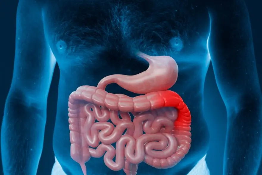
Insights into Ulcerative Colitis (UC): Understanding the Condition and Its Implications
Ulcerative colitis (UC) is more than just a gastrointestinal disorder; it profoundly impacts daily life. In this article, we'll delve deeper into UC, its symptoms, diagnosis, treatment options, and strategies for living well with this condition. Read on to learn more! What is Ulcerative Colitis (UC)? Ulcerative colitis (UC) is a chronic inflammatory condition that affects your large intestine (colon) and rectum. It belongs to a group of conditions known as Inflammatory Bowel Diseases (IBD). Ulcerative colitis is characterised by inflammation and ulceration of the inner lining of your colon and rectum, leading to various gastrointestinal symptoms. What are the Types of Ulcerative Colitis? The main types of ulcerative colitis include: Ulcerative Proctitis: This type of ulcerative colitis involves inflammation limited to your rectum (the lowest part of your large intestine). Left-sided Colitis: In left-sided colitis, the inflammation extends beyond your rectum and involves the left side of the colon (the sigmoid colon and possibly the descending colon). Pancolitis or Total Colitis: This type of ulcerative colitis affects your entire colon, from the rectum to the cecum (the beginning of the colon). You may be at a higher risk of developing complications such as colon cancer. Fulminant Colitis: Fulminant colitis is a severe form of ulcerative colitis characterised by a rapid onset of symptoms, including severe bloody diarrhoea, abdominal pain, dehydration, fever, and rapid heart rate. How Common is Ulcerative Colitis? Ulcerative colitis is more prevalent in developed countries, particularly in North America and Europe, compared to developing regions. Ulcerative colitis has an annual prevalence of 156 to 291 cases per 100,000. What are the Symptoms of Ulcerative Colitis? Common ulcerative colitis symptoms include: Abdominal pain and cramping Persistent diarrhoea with blood or mucus Rectal bleeding Urgency to have bowel movements ( even when bowels are empty: also known as tenesmus) Fatigue Weight loss Loss of appetite Fever Joint pain, skin rashes, or eye inflammation It is worth noting that ulcerative colitis symptoms in females are similar to men. What Causes Ulcerative Colitis? The exact ulcerative colitis causes remain unknown, but it is believed to result from various factors. Potential ulcerative colitis causes include: Ulcerative colitis is considered an autoimmune disease, where your immune system mistakenly attacks healthy cells in your digestive tract. Therefore, ulcerative colitis can be caused due to overaction of your immune system. Alterations in the composition and function of your gut microbiota (the community of microorganisms residing in your gastrointestinal tract) have been implicated in ulcerative colitis pathogenesis. Defects in your intestinal epithelial barrier, which normally protects your intestinal lining from harmful substances and pathogens, can cause ulcerative colitis. While not fully understood, medications (e.g., antibiotics, nonsteroidal anti-inflammatory drugs) and microbial infections may trigger or exacerbate ulcerative colitis symptoms in some cases. What are the Risk Factors of Ulcerative Colitis? Here are some of the risk factors: Having a first-degree relative (parent, sibling, or child) with UC colitis increases the risk of developing the disease given the autoimmune nature of the disease. Multiple genetic variants such as ABCB1, IL10RA, IL10RB and PTPN2 increase the risk ulcerative colitis susceptibility. Ulcerative colitis most commonly develops during late adolescence or early adulthood, with a second peak in incidence occurring between ages 50 and 80. Therefore, increasing age is a risk factor. Factors such as your diet, smoking, stress etc may influence the development and progression of ulcerative colitis. What are the Complications of Ulcerative Colitis? Complications of ulcerative colitis include: Colon cancer Severe inflammation can lead to a dangerous dilation of the colon. Perforated colon Chronic bleeding and inflammation can lead to iron deficiency, causing anaemia Bone thinning Ulcerative colitis may cause primary sclerosing cholangitis or autoimmune hepatitis How is Ulcerative Colitis Diagnosed? Ulcerative colitis diagnosis involves the following: Evaluation of your medical history Physical examination and abdominal palpation Blood tests may be performed to check for signs of inflammation (e.g., elevated C-reactive protein or Erythrocyte Sedimentation Rate - ESR) and to evaluate for anaemia or nutritional deficiencies Stool samples may be analysed for the presence of blood, mucus, or infectious agents Colonoscopy is the gold standard for diagnosing ulcerative colitis. A thin, flexible tube with a camera (colonoscope) is inserted into your rectum and advanced through your colon to examine the lining for signs of inflammation and ulceration In cases where a full colonoscopy is not feasible, flexible sigmoidoscopy may be performed to visualize the lower part of the colon and rectum Upper endoscopy or capsule endoscopy may be performed in cases where ulcerative colitis involvement is suspected beyond the colon and rectum How is Ulcerative Colitis Treated? Common ulcerative colitis treatment approaches include: Medications: Aminosalicylates, Corticosteroids and Immunosuppressants Biologic Therapies: Anti-tumour necrosis factor (TNF) agents (e.g., infliximab, adalimumab), anti-integrin agents (e.g., vedolizumab), or anti-interleukin 12/23 agents (e.g., ustekinumab) are reserved for moderate to severe ulcerative colitis that doesn't respond to other treatments JAK Inhibitors: Janus kinase (JAK) inhibitors (e.g., tofacitinib) may be prescribed for moderate to severe ulcerative colitis therapy in adults Rectal Therapies Rectal 5-aminosalicylate: This can be used to treat inflammation localised in the rectum or lower part of the colon. Corticosteroid Enemas: If you have left-sided ulcerative colitis or proctitis, rectal corticosteroid formulations (e.g., hydrocortisone or budesonide enemas) may be prescribed. Colectomy: Surgical removal of the colon (total colectomy) may be necessary in cases of severe ulcerative colitis. What is the Outlook for Ulcerative Colitis? The outlook for ulcerative colitis varies widely among patients and depends on factors such as disease severity, extent of inflammation, response to ulcerative colitis treatment, and presence of complications. With proper management, many patients with ulcerative colitis can achieve remission and lead fulfilling lives, although the condition may require lifelong monitoring and treatment. Does Ulcerative Colitis Ever Go Away? Ulcerative colitis is a chronic condition with no known cure. While symptoms can be managed with treatment, ulcerative colitis typically follows a relapsing-remitting course, with periods of remission and flare-ups. What Should I Ask My Doctor Related to Ulcerative Colitis? When discussing ulcerative colitis with your doctor, consider asking questions such as: What is the extent and severity of my ulcerative colitis? What are the potential risks and benefits of each treatment? How can I manage ulcerative colitis symptoms? Are there any lifestyle modifications or dietary changes that may help? How often should I follow up with you for monitoring and adjustments to my treatment plan? What are the signs of complications I should watch for? Conclusion Understanding the biology of ulcerative colitis is essential for effectively managing this chronic condition. With proper treatment, lifestyle adjustments, and regular monitoring, one can achieve remission and lead a healthy life despite its challenges. If one is experiencing symptoms suggestive of ulcerative colitis, do not delay and get in touch with Metropolis Healthcare for a comprehensive blood test to evaluate the C- reactive protein levels and ESR. Metropolis Healthcare offers at-home testing by expert phlebotomists with prompt and reliable digital reports at zero extra charges! Book your test today!

Exploring Varicocele: The Symptoms, Diagnosis, And Treatment
What is a varicocele? When the veins within your scrotum, a pouch of skin and muscular tissues that contain the testicles, enlarge, the condition is called varicocele. These veins carry the deoxygenated blood from your testicles. However, when the blood fails to circulate properly and pools in the veins, it leads to varicocele. Can varicoceles affect fertility? A varicocele is likely to affect the development of a testicle and cause low sperm production. These varicocele-related problems may lead to infertility. How common are varicoceles? Varicocele is a common condition. Around 15% of the healthy men population develop varicocele, and nearly 35% with primary infertility have varicocele. What are the symptoms of a varicocele? In most cases, the left side of the scrotum develops varicocele. Although you may not experience any signs or symptoms if you have varicocele, the possible varicocele symptoms may include the following: A dull pain in your testicles or scrotum that feels better when you lie back Scrotal or testicular swelling due to varicocele Varicocele-related testicular atrophy (shrinkage of your testicles) Inability to father a child (infertility) after trying for at least one year Formation of a lump on the varicocele-affected testicle What is the main cause of a varicocele? Read to understand what causes varicocele. There are two testicular arteries on either side of your scrotum. These arteries carry the oxygen-rich blood to the testicles. Likewise, there are two veins in your testicles that transport the deoxygenated blood from your testicles back to the heart. Also, there is a nexus of small veins within your scrotum. It is called the pampiniform plexus. This network takes oxygen-depleted blood from your testicles to the chief testicular vein. The enlargement of the pampiniform plexus is one of the varicocele causes. While the exact varicocele cause is not yet known, when the valves in the testicular vein do not work properly, the blood may pool inside the veins. It leads to swelling of the veins over time, causing varicocele. What are the complications of a varicocele? Varicoceles often hamper your body's ability to regulate your testicular temperature, resulting in toxin build-up and oxidative stress. The following complications are associated with varicocele: Male hypogonadism (low testosterone): Testosterone is a male hormone produced by your testicles. It is responsible for the development of male characteristics when you hit puberty. A low testosterone level may lead to shrinkage of your testicles while reducing your muscle mass and sexual urge and causing depression. It is one of the complications of varicocele. Azoospermia: Larger varicoceles may also cause azoospermia (the absence of sperm in the ejaculated semen). It is likely to cause infertility in males. How is a varicocele diagnosed? Your doctor will diagnose a varicocele by examining your scrotum physically - in a stand-up and lie-down position. When you are standing up, your doctor is likely to ask you to take a deep breath, hold it, and put pressure on your abdomen. This process is called the Valsalva manoeuvre. It makes varicocele diagnosis easy. Your order may ask you to test the following test done: Pelvic ultrasound: This is an imaging test that provides a clear view of the testicular veins and is one of the widely used tests to diagnose varicocele. Semen analysis: This analysis helps your doctor get an idea of your sperm health. Blood test: It is ordered to check the levels of your testosterone and follicle-stimulating hormone (FSH). If you have varicocele, your testosterone levels are usually low. Once a varicocele is diagnosed, your doctor will grade varicocele in the following way: Grade 0 Varicocele - It is when your doctor cannot see or feel a varicocele during the physical assessment but on a pelvic ultrasound plate. Grade I Varicocele: A varicocele is identified as Grade I when your doctor cannot see the enlargement but during the Valsalva manoeuvre test. Grade II Varicocele: It is a varicocele that your doctor can feel even without performing the Valsalva manoeuvre test. However, it still cannot be seen. Grade III Varicocele: This type of varicocele can be easily felt and seen. What is varicocele surgery? A varicocele treatment surgery is also known as varicocelectomy. Your doctor recommends varicocele surgery if your varicocele is severe, painful, or affects your ability to be a father. During this varicocele surgery, a specialised surgeon will operate and trim the affected veins and seal the ends while redirecting blood flow to the healthy scrotal veins. How long does it take to recover from varicocele treatment? Usually, it takes around six weeks to fully recover after a varicocele treatment surgery. What happens if a varicocele is left untreated? Varicocele treatment depends on its grade. If your varicocele is small and does not cause any symptoms, your doctor is unlikely to recommend a treatment. However, if your varicocele is large and causes discomfort, leaving it untreated can lead to permanent damage to your testicles. What is the outlook for varicocele? Varicoceles may not cause any symptoms in most people. Some may feel mild pain or discomfort while performing certain physical activities. However, varicoceles are unlikely to cause long-standing health issues. If you are experiencing fertility issues, get in touch with your doctor to understand the treatment options, including varicocele therapy and varicocele surgery. When to see a doctor? If you are experiencing varicocele symptoms, such as infertility, it is important to consult a doctor at the earliest. They can diagnose varicocele and suggest the most suitable varicocele treatment for you. Additionally, after receiving the varicocele treatment, it is essential to schedule follow-up appointments with your doctor. It will allow them to monitor your health and order any necessary tests to ensure that your varicocele treatment is effective. Conclusion Varicocele is a common health condition that affects people assigned male at birth (AMAB) at various ages and stages of life. Although some may not experience any varicocele symptoms, others may experience mild symptoms of varicocele. The decision to treat a varicocele depends on the grade of varicocele. In most cases, wearing supportive underwear or a jockstrap or using over-the-counter (OTC) pain medication can provide relief from minor symptoms of varicocele.

A Comprehensive Guide to Amnesia
What is amnesia? Amnesia is a condition characterised by partial or complete loss of memory, often caused by brain injury, trauma, psychological factors, or neurological disorders. What are the different types of amnesia? There are two primary types of amnesia. Retrograde amnesia occurs when one is unable to recall memories from their past, while anterograde amnesia is characterised by the inability to form new memories while still retaining old ones. Other types of amnesia are: Post-traumatic amnesia, which emerges following an injury and may encompass various types of memory loss. Transient global amnesia, a temporary condition marked by both anterograde and retrograde amnesia, typically lasting less than 24 hours. Infantile amnesia, a common phenomenon where memories from infancy are rarely recalled in adulthood. Dissociative amnesia, arising from mental health-related factors. What are the symptoms of amnesia? The symptoms of amnesia vary depending on its type and underlying cause. Some common symptoms include: Memory loss Confusion Difficulty recognising familiar faces or places Challenges in learning new information Formation of false memories Trouble recalling names and faces Forgetting directions and navigational skills Missing out on planned future events due to forgetfulness. What causes amnesia? Various regions of the brain play roles in memory function, and damage or disease affecting these areas can impact memory that can cause amnesia. Potential amnesia causes are as follows: Stroke Encephalitis or inflammation of the brain, which may result from viral infections or autoimmune responses Reduced oxygen supply to the brain, possibly due to conditions like heart attacks or respiratory distress Brain tumours Chronic alcohol abuse Certain medications such as benzodiazepines (tranquilisers) Seizures Alzheimer’s disease and other types of dementia Severe head injuries Emotional trauma How is amnesia diagnosed? Diagnosis of amnesia requires a comprehensive evaluation to differentiate it from other causes of memory loss like Alzheimer's disease, dementia, depression, or brain tumours. The evaluation begins with a detailed medical history, often provided by a family member, friend, or caregiver. Questions may cover the type and duration of memory loss, triggers such as head injuries or strokes, family medical history, substance use, and other associated symptoms like confusion or personality changes. A physical examination, including a neurological assessment, checks reflexes, sensory function, and balance. Cognitive tests assess various aspects of thinking, judgement, and memory, including general knowledge, personal information, and the ability to recall words or events. The results of the evaluation help determine the severity of memory loss and guide recommendations for treatment and support. Timely diagnosis helps in better and effective amnesia treatment among the patients. What tests will be done to diagnose amnesia? Diagnostic tests that your healthcare provider may ask you to start the amnesia treatment are: Imaging tests such as MRI and CT scans to assess for brain damage or structural changes. Blood tests to screen for infections, nutritional deficiencies, or other underlying issues. Electroencephalogram (EEG) to detect any abnormal seizure activity in the brain. How amnesia is treated/is there a cure? Permanent amnesia differs from temporary memory loss episodes, and while there's no specific treatment for amnesia itself, addressing the underlying cause can be beneficial. Amnesia treatment options are as follows: Occupational therapy: Assists in learning new information and compensating for lost memories. Technological aids: Smart devices like smartphones or tablets can aid memory recall and organisation. Medications or supplements: Healthcare providers may prescribe medications to support cognitive functions and memory retention. Detoxification: Resolving chemically induced amnesia may involve detox programs. Rest: Resting is crucial for recovery, especially in cases of amnesia caused by concussion. Abstinence: Ceasing alcohol consumption can aid recovery from alcohol-induced amnesia. Emotional support: Counselling and emotional support are essential for individuals dealing with alcoholism and its associated memory issues. Dietary adjustments: Addressing nutritional deficiencies through dietary changes can support recovery from alcohol-induced amnesia. Can amnesia be prevented? You can prevent amnesia by being careful: Limit alcohol and drug consumption to avoid potential memory impairments. Wear protective headgear during high-risk sports activities to prevent head injuries. Always use seat belts while travelling in vehicles to minimise the risk of head trauma. Promptly treat infections to prevent them from spreading to the brain. Older individuals, undergo annual eye examinations and consult with healthcare providers or pharmacists about medications that may cause dizziness, reducing the risk of falls. Engage in lifelong mental stimulation through activities such as taking classes, travelling to new places, reading, and playing mentally challenging games. Maintain regular physical activity to support overall brain health. Follow a heart-healthy diet rich in fruits, vegetables, whole grains, and lean proteins to lower the risk of strokes and cardiovascular issues that can lead to memory problems. Ensure adequate hydration, as even mild dehydration can negatively impact brain function, particularly in women. What can I expect if I have amnesia? If you have amnesia, you can experience: Memory loss, either partial or complete. Confusion about past events or people. Difficulty forming new memories. Challenges with daily tasks due to memory impairment. Potential emotional distress or frustration. What are the risk factors for amnesia? The risk factors for amnesia include: Brain surgery, head injury, or trauma Stroke Alcohol abuse Seizures Migraine history Cardiovascular disease risk factors like high blood pressure or high cholesterol Emotional stress Chronic stress Ageing Chronic sleep deprivation Medications Prolonged alcohol use Chemotherapy Dialysis Extreme dieting Severe malnutrition Genetic factors What are the complications of amnesia? Even mild amnesia can significantly impact one's quality of life, making it difficult to carry out daily tasks and engage in social activities due to memory difficulties. In certain instances, lost memories may be irretrievable. Individuals with severe amnesia may need constant supervision. Conclusion In conclusion, amnesia is a complex condition characterised by memory loss, which can stem from various causes such as brain injury, trauma, or neurological disorders. While there is no cure for amnesia, proper diagnosis and treatment can help manage symptoms and improve quality of life. Regular check-ups and diagnostic tests play a crucial role in the early detection and management of amnesia. For convenient and reliable healthcare services, contact us at Metropolis Healthcare/Labs. We offer an at-home testing facility and you can access your reports online. Schedule your appointment with Metropolis for your medical test to cure your health related problems in time.


 WhatsApp