Latest Blogs
Patent Ductus Arteriosus (PDA): Symptoms, Causes, and Effective Treatment
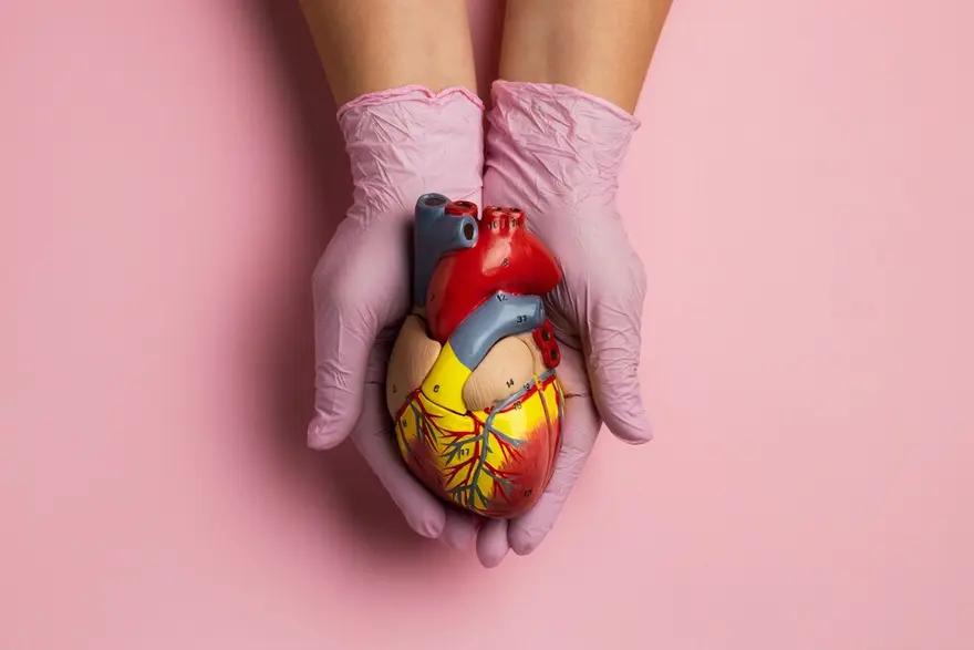
What is patent ductus arteriosus (PDA)? A congenital heart defect, Patent Ductus Arteriosus (PDA) is a lasting opening between the heart's major blood vessels. During foetal development, a ductus arteriosus naturally exists but typically closes after birth. If this persists, it becomes a patent ductus arteriosus. What does the ductus arteriosus do? While a minor patent ductus arteriosus may be benign, an untreated large one can lead to the improper flow of oxygen-poor blood. This misdirection poses risks, potentially weakening the heart muscle and triggering complications like heart failure. What happens in babies with patent ductus arteriosus? Babies with patent ductus arteriosus (PDA) have an ongoing connection between major heart vessels, causing excess blood flow away from the lungs. This condition arises when the fetal ductus arteriosus fails to close post-birth. Symptoms in each infant vary based on the opening's size and may include rapid breathing, poor weight gain, and feeding difficulties. Who does PDA affect? PDA predominates among premature infants and afflicts twice as many girls as boys. It's prevalent in babies with neonatal respiratory distress syndrome, genetic disorders like Down syndrome, and those whose mothers contracted rubella (German measles) during pregnancy. How common is PDA? Patent Ductus Arteriosus (PDA) is a common heart condition in newborns, making up 5–10% of congenital heart diseases in term infants. It is more prevalent among premature infants and is twice as common in girls compared to boys. How does PDA affect my baby? The impact of Patent Ductus Arteriosus (PDA) on your baby involves abnormal blood flow between the heart's major vessels. Although a small PDA may not pose immediate problems, a larger one can lead to improper circulation of oxygen-poor blood, potentially weakening the heart muscle and causing complications such as heart failure. What causes patent ductus arteriosus? The causes of Patent Ductus Arteriosus (PDA) remain unknown, with notable associations: Premature infants may have underdeveloped muscles in the ductus arteriosus, hindering natural closure. Genetic abnormalities, such as Down Syndrome, are linked to PDA. In premature births: Muscular underdevelopment in the ductus arteriosus can prevent self-closure. Mothers with German measles during pregnancy are more likely to have babies with heart defects, including PDA. What are patent ductus arteriosus symptoms? Symptoms of patent ductus arteriosus vary based on the type: Small PDAs may be asymptomatic, except for the presence of a heart murmur. Larger PDAs may manifest as: Rapid breathing. Shortness of breath (dyspnea). Sweating during feedings. Fatigue or tiredness. Feeding and eating difficulties. Poor weight gain or growth. Fast pulse or heart rate. How do healthcare providers diagnose PDA? Doctors diagnose patent ductus arteriosus (PDA) through various methods. They typically begin with a physical examination, listening for characteristic heart murmurs and reports from echocardiography and chest x-ray reports. In certain cases, cardiac catheterization provides direct visualisation of the ductus arteriosus and evaluates its size and blood flow patterns. What tests do providers use to diagnose patent ductus arteriosus? Healthcare providers diagnose Patent Ductus Arteriosus (PDA) through a multi-step approach, including: Auscultation: Listening for a heart murmur using a stethoscope. Pulse and Blood Pressure Evaluation: Checking pulse rate and blood pressure. Clinical Assessment: Examining for signs of lung congestion and conducting a thorough physical examination. Echocardiogram: Echocardiogram helps to diagnose the problem by checking the heart's structure and blood flow. Additional Tests: Depending on the situation, health providers may ask you to get supplementary tests done like chest X-rays, electrocardiograms (EKG), and blood tests to get more comprehensive diagnostic information. Do healthcare providers diagnose PDA in adults? In adults, healthcare providers sometimes find patent ductus arteriosus (PDA). It's not common but can be discovered during regular check-ups or echocardiography tests. A distinct continuous murmur often signals the presence of PDA, heard at the upper left side of the chest. How do healthcare providers treat PDA? PDA is treated by the following ways: Monitoring: Small PDAs may be regularly observed without immediate intervention. Medications: Indomethacin or ibuprofen might be prescribed to help ductus arteriosus closure. Surgery: Larger or persistent PDAs may require surgical intervention, such as tying off the ductus arteriosus or employing other surgical techniques to close the opening. Catheterization: Minimally invasive procedures like catheterization may be utilised to place a device that closes the PDA. What medications do providers use to treat PDA? Healthcare providers may employ medications to address Patent Ductus Arteriosus (PDA), with common choices being: Indomethacin: A nonsteroidal anti-inflammatory drug (NSAID) that aids ductus arteriosus closure by reducing prostaglandin production. Ibuprofen: Another NSAID, similar to indomethacin, used to stimulate the closure of the ductus arteriosus. Prostaglandins may be used to maintain ductus arteriosus patency until surgical ligation is performed. Intravenous (IV) indomethacin or IV ibuprofen is used for treating PDA in neonates and premature infants. What are other types of patent ductus arteriosus treatment? Healthy Lifestyle: Maintaining overall cardiovascular health through a healthy diet and avoiding smoking is beneficial. Premature Babies: NSAIDs may be administered to aid ductus closure in premature infants with PDA. Children and Adults: Surgery or procedures may be necessary for larger or problematic PDAs. Monitoring and Checkups: Regular health checkups are crucial for early detection of complications. Are there other complications from PDA? If left untreated, Patent Ductus Arteriosus (PDA) can result in various complications, including: Heart Failure Endocarditis Pulmonary Edema Pulmonary Hypertension Blood Vessel Damage Other potential complications involve: Injury to Recurrent Laryngeal Nerve Occlusion of Descending Aorta Injury to Pulmonary Artery and Phrenic Nerve Heart Murmur Lung Infections (Pneumonia) Difficulty Feeding and Poor Growth Are there conditions that put my baby at higher risk for PDA? Factors that can increase the risk of Patent Ductus Arteriosus (PDA) in infants include premature birth, specific genetic conditions, a family history of congenital heart issues, foetal distress during pregnancy, maternal or foetal infections (such as rubella), and pregnancy-related risks like smoking or certain medications. What can I expect if my baby has a PDA? Kids with an open PDA face an increased risk of bacterial endocarditis, a heart infection. Antibiotics are necessary before dental procedures to prevent this. With prompt and suitable treatment, the outlook for infants with PDA is usually positive. Is patent ductus arteriosus curable? Yes, patent Ductus Arteriosus (PDA) is typically manageable through medical or surgical interventions to close the opening or address complications. Small PDAs may close on their own, especially in premature infants How do I take care of my baby with patent ductus arteriosus? You can take care of your baby by taking the following steps; Medical Monitoring Follow Treatment Plan Antibiotic Prophylaxis Healthy Lifestyle Regular Follow-ups Symptom Observation When to see a doctor? If your child experiences a fever, chills, cough, or congestion sounds, or if you notice redness, swelling, or pus draining from the incision, it's essential to contact your doctor promptly. Conclusion In conclusion, Patent Ductus Arteriosus (PDA) is a manageable condition with various treatment options available, including medical intervention and surgical procedures. Timely diagnosis, regular monitoring, and adherence to prescribed treatments contribute to positive outcomes. Visit us at Metropolis Labs and get yourself tested to get accurate test results and reliable health check-up services. You can also book a test from home, where the experts will collect the sample by coming to your home, without stepping out of your home.
Insights into Ulcerative Colitis (UC): Understanding the Condition and Its Implications
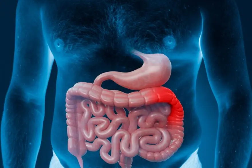
Ulcerative colitis (UC) is more than just a gastrointestinal disorder; it profoundly impacts daily life. In this article, we'll delve deeper into UC, its symptoms, diagnosis, treatment options, and strategies for living well with this condition. Read on to learn more! What is Ulcerative Colitis (UC)? Ulcerative colitis (UC) is a chronic inflammatory condition that affects your large intestine (colon) and rectum. It belongs to a group of conditions known as Inflammatory Bowel Diseases (IBD). Ulcerative colitis is characterised by inflammation and ulceration of the inner lining of your colon and rectum, leading to various gastrointestinal symptoms. What are the Types of Ulcerative Colitis? The main types of ulcerative colitis include: Ulcerative Proctitis: This type of ulcerative colitis involves inflammation limited to your rectum (the lowest part of your large intestine). Left-sided Colitis: In left-sided colitis, the inflammation extends beyond your rectum and involves the left side of the colon (the sigmoid colon and possibly the descending colon). Pancolitis or Total Colitis: This type of ulcerative colitis affects your entire colon, from the rectum to the cecum (the beginning of the colon). You may be at a higher risk of developing complications such as colon cancer. Fulminant Colitis: Fulminant colitis is a severe form of ulcerative colitis characterised by a rapid onset of symptoms, including severe bloody diarrhoea, abdominal pain, dehydration, fever, and rapid heart rate. How Common is Ulcerative Colitis? Ulcerative colitis is more prevalent in developed countries, particularly in North America and Europe, compared to developing regions. Ulcerative colitis has an annual prevalence of 156 to 291 cases per 100,000. What are the Symptoms of Ulcerative Colitis? Common ulcerative colitis symptoms include: Abdominal pain and cramping Persistent diarrhoea with blood or mucus Rectal bleeding Urgency to have bowel movements ( even when bowels are empty: also known as tenesmus) Fatigue Weight loss Loss of appetite Fever Joint pain, skin rashes, or eye inflammation It is worth noting that ulcerative colitis symptoms in females are similar to men. What Causes Ulcerative Colitis? The exact ulcerative colitis causes remain unknown, but it is believed to result from various factors. Potential ulcerative colitis causes include: Ulcerative colitis is considered an autoimmune disease, where your immune system mistakenly attacks healthy cells in your digestive tract. Therefore, ulcerative colitis can be caused due to overaction of your immune system. Alterations in the composition and function of your gut microbiota (the community of microorganisms residing in your gastrointestinal tract) have been implicated in ulcerative colitis pathogenesis. Defects in your intestinal epithelial barrier, which normally protects your intestinal lining from harmful substances and pathogens, can cause ulcerative colitis. While not fully understood, medications (e.g., antibiotics, nonsteroidal anti-inflammatory drugs) and microbial infections may trigger or exacerbate ulcerative colitis symptoms in some cases. What are the Risk Factors of Ulcerative Colitis? Here are some of the risk factors: Having a first-degree relative (parent, sibling, or child) with UC colitis increases the risk of developing the disease given the autoimmune nature of the disease. Multiple genetic variants such as ABCB1, IL10RA, IL10RB and PTPN2 increase the risk ulcerative colitis susceptibility. Ulcerative colitis most commonly develops during late adolescence or early adulthood, with a second peak in incidence occurring between ages 50 and 80. Therefore, increasing age is a risk factor. Factors such as your diet, smoking, stress etc may influence the development and progression of ulcerative colitis. What are the Complications of Ulcerative Colitis? Complications of ulcerative colitis include: Colon cancer Severe inflammation can lead to a dangerous dilation of the colon. Perforated colon Chronic bleeding and inflammation can lead to iron deficiency, causing anaemia Bone thinning Ulcerative colitis may cause primary sclerosing cholangitis or autoimmune hepatitis How is Ulcerative Colitis Diagnosed? Ulcerative colitis diagnosis involves the following: Evaluation of your medical history Physical examination and abdominal palpation Blood tests may be performed to check for signs of inflammation (e.g., elevated C-reactive protein or Erythrocyte Sedimentation Rate - ESR) and to evaluate for anaemia or nutritional deficiencies Stool samples may be analysed for the presence of blood, mucus, or infectious agents Colonoscopy is the gold standard for diagnosing ulcerative colitis. A thin, flexible tube with a camera (colonoscope) is inserted into your rectum and advanced through your colon to examine the lining for signs of inflammation and ulceration In cases where a full colonoscopy is not feasible, flexible sigmoidoscopy may be performed to visualize the lower part of the colon and rectum Upper endoscopy or capsule endoscopy may be performed in cases where ulcerative colitis involvement is suspected beyond the colon and rectum How is Ulcerative Colitis Treated? Common ulcerative colitis treatment approaches include: Medications: Aminosalicylates, Corticosteroids and Immunosuppressants Biologic Therapies: Anti-tumour necrosis factor (TNF) agents (e.g., infliximab, adalimumab), anti-integrin agents (e.g., vedolizumab), or anti-interleukin 12/23 agents (e.g., ustekinumab) are reserved for moderate to severe ulcerative colitis that doesn't respond to other treatments JAK Inhibitors: Janus kinase (JAK) inhibitors (e.g., tofacitinib) may be prescribed for moderate to severe ulcerative colitis therapy in adults Rectal Therapies Rectal 5-aminosalicylate: This can be used to treat inflammation localised in the rectum or lower part of the colon. Corticosteroid Enemas: If you have left-sided ulcerative colitis or proctitis, rectal corticosteroid formulations (e.g., hydrocortisone or budesonide enemas) may be prescribed. Colectomy: Surgical removal of the colon (total colectomy) may be necessary in cases of severe ulcerative colitis. What is the Outlook for Ulcerative Colitis? The outlook for ulcerative colitis varies widely among patients and depends on factors such as disease severity, extent of inflammation, response to ulcerative colitis treatment, and presence of complications. With proper management, many patients with ulcerative colitis can achieve remission and lead fulfilling lives, although the condition may require lifelong monitoring and treatment. Does Ulcerative Colitis Ever Go Away? Ulcerative colitis is a chronic condition with no known cure. While symptoms can be managed with treatment, ulcerative colitis typically follows a relapsing-remitting course, with periods of remission and flare-ups. What Should I Ask My Doctor Related to Ulcerative Colitis? When discussing ulcerative colitis with your doctor, consider asking questions such as: What is the extent and severity of my ulcerative colitis? What are the potential risks and benefits of each treatment? How can I manage ulcerative colitis symptoms? Are there any lifestyle modifications or dietary changes that may help? How often should I follow up with you for monitoring and adjustments to my treatment plan? What are the signs of complications I should watch for? Conclusion Understanding the biology of ulcerative colitis is essential for effectively managing this chronic condition. With proper treatment, lifestyle adjustments, and regular monitoring, one can achieve remission and lead a healthy life despite its challenges. If one is experiencing symptoms suggestive of ulcerative colitis, do not delay and get in touch with Metropolis Healthcare for a comprehensive blood test to evaluate the C- reactive protein levels and ESR. Metropolis Healthcare offers at-home testing by expert phlebotomists with prompt and reliable digital reports at zero extra charges! Book your test today!
Exploring Varicocele: The Symptoms, Diagnosis, And Treatment
What is a varicocele? When the veins within your scrotum, a pouch of skin and muscular tissues that contain the testicles, enlarge, the condition is called varicocele. These veins carry the deoxygenated blood from your testicles. However, when the blood fails to circulate properly and pools in the veins, it leads to varicocele. Can varicoceles affect fertility? A varicocele is likely to affect the development of a testicle and cause low sperm production. These varicocele-related problems may lead to infertility. How common are varicoceles? Varicocele is a common condition. Around 15% of the healthy men population develop varicocele, and nearly 35% with primary infertility have varicocele. What are the symptoms of a varicocele? In most cases, the left side of the scrotum develops varicocele. Although you may not experience any signs or symptoms if you have varicocele, the possible varicocele symptoms may include the following: A dull pain in your testicles or scrotum that feels better when you lie back Scrotal or testicular swelling due to varicocele Varicocele-related testicular atrophy (shrinkage of your testicles) Inability to father a child (infertility) after trying for at least one year Formation of a lump on the varicocele-affected testicle What is the main cause of a varicocele? Read to understand what causes varicocele. There are two testicular arteries on either side of your scrotum. These arteries carry the oxygen-rich blood to the testicles. Likewise, there are two veins in your testicles that transport the deoxygenated blood from your testicles back to the heart. Also, there is a nexus of small veins within your scrotum. It is called the pampiniform plexus. This network takes oxygen-depleted blood from your testicles to the chief testicular vein. The enlargement of the pampiniform plexus is one of the varicocele causes. While the exact varicocele cause is not yet known, when the valves in the testicular vein do not work properly, the blood may pool inside the veins. It leads to swelling of the veins over time, causing varicocele. What are the complications of a varicocele? Varicoceles often hamper your body's ability to regulate your testicular temperature, resulting in toxin build-up and oxidative stress. The following complications are associated with varicocele: Male hypogonadism (low testosterone): Testosterone is a male hormone produced by your testicles. It is responsible for the development of male characteristics when you hit puberty. A low testosterone level may lead to shrinkage of your testicles while reducing your muscle mass and sexual urge and causing depression. It is one of the complications of varicocele. Azoospermia: Larger varicoceles may also cause azoospermia (the absence of sperm in the ejaculated semen). It is likely to cause infertility in males. How is a varicocele diagnosed? Your doctor will diagnose a varicocele by examining your scrotum physically - in a stand-up and lie-down position. When you are standing up, your doctor is likely to ask you to take a deep breath, hold it, and put pressure on your abdomen. This process is called the Valsalva manoeuvre. It makes varicocele diagnosis easy. Your order may ask you to test the following test done: Pelvic ultrasound: This is an imaging test that provides a clear view of the testicular veins and is one of the widely used tests to diagnose varicocele. Semen analysis: This analysis helps your doctor get an idea of your sperm health. Blood test: It is ordered to check the levels of your testosterone and follicle-stimulating hormone (FSH). If you have varicocele, your testosterone levels are usually low. Once a varicocele is diagnosed, your doctor will grade varicocele in the following way: Grade 0 Varicocele - It is when your doctor cannot see or feel a varicocele during the physical assessment but on a pelvic ultrasound plate. Grade I Varicocele: A varicocele is identified as Grade I when your doctor cannot see the enlargement but during the Valsalva manoeuvre test. Grade II Varicocele: It is a varicocele that your doctor can feel even without performing the Valsalva manoeuvre test. However, it still cannot be seen. Grade III Varicocele: This type of varicocele can be easily felt and seen. What is varicocele surgery? A varicocele treatment surgery is also known as varicocelectomy. Your doctor recommends varicocele surgery if your varicocele is severe, painful, or affects your ability to be a father. During this varicocele surgery, a specialised surgeon will operate and trim the affected veins and seal the ends while redirecting blood flow to the healthy scrotal veins. How long does it take to recover from varicocele treatment? Usually, it takes around six weeks to fully recover after a varicocele treatment surgery. What happens if a varicocele is left untreated? Varicocele treatment depends on its grade. If your varicocele is small and does not cause any symptoms, your doctor is unlikely to recommend a treatment. However, if your varicocele is large and causes discomfort, leaving it untreated can lead to permanent damage to your testicles. What is the outlook for varicocele? Varicoceles may not cause any symptoms in most people. Some may feel mild pain or discomfort while performing certain physical activities. However, varicoceles are unlikely to cause long-standing health issues. If you are experiencing fertility issues, get in touch with your doctor to understand the treatment options, including varicocele therapy and varicocele surgery. When to see a doctor? If you are experiencing varicocele symptoms, such as infertility, it is important to consult a doctor at the earliest. They can diagnose varicocele and suggest the most suitable varicocele treatment for you. Additionally, after receiving the varicocele treatment, it is essential to schedule follow-up appointments with your doctor. It will allow them to monitor your health and order any necessary tests to ensure that your varicocele treatment is effective. Conclusion Varicocele is a common health condition that affects people assigned male at birth (AMAB) at various ages and stages of life. Although some may not experience any varicocele symptoms, others may experience mild symptoms of varicocele. The decision to treat a varicocele depends on the grade of varicocele. In most cases, wearing supportive underwear or a jockstrap or using over-the-counter (OTC) pain medication can provide relief from minor symptoms of varicocele.
A Comprehensive Guide to Amnesia
What is amnesia? Amnesia is a condition characterised by partial or complete loss of memory, often caused by brain injury, trauma, psychological factors, or neurological disorders. What are the different types of amnesia? There are two primary types of amnesia. Retrograde amnesia occurs when one is unable to recall memories from their past, while anterograde amnesia is characterised by the inability to form new memories while still retaining old ones. Other types of amnesia are: Post-traumatic amnesia, which emerges following an injury and may encompass various types of memory loss. Transient global amnesia, a temporary condition marked by both anterograde and retrograde amnesia, typically lasting less than 24 hours. Infantile amnesia, a common phenomenon where memories from infancy are rarely recalled in adulthood. Dissociative amnesia, arising from mental health-related factors. What are the symptoms of amnesia? The symptoms of amnesia vary depending on its type and underlying cause. Some common symptoms include: Memory loss Confusion Difficulty recognising familiar faces or places Challenges in learning new information Formation of false memories Trouble recalling names and faces Forgetting directions and navigational skills Missing out on planned future events due to forgetfulness. What causes amnesia? Various regions of the brain play roles in memory function, and damage or disease affecting these areas can impact memory that can cause amnesia. Potential amnesia causes are as follows: Stroke Encephalitis or inflammation of the brain, which may result from viral infections or autoimmune responses Reduced oxygen supply to the brain, possibly due to conditions like heart attacks or respiratory distress Brain tumours Chronic alcohol abuse Certain medications such as benzodiazepines (tranquilisers) Seizures Alzheimer’s disease and other types of dementia Severe head injuries Emotional trauma How is amnesia diagnosed? Diagnosis of amnesia requires a comprehensive evaluation to differentiate it from other causes of memory loss like Alzheimer's disease, dementia, depression, or brain tumours. The evaluation begins with a detailed medical history, often provided by a family member, friend, or caregiver. Questions may cover the type and duration of memory loss, triggers such as head injuries or strokes, family medical history, substance use, and other associated symptoms like confusion or personality changes. A physical examination, including a neurological assessment, checks reflexes, sensory function, and balance. Cognitive tests assess various aspects of thinking, judgement, and memory, including general knowledge, personal information, and the ability to recall words or events. The results of the evaluation help determine the severity of memory loss and guide recommendations for treatment and support. Timely diagnosis helps in better and effective amnesia treatment among the patients. What tests will be done to diagnose amnesia? Diagnostic tests that your healthcare provider may ask you to start the amnesia treatment are: Imaging tests such as MRI and CT scans to assess for brain damage or structural changes. Blood tests to screen for infections, nutritional deficiencies, or other underlying issues. Electroencephalogram (EEG) to detect any abnormal seizure activity in the brain. How amnesia is treated/is there a cure? Permanent amnesia differs from temporary memory loss episodes, and while there's no specific treatment for amnesia itself, addressing the underlying cause can be beneficial. Amnesia treatment options are as follows: Occupational therapy: Assists in learning new information and compensating for lost memories. Technological aids: Smart devices like smartphones or tablets can aid memory recall and organisation. Medications or supplements: Healthcare providers may prescribe medications to support cognitive functions and memory retention. Detoxification: Resolving chemically induced amnesia may involve detox programs. Rest: Resting is crucial for recovery, especially in cases of amnesia caused by concussion. Abstinence: Ceasing alcohol consumption can aid recovery from alcohol-induced amnesia. Emotional support: Counselling and emotional support are essential for individuals dealing with alcoholism and its associated memory issues. Dietary adjustments: Addressing nutritional deficiencies through dietary changes can support recovery from alcohol-induced amnesia. Can amnesia be prevented? You can prevent amnesia by being careful: Limit alcohol and drug consumption to avoid potential memory impairments. Wear protective headgear during high-risk sports activities to prevent head injuries. Always use seat belts while travelling in vehicles to minimise the risk of head trauma. Promptly treat infections to prevent them from spreading to the brain. Older individuals, undergo annual eye examinations and consult with healthcare providers or pharmacists about medications that may cause dizziness, reducing the risk of falls. Engage in lifelong mental stimulation through activities such as taking classes, travelling to new places, reading, and playing mentally challenging games. Maintain regular physical activity to support overall brain health. Follow a heart-healthy diet rich in fruits, vegetables, whole grains, and lean proteins to lower the risk of strokes and cardiovascular issues that can lead to memory problems. Ensure adequate hydration, as even mild dehydration can negatively impact brain function, particularly in women. What can I expect if I have amnesia? If you have amnesia, you can experience: Memory loss, either partial or complete. Confusion about past events or people. Difficulty forming new memories. Challenges with daily tasks due to memory impairment. Potential emotional distress or frustration. What are the risk factors for amnesia? The risk factors for amnesia include: Brain surgery, head injury, or trauma Stroke Alcohol abuse Seizures Migraine history Cardiovascular disease risk factors like high blood pressure or high cholesterol Emotional stress Chronic stress Ageing Chronic sleep deprivation Medications Prolonged alcohol use Chemotherapy Dialysis Extreme dieting Severe malnutrition Genetic factors What are the complications of amnesia? Even mild amnesia can significantly impact one's quality of life, making it difficult to carry out daily tasks and engage in social activities due to memory difficulties. In certain instances, lost memories may be irretrievable. Individuals with severe amnesia may need constant supervision. Conclusion In conclusion, amnesia is a complex condition characterised by memory loss, which can stem from various causes such as brain injury, trauma, or neurological disorders. While there is no cure for amnesia, proper diagnosis and treatment can help manage symptoms and improve quality of life. Regular check-ups and diagnostic tests play a crucial role in the early detection and management of amnesia. For convenient and reliable healthcare services, contact us at Metropolis Healthcare/Labs. We offer an at-home testing facility and you can access your reports online. Schedule your appointment with Metropolis for your medical test to cure your health related problems in time.
Understanding Brain Hemorrhage: Symptoms, Causes, Treatment, Types
What is brain haemorrhage? Brain haemorrhage, also known as intracranial haemorrhage, refers to bleeding that occurs within or around the brain tissue. What happens during a brain haemorrhage? When a brain haemorrhage happens, it interrupts the oxygen supply to the brain and results in blood leaking into the space between the skull and the brain. This blood accumulation forms pools within the skull, exerting pressure that obstructs the flow of oxygen to the brain tissues. What causes bleeding in the brain? Intracranial haemorrhage or brain haemorrhage causes include bleeding within or surrounding the brain. This can frequently be attributed to head trauma, such as that sustained in car accidents, falls, sports-related injuries, or bicycle accidents. Additional factors contributing to brain haemorrhages include: Usage of blood thinners or aspirin, which heightens the risk even with minor head injuries. Untreated high blood pressure leads to the weakening of blood vessel walls. Presence of brain tumours with thin-walled, fragile vessels prone to rupture and bleeding. Aneurysms are characterised by weakened spots in blood vessel walls that swell. Arteriovenous malformations (AVMs), where abnormal connections between arteries and veins weaken blood vessels in and around the brain. Cerebral amyloid angiopathy is a condition often observed in older adults with hypertension, causing irregularities in blood vessels. Various diseases may trigger spontaneous blood leakage into the brain. What are the symptoms of brain bleeding? Brain haemorrhage symptoms may emerge shortly after a head injury or gradually over time. These symptoms include: A sudden, severe headache, often described as a "thunderclap" headache Weakness, numbness, or tingling, typically affecting one side of the body Nausea or vomiting, potentially indicating a brain bleed Dizziness, which can also be a sign of a brain bleed Confusion Difficulty speaking or understanding speech Loss of movement on the side of the body opposite to the head injury Drowsiness and progressive loss of consciousness, suggestive of an intracranial hematoma Unequal pupil size, a potential indicator of an intracranial hematoma Slurred speech, which may signify an intracranial hematoma Fatigue and sleepiness, which could also be symptoms of a brain bleed What are the types of brain bleeds? Bleeding in the brain can occur either inside the brain tissue or outside it, involving the protective layers covering the brain. Epidural bleed: Blood accumulates between the skull and the outer layer known as the dura mater. If left untreated, it can result in increased blood pressure, breathing difficulties, brain damage, or death. Typically caused by injury, often associated with a skull fracture. Subdural bleed: Blood leakage happens between the dura mater and the thin layer beneath it called the arachnoid mater. There are two types: Acute subdural bleed: Develops rapidly and has a high death rate, usually following head trauma from falls, car accidents, sports injuries, whiplash, or similar incidents. Chronic subdural bleed: Forms gradually and is less deadly, often caused by mild head injuries in elderly individuals, those on blood thinning medications, or individuals with brain shrinkage due to dementia or alcohol use disorder. Subarachnoid bleed: Blood collects below the arachnoid mater and above the delicate inner layer, the pia mater. If untreated, it can lead to permanent brain damage and death, often caused by brain aneurysms or other vascular issues. A sudden, severe headache is a common warning sign. Intracerebral haemorrhage: Blood pools within the brain tissue, usually resulting from untreated high blood pressure. It is the second most common cause of stroke and the deadliest. How is a brain haemorrhage treated? Brain haemorrhage treatment initiates with supporting the body's functions, focusing on: Ensuring an open airway for breathing and adequate oxygen intake. Supporting blood circulation and maintaining blood pressure. Monitoring intracranial pressure, which can dangerously increase due to bleeding. Surgical intervention may be necessary for the treatment of brain haemorrhage, aiming to achieve one or more of the following: Halting the bleeding. Alleviating pressure within the skull. Repairing the affected area. Depending on the location and severity of the haemorrhage, surgery may involve minimally invasive techniques or craniotomy. Medications play a crucial role in brain haemorrhage treatment and may include: Blood pressure medications to reduce hypertension. Antiseizure medications to prevent or stop seizures. Symptomatic relief medications such as pain and anti-nausea drugs. Reversal agents for blood thinners. Medications to regulate blood sugar levels. Corticosteroids to decrease swelling. Rehabilitation post-treatment is vital for recovery and may encompass: Physical therapy. Occupational therapy. Speech and language therapy. Psychotherapy is tailored to individual needs. What are the complications of brain haemorrhage? Depending on the haemorrhage's location and resulting damage, certain complications may become permanent, such as: Paralysis Numbness or weakness in specific body regions Difficulty swallowing (dysphagia) Vision loss Impaired speech comprehension or expression Confusion or memory deficits Personality changes or emotional disturbances Can brain haemorrhages be prevented? Yes, brain haemorrhage can be prevented by the following: Manage high blood pressure Prevent head trauma Consider corrective surgery Exercise caution with anticoagulant drugs Maintain a nutritious diet Conclusion In conclusion, understanding the risks, symptoms, and preventative measures for brain haemorrhage is crucial for maintaining overall health and well-being. By managing brain haemorrhage symptoms like high blood pressure, avoiding head trauma, considering corrective surgeries, exercising caution with anticoagulant drugs, and maintaining a nutritious diet, you can keep this disease away. Metropolis Labs provides a convenient and reliable solution for at-home testing and we also have our labs pan India.
Understanding Gangrene: Symptoms, Causes, Treatment & Types
Gangrene is a serious medical condition characterised by tissue death with a high mortality rate. It can affect any part of your body, leading to severe complications if left untreated. So, let's know how you prevent gangrene and what exactly causes gangrene. What is Gangrene? Gangrene is a condition which occurs when tissues in your body die due to a lack of blood supply, a condition known as ischemia. Without adequate blood supply, tissues cannot survive, resulting in gangrene. What are the Types of Gangrene? There are several gangrene types. They are: Dry Gangrene: Typically occurs when blood flow to the affected area is obstructed without significant bacterial infection. The affected tissue becomes dry, shrinks, and darkens in colour. Wet Gangrene: This gangrene type occurs when there is a bacterial infection alongside the lack of blood supply. It results in swelling, blistering, and tissue decay, with a foul odour due to bacterial decomposition. Gas Gangrene: Caused by bacterial infection with organisms such as Clostridium perfringens, gas gangrene leads to the release of gases within tissues, causing swelling, discolouration, and severe pain. Due to this gas accumulation, the affected area may produce a crackling sensation upon palpation. Internal Gangrene: Unlike the other gangrene types, internal gangrene affects internal organs or tissues. It can occur due to conditions such as bowel obstruction, strangulated hernia, or arterial occlusion, leading to tissue death within the affected organ. Fournier's Gangrene: It is a rare but severe form of gangrene that affects your genital and perineal regions. What are the Symptoms of Gangrene? Gangrene symptoms vary depending on the type, severity and gangrene causes. Common gangrene symptoms include: Discolouration of the affected area, ranging from pale to blue, black, or bronze Severe pain or numbness in the affected area Swelling, blisters, or a foul-smelling discharge Ulcers or sores that fail to heal Skin that appears shiny and stretched Systemic gangrene symptoms include fever, rapid heart rate, and confusion in severe cases How Do You Diagnose Gangrene? Diagnosing gangrene typically involves a combination of a medical history review, physical examination, and diagnostic tests. Your doctor will assess gangrene signs such as skin discolouration, pain, and tissue changes. He may also evaluate your medical history, including any underlying conditions like diabetes or vascular diseases. Diagnostic tests may include imaging studies, blood tests, and bacterial culture tests to check for infection in the blood. Who’s at Risk for Developing Gangrene? Several factors increase the risk of developing gangrene: If you have conditions affecting blood flow, such as diabetes, peripheral artery disease, or atherosclerosis, you will be at higher risk of gangrene due to compromised circulation. Injuries due to major accidents that cause severe tissue damage can also lead to gangrene. People with weakened immune systems, such as those undergoing chemotherapy or with HIV/AIDS, are more susceptible to infections that can result in gangrene. Additionally, if you have a history of smoking, obesity, or chronic health conditions like kidney disease or hypertension, you may have an elevated risk of gangrene. How Does a Person Get Gangrene? Usually, severe injury from accidents or surgical complications creates open wounds through which bacteria can enter the body. If these bacteria infect tissues and are not treated promptly, gangrene may develop, leading to oxygen and nutrient deprivation of the tissues and, eventually, causing tissue death. What Tests Diagnose Gangrene? Some common tests for gangrene detection include the following: X-rays, CT scans, or MRI scans to assess blood flow and identify tissue damage. These imaging techniques help determine the extent of tissue involvement and aid in treatment planning. Blood tests, including Complete Blood Count (CBC) and blood cultures, are conducted to evaluate for signs of infection, such as elevated white blood cell count or the presence of bacteria in the bloodstream. Doppler ultrasound measures blood flow through arteries and veins, helping to identify any blockages or impaired circulation contributing to gangrene development. In some cases, a tissue biopsy may be performed to confirm the diagnosis of gangrene and determine the extent of tissue necrosis. What are the Treatments for Gangrene? The gangrene treatment depends on the underlying gangrene causes and gangrene symptoms. For instance: Surgical Debridement: This involves the surgical removal of dead or infected tissue to prevent the spread of gangrene and promote healing. In severe cases, amputation of the affected limb or area may be necessary to save the patient's life. Antibiotic Therapy: Antibiotics are administered to combat bacterial infection associated with gangrene, particularly in cases of wet or gas gangrene. Intravenous antibiotics are often prescribed to ensure effective delivery and penetration into infected tissues. Hyperbaric Oxygen Therapy (HBOT): HBOT involves breathing pure oxygen in a pressurised chamber, which helps improve oxygen delivery to tissues, promote healing, and inhibit bacterial growth associated with gangrene. Wound Care: Proper wound care, including cleaning, dressing changes, and protection from further injury or infection, is essential for gangrene treatment and facilitating tissue healing. What Happens If Gangrene is Left Untreated? If left untreated, gangrene can lead to severe complications, including sepsis, severely low blood pressure, organ failure, and even death. Without prompt medical intervention, gangrene can spread rapidly and become life-threatening. How Can You Prevent Gangrene? To prevent gangrene: Manage underlying health conditions like diabetes and peripheral artery disease. Practice proper care and hygiene when wounded Avoid tobacco use Maintain a healthy lifestyle with regular exercise and a balanced diet with low cholesterol What is the Long-Term Outlook for Gangrene? The long-term outlook for gangrene depends on various factors, including the type and extent of gangrene, gangrene causes, promptness of treatment, and your overall health. With timely and appropriate treatment, many people can recover fully. Dry gangrene has a much better prognosis than wet gangrene. Overall, if treatment is started early, only 15-20% of patients may require surgical intervention for dead tissue removal or amputation for gangrene treatment. When to See a Doctor? You should see a doctor promptly if you experience any symptoms suggestive of gangrene, such as skin discolouration, severe pain, or non-healing wounds. Seek immediate medical attention if you notice signs of infection, such as fever, dizziness, vomiting, breathing difficulties, swelling, or foul-smelling discharge from a wound. Conclusion By recognising the causes of gangrene, identifying symptoms early, and seeking timely medical intervention, you can mitigate the risk of complications and improve prognosis. Awareness, preventive measures, and access to appropriate healthcare are crucial in combating this serious medical condition. This is why Metropolis Healthcare provides you with comprehensive and reliable blood tests and bacterial culture tests to help you identify the presence of gangrene. Metropolis Healthcare is the number one choice for top doctors and hospitals for diagnostic services across the country. So do not delay and book your appointment today!
Exploring Bipolar Disorder: Symptoms, Causes, Treatment, and Types
What is Bipolar Disorder? Previously known as manic depression, bipolar disorder is a mental health condition in which a person experiences bouts of high (mania) and low mood (depression). These bipolar disorder-related mood swings affect their emotions, temper, behaviour, thinking abilities, and energy levels. A person can experience episodes of bipolar disorder either once in a while or multiple times in a year. While bipolar disorder is a lifelong ailment, with the right treatment plan and support, you can control your mood swings and other bipolar disorder symptoms and lead a happy and productive life. What are the Different Bipolar Disorder Types? Bipolar disorder is broadly divided into three categories, depending on different bipolar disorder diagnoses. These include the following - Bipolar Disorder I - If a person experiences at least one episode of extreme highs (mania) that lasts for more than 7 days, it is called bipolar disorder I. If left untreated, the high bouts can last up to 3 to 6 months. In most people, mania is preceded or followed by a depressive mood that can stay for around 6 to 12 months without proper treatment. Bipolar Disorder II - With bipolar disorder II, one experiences at least one major episode of depression and hypomania. Most people with bipolar disorder type II are unlikely to have a maniac episode. Cyclothymic Bipolar Disorder - Also known as cyclothymia, in this type of bipolar disorder, you are likely to experience frequent episodes of depression and hypomania. The symptoms usually last for a minimum of 2 years. Although the symptoms of cyclothymia are milder than bipolar I and II, it can grow into bipolar disorder. Other Bipolar Disorder Types Mixed Bipolar Disorder - If one experiences depression and mania simultaneously, it is most likely to be a mixed bipolar disorder state. With this bipolar disorder type, one may experience feelings of despair or hopelessness while also feeling extremely energised or excited at the same time. Rapid Cycling Bipolar Disorder - If one experiences bouts of mania, depression, hypomania, or mixed within 1 year, it could be a cycling bipolar disorder. The switch from depression to mania may happen on a daily, weekly, or monthly basis. What are the Common Symptoms of Bipolar Disorder? Bipolar disorder can affect anyone, regardless of gender. However, according to a study, depressive episodes are one of the first and dominant bipolar disorder symptoms in females in comparison to males. According to the National Alliance of Mental Illness (NAMI), the symptoms of bipolar disorder are likely to vary from one person to another. However, the common bipolar disorder symptoms include the following: Symptoms of mania when you have bipolar disorder are: Unusual sense of happiness and positivity Feeling restless and overly active or energetic Having multiple lines of thought at once Lack of focus Decreased need for sleep Becoming more social than usual On the other hand, the hypomania bipolar disorder symptoms are more or less similar but milder and more manageable than mania. Symptoms of depression with bipolar disorder include: Feeling depressive, sad, and negative Exhaustion, less energy Feeling clueless and worthless Feeling of guilt Difficulty making decisions and concentrating Feeling irritated Excessive or no sleep Suicidal thoughts Symptoms of psychosis bipolar disorder are: Delusions Hallucinations What are the Causes of Bipolar Disorder? According to the National Institute of Mental Health (NIMH), the cause of bipolar disorder is not clearly known. A combination of different factors may lead to bipolar disorder. However, some common causes of bipolar disorder include the following: Genetics: If a close family member or relative has bipolar disorder, one has the likelihood of acquiring this condition increases. Biological: Hormonal or neurotransmitter imbalances affecting the brain can also be one of the bipolar disorder causes. Environmental: Events of mental stress, trauma, abuse, or loss of someone or something important may also trigger the initial symptoms of bipolar disorder. What are Bipolar Disorder Risk Factors? The risk factors and causes of bipolar disorder are mostly the same as the causes, including: If a sibling or parent (first-degree relative) has bipolar disorder, one may be at risk of having this condition. Events that lead to extreme stress, trauma, or loss of a loved one can also lead to bipolar disorder. If one is indulged in excessive alcoholism or drug abuse, the person is most likely to be at risk of developing bipolar disorder. How is Bipolar Disorder Diagnosed? To diagnose bipolar disorder, the doctor may perform the following assessment test: Physical exam to understand the reason behind bipolar disorder Psychiatric assessment to understand the bipolar disorder type Mood charting to assess the mood swings in relation to bipolar disorder A comparison of the symptoms to the Diagnostic and Statistical Manual of Mental Disorders (DSM-5 - a manual published by the American Psychiatric Association) criteria for bipolar disorder and similar conditions What are the Treatments for Bipolar Disorder? Treatment of bipolar disorder aims to help a person or caregiver manage the person's mood and minimise the intensity of the symptoms. Here is how: Medications The doctor is likely to prescribe medications to help manage the mood swings. Bipolar disorder medications may include the following: Mood stabilisers, such as valproic acid lithium, divalproex sodium, lamotrigine, and carbamazepine, may be useful to calm down during an episode of bipolar disorder. Antipsychotics, including risperidone, olanzapine, quetiapine, ziprasidone, lurasidone, aripiprazole, and asenapine, are some of the proven medications to treat bipolar disorder. Antidepressants, like Selective Serotonin Reuptake Inhibitors (SSRIs) and Serotonin-Norepinephrine Reuptake Inhibitors (SNRIs), may be effective in managing depression with bipolar disorder. Antidepressant-antipsychotics, such as a combination of fluoxetine and olanzapine, may provide relief by stabilising the bipolar disorder-related mood swings. Anti-anxiety medications, like benzodiazepines, can help lower the anxiety level and improve sleep when dealing with bipolar disorder. Psychotherapy The doctor may also recommend the following, depending on the individual needs. These include: Cognitive behavioural therapy (CBT) for bipolar disorder management Family-focused therapy for coping with bipolar disorder Are There Any Alternative Treatments for Bipolar Disorder? Some of the other bipolar disorder treatment plans include the following: Self-management strategies: Educate yourself about bipolar disorder. It will help you understand the condition so that you can recognise the early symptoms. Complementary bipolar disorder therapies: These include meditation, lifestyle modifications, exercise, and self-care. What is the Outlook for Bipolar Disorder? As bipolar disorder is a lifelong condition, one needs to be under continued treatment. With proper treatment and care, a person can live a fulfilling, happy, and productive life. Bipolar Disorder and Suicide: How Are These Related? Bipolar disorder can lead to intense episodes of highs and lows. At times, it could be a combination of these extremes, at the same time, and hard to manage. When a person has bipolar disorder and experiences depression, the mood may dip even more. This drop can make one prone to having suicidal thoughts. When one is on the other extreme of the mood and feels super high, there is a tendency to take risks increases, or one may hallucinate. If one is in this state of bipolar disorder, the person may end up hurting himself. So, here is a list of scenarios that may increase the chances of suicide of a person with bipolar disorder: When one is in the early period of the diagnoses If one has a history of suicide in your family If one has attempted suicide previously If one is younger than 35 years If one experiences severe episodes of depression If one has left your condition untreated Bottom Line Bipolar disorder is a condition that lasts for a lifetime. However, with long-term and continuous bipolar disorder treatment like medication and talk therapy, one can manage the symptoms and lead a healthy and purposeful life. Therefore, it is crucial to visit the healthcare team regularly to keep track of bipolar disorder treatment plans and symptoms. Remember that your healthcare providers and loved ones are there to support you. At Metropolis Healthcare, we ensure your wellness. Get yourself tested and keep your blood parameters in control seamlessly. Get in touch with us for home testing.
 Home Visit
 Upload


 WhatsApp